ISSN 0854-8587
Morphological Characteristics of in vitro Cultured Cells Derived
from Tumor in Domestic Animals
BAMBANG PONTJO PRIOSOERYANTO1*, HERNOMOADI HUMINTO1, I WAYAN TEGUH WIBAWAN1,
RISA TIURIA1, SUSUMU TATEYAMA2
1Faculty of Veterinary Medicine, Bogor Agricultural University, Kampus Darmaga, Bogor 16680
2Department of Veterinary Pathology, Faculty of Agriculture, Miyazaki University, Miyazaki, Japan
Diterima 19 November 2001/Disetujui 5 Februari 2002
Twenty-two tumors derived from dog, cat, sheep, chicken, Guinea pig and cow were processed for cell culture. Cultured cells derived from mammary gland tumors could be grouped into three types; spindle, large elongated and polygonal shaped cells. The polygonal cells formed pavement-like colonies and blister-like structure or domes. The cultured cell from mixed tumor and adenoma complex showed various cell morphologies, while the cells derived from adenoma and adenocarcinoma revealed a monotonous appearance. Mesothelioma-derived cultured cells were seen as an attached cell with large and long cytoplasmic processes and the other was small to medium floating cells. The cultured cells from canine transmissible venereal tumor (CTVT) shown as floating and attached cells and cells from melanoma were elongated to spindle and round to oval shape cells with dark cytoplasmic granules. The cells derived from liposarcoma showed numerous cytoplasmic vacuoles, while the pattern of cells in monolayer derived from basal cell tumors, which could be grouped into three types revealed similarity with the histological appearance of the paraffin section. All these findings indicated that the tissue culture system might useful for the precise understanding of the morphology, proliferation and differentiation of some tumors in domestic animals.
___________________________________________________________________________
_________________
* Penulis untuk korespondensi, Tel. 421807, Fax. +62-251-421807, E-mail: [email protected]
INTRODUCTION
Tumor is one of the most important disease problems to be solved in the field of medical sciences. The cause of tumor is complex; tumor risk is related to exposure to carcinogen-esis agents, environmental co-carcinogens and predisposing host factors. Neoplasia in human and domestic animals ex-hibits various morphology causing great difficulties in rou-tine histological diagnosis (Destexhe et al. 1993, Priosoeryanto
et al. 1997, Sari et al.1997).
Little is known about the nature of cellular changes that accompany tumor progression. Since studying changes in a population of tumor cell in the intact animal is difficult, cell culture technique offers a possible way out of these limita-tions. The problem has been to develop system in culture that is reasonable facsimiles of the changes occurring in the or-ganism. Dynamic changes in cellular growth properties have most obvious relevance to progression. Cell line can be the most suitable tool because it is applicable to wide variety of experiments, such as proliferation, differentiation and behav-ior of the tumor cell as well as production of monoclonal an-tibodies specific to neoplasm (Tannock & Hill 1998).
Cell culture from normal, neoplastic and embryonic tis-sues is a valuable tool in the medical science, and its potential value in virology, biotechnology and molecular biology have been recognized (Paul 1961, Lovel-Badge 1987, Madewell & Theillen 1987, Priosoeryanto et al. 1995a, Tateyama et al. 1995). Moreover, the reproducibility and predictability of cell culture techniques may provide some advantage for the
research on tumors such as anti-tumor drugs, hormone recep-tors, growth facrecep-tors, specific carcinogens or carcinogenic in-fluence and morphogenesis of tumor cells (Broder 1991, Tannock & Hill 1998).
In this study, the establishment of tissue culture derived from several tumors in domestic animals was described in
order to examine their growth behavior and morphology in
vitro. The method of Hiratsuka et al. (1981) was applied with
a slight modification described by Tateyama et al. (1990a) in which mammary epithelial cells were cultured using mixed mechanical and enzyme disruption. The present study should be useful for investigation on cell growth, differentiation and proliferation as well as comparative studies of tumors.
MATERIALS AND METHODS
Tumor Tissues. Tumor tissues were obtained from bi-opsy or necrbi-opsy materials of 14 dogs, 4 cats, one guinea pig, one sheep, one chicken and one cow. Representative parts of the tissues were processed for routine histopathology, elec-tron microscopy and cell culture. Histopathological diagnoses of the tumors were performed based on the standard criteria according to Moulton (1990), Goldschmidt and Shoter (1992), Jones et al. (1997).
Cell Dissociation. Pieces of fresh tumor tissue about 1 cm3 in size were stored in Dulbecco’s modified eagle’s
medium and ham’s nutrient mixture F-12 (DME/F-12) (Sigma, St. Louis, MO., USA), containing 10% fetal calf serum (FCS), 100 IU/ml penicillin and 100 ug/ml streptomycin, for 1-2 h at 40C for sterilization. The tissue was minced finely with
collagenase, Wako, Osaka, Japan) and incubated for 8-16 h in a 95% air/ 5% CO2 atmosphere at 370C. The cell
suspen-sion were filtered with a stainless steel mesh cloth (size 80 um) and centrifuged at 1,000 rpm for 10 min. The superna-tant was discarded and the pellet was washed gently by pipetting in 5 ml of culture medium and left to stand for 10 min. The resulting supernatant was used as the S-1 fraction. The pellet was then resuspended in 5 ml of culture medium and left to stand for 10 min, after which the resulting super-natant was used as the S-2 fraction. The pellet was treated again, following the same procedure for the S-3 and S-4 frac-tions (Priosoeryanto et al. 1995bc).
Cell Culture. The dissociated cells were plated and vi-ability was estimated by a Trypan Blue dye exclusion test. The cells were plated at a density of 1.0 x 10⁵ cells/ml in a
50-mm dish, together with 18 x 18 50-mm2 coverslips for electron
microscopic study in DME/F-12 medium supplemented with 10% FCS, 100 IU/ml penicillin and 100 ug/ml streptomycin, 20 ng/ml epidermal growth factor, 10 ug/ml insulin and 1 ug/ ml hydrocortisone. The culture dishes were maintained in a 95% air/ 5% CO2 atmosphere at 370C and observed daily
us-ing a phase-contrast microscope (Nikon, DIAPHOT-TMD, Tokyo, Japan).
Subculture was made by washing with 0.2 mM ethylene-diamine tetraacetic acid in phosphate-buffered saline (EDTA-PBS) and digestion with 0.05% trypsin in EDTA-PBS,
fol-lowed by incubation in a 95% air/ 5% CO2 atmosphere at
370C for 7 min. Then DME/F-12 medium containing 10%
FCS was added to stop the trypsinization. The viable cells
were adjusted to 5.0 x 10⁵ cells/ml and plated in a 50-mm
dish. The cells were stocked in medium consisting of DME/ F-12 containing 10% FCS and 10% dimethylsulfoxide (DMSO) and stored at -800C and then in liquid nitrogen and
were kept until used (Freshney 1992, Priosoeryanto et al.
1995bc).
Collagen Gel Culture. Only cells derived from mammary tumors were cultured on the surface or within the collagen gel. Collagen gel was reconstituted according to the manufacturer’s recommendations. Briefly, eight volumes of acid-soluble type I collagen solution from porcine tendon (Cellmatrix IA, 3mg/ml, pH 3.0, Nitta, Osaka, Japan) were mixed with two volumes of 5 times concentrated DME/F-12
and one volume of reconstituted buffer (2.2 g NaHCO3 in
100 ml of 0.05 N NaOH and 200 mM N-2-hydroxyethylpiperazine-N’-2-ethanesulfonic acid/ HEPES), and kept on ice. For fixed or floating collagen gel cultures, 1.5 ml of the collagen gel solution was spread onto 35-mm dishes. After polymerization at 370C for 15 min, the gel was
overlaid with 2 ml of cell suspension. For gel-embedded cul-tures, the cell suspension was centrifuged at 1,000 rpm for 5 min, and 1.5 ml of collagen solution was added to the cell precipitate. The cell suspension with collagen gels was spread
onto a 35-mm dish, incubated at 370C for 15 min, and then
overlaid with 2 ml of DME/F-12 supplemented with 10% fe-tal calf serum (FCS), and observed daily by phase-contrast microscope (Priosoeryanto et al. 1995c).
Light Microscopy. Coverslips with attached cells were washed in phosphate buffered saline (PBS), fixed for 30 min. in methanol Carnoy’s solution and finally were stained with hematoxylin-eosin (HE) and other special staining as required (Priosoeryanto et al. 1995bc).
RESULTS
In general, within 24 h after plating on the plastic dishes during the primary culture, the cells began to emerge from the attached organoids or cell clumps. Passage time and cul-ture period of tumor cells are summarized in Table 1.
Culture of Mammary Tumors. Between 3-12 days of culture, primary culture of mammary tumors from 5 dogs and one cat showed several types of cell shapes, which could be grouped generally into 3 types. The first was spindle shaped (Figure 1a), the second was large elongated spindle (Figure 1b) and the third was polygonal shaped cell forming typical growth pattern of colonies called “pavement-like appearance” (Figure 1c). Several blister-like structures or “domes” ap-peared in confluent culture. In the course of growth, the colo-nies fused each other and formed cell sheets. Some large and small spindle shape cells were observed in the space among colonies consisting of epithelial cells. In the culture from be-nign mixed tumors, several cell morphologies as described
Table 1. Passage times and culture periods of tumor cells
above were also seen, while the cultured cells derived from solid carcinoma showed monotonous appearance of the colo-nies representing round to oval cells with large nuclei and one or more prominent nucleoli (Figure 1d). The cell mor-phology of the cultured cells depended on the tumor type. Cultured cells derived from benign mixed tumor, adenoma complex and malignant mixed tumor showed various mor-phologies, whereas cells from adenoma and adenocarcinoma were more uniform. The morphologic characteristics of the cultured cells are summarized in Table 2.
The mammary tumors cells grew on the surface or fixed collagen gel were appeared as a rounded to elongated struc-tures, while the cells on the floating collagen gel were rounded to oval. The growth pattern of cells embedded within the collagen gel was characterized by the oval to elongated three-dimensional structures and they grew actively with branch-ing arrangement and formed duct-like structures (Figure 2).
Cultured of Other Tumors. A few days after primary culture (depending on the tumor types), some cell clumps began to attach to the plastic dishes. These small colonies progressively became larger when allowed to age a few days after confluence and tended to unite forming large aggregates (Table 2).
The cultured cell derived from canine transmissible vene-real tumor (CTVT) showed two cell appearances, floating and attached cells. The number of floating and attached cells in-creased slowly. In the second passage, some large attached cells with numerous vacuoles were encountered, and then the cultured cells stopped growing and degenerated.
In malignant melanoma, the cultured cells were elongated to spindle and round to oval shaped cells. The elongated to spindle cells were slender with round to oval vesicular nuclei and large nucleoli, while the round to oval cells had one or more nucleoli. In some cells, dark granules considered as melanin granules were encountered on their cytoplasms (Fig-ure 3). Both types of cells were adhered weakly to the dish wall.
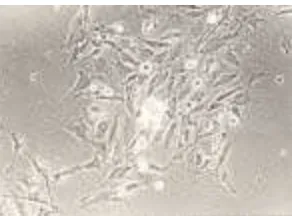
When allowed to age a few days after confluence was at-tained, the cultured cells derived from liposarcoma showed stellate to elongated shaped cells containing many vacuoles considered to be intracytoplasmic lipid droplets. These cells had round nuclei, a finely granular chromatin pattern with
a
c d
b
Figure 1. Phase-contrast microscopy observation of the cultured cells de-rived from canine mammary gland tumors. a. Spindle shape cells, b. Large elongated spindle, c. Polygonal shape cells with pave-ment-like colony, d. The cultured cells derived from solid carci-noma showing monotonous appearance of the colony x200.
Figure 2. The cultured cell derived from mammary gland tumor embedded within the collagen gel matrix show three dimensional colonies and duct-like structure. Phase-contrast x300.
Table 2. Morphology of the cultured cells
Animals Tumor type Cell morphology
Small round to oval Small and large round to oval Round to oval and some elongated Round to oval with some elongated spindle
Small and large round to oval Small round to oval Oval to elongated Large elongated and round floating
Oval to elongated
Round to oval with some marked keratinised
Small and large round to oval Elongated to spindle
Small and large round to oval with some spindle
Elongated to spindle with dark cytoplasmic granules
Attached cell with large and long cytoplasmic processes and the other was small to medium floating cells
Small and large round to oval Elongated to spindle
Round to oval with some marked keratinised
Round to oval with some marked keratinised
peripheral margination, microfilaments and one or more prominent nucleoli.
There were two types of cells derived from pleural effu-sion of mesothelioma, one was attached cell with large and long cytoplasmic processes and the other was small to me-dium floating cell (Figure 4). The number of the floating cells was dominant when they let to age; and they weakly con-nected with the attached cells.
In the cells derived from basal cell tumors, the typical pattern of cells in monolayer was round to oval surrounded by spindle to elongated cells (Figure 5a). Judge from the pat-tern of cell growth, they can be classified into 3 categories: (i) the migratory zone in the periphery of the organoids, in which the cell density was high, clearly spindle-shaped and was ar-ranged in more or less distinct bands radiating from the organoids, (ii) single cell, which occurred throughout the cul-tures and showed distinct polymorphism, from spindle shape or an irregular shape to rounded or polygonal shape with dis-tinct cell borders and no processes, and (iii) cell clumps or organoids, which varied considerably in size and density and occurred throughout the cultures. Migration of cells from the edge of some organoids was seen. The culture morphology somehow reflects the histological pattern of the original tu-mor tissue (Figure 5b), based on some similarity pattern among cell colonies of the tumor tissue in paraffin sections which showed epithelial cells surrounded by spindle shape cells con-sidered as fibroblast.
DISCUSSION
The common features of growth pattern for cell culture derived from mammary tumors were small and large round to oval shaped cells in the center, and surrounded by elon-gated to spindle shaped cells. These findings were also previ-ously reported (Tateyama et al. 1990b). The arrangement of this growth pattern seemed to somehow reflect the in vivo texture of the mammary tissue. It is well known that myoepi-thelial cells are situated in the basement area and the glandu-lar epithelial cells in the organoids (Namba et al. 1982). The cell populations that grow from organoids may contain glan-dular epithelial and myoepithelial cells (McGrath & Soule 1983, Tateyama et al. 1990a), therefore the polygonal cells might be glandular epithelial cells, while the large elongated to spindle-shaped cells are considered as myoepithelial cells (Tateyama et al. 1990ab, Priosoeryanto et al. 1995b). Obser-vation of fibroblast or myofibroblast is also necessary since they are also present in the canine mammary carcinoma, and plays some significant roles (Hayden et al. 1986, Tateyama
et al. 1988).
The differences in appearance of the mammary gland cul-tured cells were depended on the tumor type, these might be due to the in vivo condition. In the mixed tumors type, they accompanied by other cell types originate from their parental organ (in this case alveolar epithelial and myoepithelial cells), while in malignant tumors type the cells can proliferate by
themselves (Tateyama et al. 1990b). In a previous paper
(Kawano et al. 1988) the pure epithelial cell obtained from milk could not proliferate when embedded in gel, due to the absence of fibroblast which is suggested to play an important role in the proliferation and differentiation of epithelial cells (Tateyama et al. 1990b).
There is an indication that neoplastic mammary cells also grow better in collagen gel matrix (Kano-Sueko 1983), there-fore, at least certain mammary carcinomas seem to favor the collagen gel matrix for their growth (Tateyama et al. 1990ab, Priosoeryanto et al. 1995b). The collagen culture may offer advantages as substitute of in vivo in observing cell morphol-ogy whereas plate culture for continuous subculture (Tateyama et al. 1990b).
Figure 3. The cultured cells derived from malignant melanoma revealed an elongated to spindle and round to oval shape with dark granules on their cytoplasm. Phase-contrast, x200.
a b
Figure 4. Morphology of the cultured cell derived from mesothelioma. a. At primary culture the cells displayed attached spindle with long cytoplasmic processes shape cells and floating cells, b. At the second passage the floating cells with small to medium in size were predominant. Phase-contrast x150.
a b
The cell origin of the CTVT is not precisely known (Nielsen & Kennedy 1990). There are some arguments that CTVT is originated from several cells, such as lymphocytes, histiocytes, reticular cells and mature end cells of the reticu-loendothelial series (Kaalund-Jorgensen & Thomsen 1937, Mulligan 1949, Bloom et al. 1951). The present tissue cul-ture study also expressed several cell morphologies. It has been reported that when this tumor is grown in cell culture, the cell population changes from a mixture of tumor cells and a few fibroblast to a culture of pure fibroblast; the tumor cells die out more rapidly than their ability to divide, and fibro-blast take over (Bloom et al. 1951, Yang et al. 1973).
Histopathologically, melanomas in domestic animals can be classified into epitheloid, spindle and mixed cell types. Our present tissue culture study of canine melanoma tumor cells expressed two types of cells. The criteria used to distin-guish between spindle and epitheloid cells are largely those used for the characterization of benign and malignant mela-noma cells (Diters et al. 1983). The appearance of the present cultured cells, suggest that the elongated to spindle cells is identical to the spindle cell classified by Pulley and Stannard (1990), while the round to oval cells are similar to that of epitheloid cells.
One major problem in the studies of the origin of primary culture of epithelial cells derived from tumor tissue has been the identification of the epithelial cells. Two approaches in addition to the use of precisely light microscopic criteria have been reported, as the demonstration of desmosomes (Russo
et al. 1975) and specific epithelial surface antigen or
interme-diate filaments (Easty et al. 1980, Priosoeryanto et al. 1992). In the present study of the cultured cells derived from basal cell tumor we use both of these criteria. The cell clumps were identified as epithelial cells, while the cells in the periphery of the cell clumps and the single cells were identified as fi-broblast.
Three of the cultured cells were able to keep grown and maintained for long period; two of them were developed and established as cell line, while the rest is still in process for establishing as another new cell line. The present study show that the tissue culture system maybe useful in the precise un-derstanding of the cell morphology, proliferation and differ-entiation of some tumors of domestic animals.
ACKNOWLWDGEMENTS
The authors wish to thanks the Directorate General of Higher Education, Ministry of National Education for fund-ing this research through the Competitive Grant VI (Hibah Bersaing) project 1997-2000 and also to the Department of Veterinary Pathology, Faculty of Agriculture, Miyazaki Uni-versity-Japan.
REFERENCES
Bloom F, Paff GH, Noback CR. 1951. The transmissible venereal tumor of the dog: Studies indicating that the tumor cells are mature end cells of reticuloendothelial origin. Amer J Path 27:119-140.
Broder S. 1991. Molecular Foundations of Oncology. Baltimore: Willian & Wilkins.
Destexhe E, Lespagnerd L, Degeyter M, Heymann R, Coignoul F. 1993. Immunohistochemical identification of myoepithelial, epithelial, and connective tissue cells in canine mammary tumors. Vet Pathol 30:146-154.
Diters RW, Dubielzig RR, Aguirre GD, Acland GM. 1983. Primary ocular melanoma in dogs. Vet Pathol 20:379-395.
Easty GC, Easty DM, Monaghan P, Ormerod MG, Neuville AM. 1980. Preparation and identification of human breast epithelial cells in cul-ture. Int J Cancer 26:577-584.
Freshney RI. 1992. Animal Cell Culture A Practical Approach. Ed ke-2. Oxford: IRL Pr.
Goldschmidt MH, Shoter FS. 1992. Skin Tumors of the Dog and Cat. Ox-ford: Pergamon Pr.
Hayden DW, Ghorial HK, Johnson KH, Buoen LC. 1986. Feline mammary sarcoma composed of cells resembling myofibroblast. Vet Pathol
23:118-124.
Hiratsuka M, Senoo T, Kimoto T, Namba M. 1981. [Cultivation of human mammary epithelial cells] [dalam bahasa Jepang]. Soshikobaiyou 7:431-437.
Jones TC, Hunt RD, King NW. 1997. Veterinary Pathology. Ed ke-6. Bal-timore: William & Wilkins.
Kaalund-Jorgensen O, Thomsen AS. 1937. Det overfor bare veneriske sarkom hos hunde. Maanedsskr. Dyrlaeger 48:561-578.
Kano-Sueko T. 1983. Factors affecting mammary cells in culture. Biochemi-cal Actions of Hormones. Vol X. Academic Pr, Inc. hlm 163-185. Kawano A, Tateyama S, Yamaguchi R, Nosaka D, Kondo F. 1988.
Mor-phology of goat milk-derived mammary epithelial cells cultured in col-lagen gel. Jpn J Vet Sci 50:1252-1258.
Lovell-Badge RH. 1987. Introduction of DNA into embryonic stem cells. Di dalam: Robertson EJ (ed). Teratocarcinomas and Embryonic Stem Cells. A practical approach. Oxford: IRL Pr. hlm 153-182.
Madewell BR, Theillen GH. 1987. Tumors of the mammary gland. Di dalam: Theillen GH, Madewell BR (ed). Veterinary Cancer Medicine. Ed ke-2. Philadelphia: Lea and Febiger. hlm 372-344.
McGrath CM, Soule HD. 1983. Renewal inhibition of human mammary cell growth in vitro: Cortisol and the recruitment of cells to terminal differentiation. J Cell Physiol 116:385-396.
Moulton JE. 1990. Tumors in Domestic Animals. Ed ke-3. Berkeley: Uni-versity of California Pr.
Mulligan RM. 1949. Neoplasma of the Dog. Baltimore. William & Wilkins. Namba M, Hyodo F, Hiratsuka M, Senoo T, Kimoto T. 1982. [Ultrastruc-ture of cul[Ultrastruc-tured human mammary cells] [dalam bahasa Jepang]. Saibou
14:62-67.
Nielsen SW, Kennedy PC. 1990. Tumors of the genital system. Di dalam: Moulton JE (ed). Tumors in Domestic Animals. Ed ke 3. Berkeley: Uni-versity of California Pr. hlm 479-517.
Paul J. 1961. Cell and Tissue Culture. Ed ke-2. Edinburgh: E & S Livingstone Ltd.
Priosoeryanto BP, Huminto H, Wibawan IWT, Bahagia S, Estuningsih S, Harlina E, Ratih D, Tateyama S, Yamaguchi R, Uchida K. 1997. In vitro and ultrastructural studies of canine tumors. Di dalam: Proceed-ing of the first ASEAN Microscopy Conference. Johor-Bahru, Malay-sia, 27-30 Nov 1997. hlm 57-59.
Priosoeryanto BP, Tateyama S, Yamaguchi R, Uchida K. 1995a. Antiproliferation and colony-forming inhibition activities of recombi-nant feline interferon (rFeIFN) on various cells in vitro. Canadian J Vet Res 59:67-69.
Priosoeryanto BP, Tateyama S, Yamaguchi R, Uchida K. 1995b. Establish-ment of a cell line (MCM-B2) derived from a benign mixed tumor of canine mammary gland. Res Vet Sci 58:272-276.
Priosoeryanto BP, Tateyama S, Yamaguchi R, Uchida K, Ogawa H, Nakai M. 1995c. A cell line (MCA-B1) derived from a canine oral acanthomatous epulis. Res Vet Sci 58:101-103.
Pulley LT, Stannard AA. 1990. Tumor of the skin and soft tissue. Di dalam: Moulton JE (ed). Tumors in Domestic Animals. Ed ke-3. Berkeley: Uni-versity of California Pr. hlm 23-87.
Russo J, Furmanski P, Rich MA. 1975. An ultrastructural study of normal human mammary epithelial cells in culture. Am J Anat 142:221-231. Sari DK, Priosoeryanto BP, Yamaguchi R, Ratih D, Tateyama S, Uchida K.
1997. Ultrastructural and pathological studies of transplantable canine lung carcinoma cell in Severe Combined Immunodeficiency (SCID) mice. Di dalam: Proceeding of the first ASEAN Microscopy Confer-ence. Johor-Bahru, Malaysia, 27-30 Nov 1997. hlm 62-63.
Tannock IF, Hill RP. 1998. The Basic Science of Oncology. Ed Ke-3. Singapore: McGraw-Hill International. Health Profession Division. Tateyama S, Furukawa H, Yamaguchi R, Nosaka D, Kondo F. 1990a. Plate
and collagen-gel cultures of normal canine mammary epithelial cells.
Res Vet Sci 49:14-19.
Tateyama S, Furukawa H, Yamaguchi R, Nosaka D, Otsuka H, Sasaki N. 1990b. Plate and collagen gel culture of neoplastic canine mammary epithelial cells. Di dalam: Proceeding of the 7th Congress of Federa-tion of Asian Veterinary AssociaFedera-tion (FAVA). Pataya, Thailand. hlm 622-634.
Tateyama S, Priosoeryanto BP, Yamaguchi R, Uchida K, Ogiwara K, Tsuchiya A. 1995. In vitro growth inhibition activities of recombinant feline interferon on all lines derived from canine tumours. Res Vet Sci
59:275-277.
Tateyama S, Shibata I,Yamaguchi R, Nosaka D, Ashizawa H. 1988. Par-ticipation of myofibroblast in feline mammary carcinoma. Jpn J Vet Sci 50:1048-1054.