Endovascular treatment with transvenous approach for
embolization of carotid cavernous istula
Abstrak
Tata laksana endovaskular terhadap istula kavernosa karotid bertujuan menutup robekan pada bagian sinus kavernosa. Hal ini dapat dilakukan baik dengan pendekatan trans-venosa atau pendekatan trans-arteri. Pilihan ini tergantung pada jenis istula dan arsitektur pembuluh darah. Kami mendeksripsikan dua pendekatan trans-venosa yang berbeda difokuskan pada pertimbangan anatomi dan aspek teknis.
Keywords: carotid-cavernous istula, coiling embolization, transvenous
pISSN: 0853-1773 • eISSN: 2252-8083 • http://dx.doi.org/10.13181/mji.v23i3.728 • Med J Indones. 2014;23:169-73
Correspondence author: Benny Young, [email protected] C a s e R e p o r t
Copyright @ 2014 Authors. This is an open access article distributed under the terms of the Creative Commons Attribution-NonCommercial-ShareAlike 4.0 International License (http://creativecommons.org/licenses/by-nc-sa/4.0/), which permits unrestricted non-commercial use, distribution, and reproduction in
any medium, provided the original author and source are properly cited.
Benny Young,Agusmanan Bojeng
Department of Radiology, University Malaysia Sarawak, Faculty of Medicine and Health Sciences, UNIMAS, Hospital Umum Sarawak, Malaysia
Generally, there are two types of carotid cavernous istula (CCF), namely direct and indirect type. Direct type is inherently resulted from trauma. Indirect istula occurs spontaneously is adural communication at cavernous sinus with meningeal branches either from internal or external carotid arteries, or both. When CCF harbored for treatment, endovascular is the mainstay option for its high eficacy and low complication risk as compared to surgery.1
The aim of endovascular technique is to seal the tear site at cavernous part. It can be achieved by catheterization into cavernous sinus (CS) to allow placement of various embolization materials. CS cannulation through transvenous route has beneit of much safer than transarterial approach. It is the preferred method in indirect type CCF.2 In the
direct type, transvenous can serve a complementary
breakthrough if trial balloon occlusion into the CS via transarterial route fail to completely obliterate the istula.1,3
We would like to describe our experience in this noble transvenous method weighting in CCF angioarchitectural background and technical hindrance that prompt us to perform transvenous technique.
CASE REPORT
Direct CCF case
A 26 year-old lady presented with right eye’s
conjunctiva chemosis, proptosis and visual
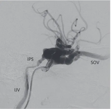
disturbance following motor-vehicle head trauma two weeks prior. Due to the nature of her eye’s problems associated with trauma, a direct type CCF was suspected. A diagnostic cerebral angiogram was immediately done which conirmed a direct type of right CCF with fast low shunt (Figure 1). A decision of endovascular treatment was taken after a board of multidisciplinary discussion from neurosurgical, eye and radiology teams. The irst
Abstract
attempt was catheterization of the affected CS from right internal carotid artery (ICA) route. Balloon-microcatheter assembly managed to reach the CS by the rent site. Trial of balloon inlation showed incomplete closing of the istula due to suboptimal conform of balloon over the tear area. We decided to abandon this transarterial way and proceeded with transvenous catheterization via inferior petrosal sinus (IPS) which clearly demonstrated angiographically. The right internal jugular vein (IJV) was approached using a 6 F guiding catheter (Figure 2). Coaxial inserted microcatheter success to catheterize the targeted CS via IPS. At this point, we were certain to pack the cavernous area with microcoils to promote complete closure of CCF. Following coils embolization, a right internal carotid artery (ICA) arteriogram showed disappearance of the CCF (Figure 3).
Indirect CCF case
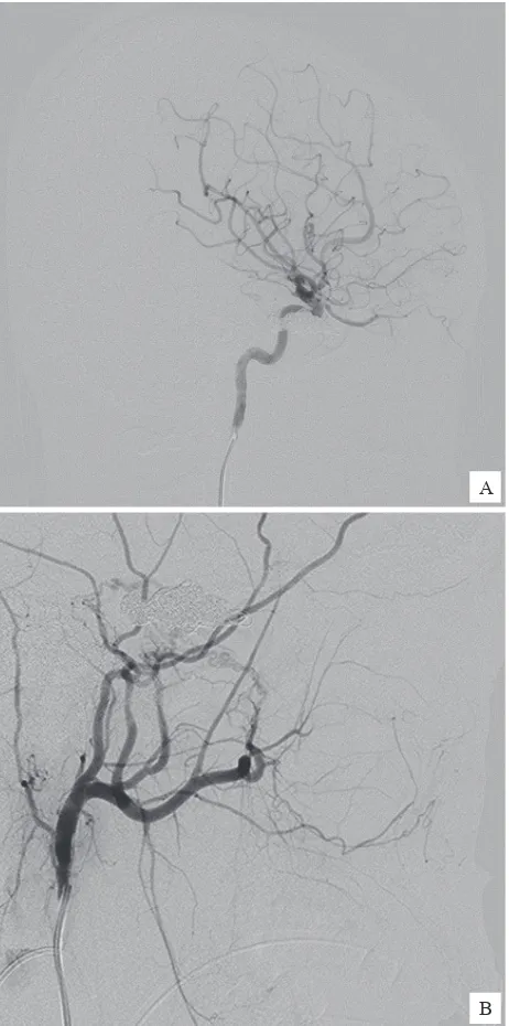
A 28-year old female presented to eye clinic with spontaneous proptosis and chemosis of the right eye for the past 3 months. There was no associated visual impairment of note. Eye tonometry revealed signiicant increased right eye’s pressure. Diagnostic angiography showed indirect right CCF, supplied by meningeal branches of both right ICA and external carotid arteries (ECA) (Figure 4a and 4b). A decision to undertake
Figure 1. Direct type of right carotid cavernous istula
demonstrated with retrograde low into superior ophthalmic vein (SOV) and inferior petrosal sinus (IPS) that eventually drain into internal jugular vein (IJV)
Figure 2. The posterior venous tributary of cavernous sinus (CS)
was approached from internal jugular vein (IJV) using guiding catheter. Later, coaxial inserted microcatheter navigated into CS through inferior petrosal sinus (IPS)
Figure 3. Complete closing of the istula after coil packing seen
from check right ICA arteriogram
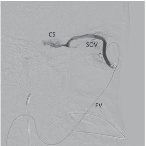
catheter (Figure 5) used to navigate the facial vein drained into IJV. Laborious catheterization was required due to intermittent venous spasm, angulation and narrowing of this pertinent vein particularly on the turningpart at the orbital rim where it continued into SOV. After successfully
advancing the microcatheter into CS via the SOV,
contrast injection done for conirmation (Figure 5). Subsequently, this right CS was packed with electrically detachable coils to cease the istula’s shunt. Final check angiogram of right internal (Figure 6a) and external arteries (Figure 6b) showed complete obliteration of the indirect right CCF after coiling embolization.
Figure 4. A) Arteriogram of right internal carotid artery showed
a low low of indirect carotid cavernous istula; B) The CCF received supplies from dural branches of internal maxillary artery displayed from right external carotid arteriogram
DISCUSSION
Clinical presentation of CCF primarily involves eye symptoms, ranging from proptosis, chemosis,
diplopia to reduced eye vision. Spontaneous
obliteration of CCF is hardly occurred for that most of CCF requires endovascular treatment. Rapid deterioration of vision acuity is the main indication for treatment in most of clinical ground practices. Other indication for immediate treatment includes cortical venous relux due to its high risk for cerebral venous hypertension and intra cranial bleed.1-3
In direct CCF, our treatment approach is a stepwise procedure, with transarterial route as the irst attempt. Technically, navigation of microcatheter into cavernous sinus’ istula area through internal carotid artery is always feasible.1 The hindrance
is to achieve accurate placement of occlusion balloon over the tear’s site for complete istula’s obliteration. If this trial is not successful, then we proceed with transvenous catheterization into CS for coils embolization. Our aim is to coil the affected compartment of CS that ultimately seal the
tear site.
In indirect type, transvenous technique is our preferential technique especially in istula that fed by internal carotid meningeal branches. It is
Figure 5. Long route of transvenous catheterization into
cavernous sinus (CS) starting from facial vein (FV) with an acute turning point before reaching superior ophthalmic vein (SOV) A
Figure 6. Post coiling embolization, angiography of right ICA
(A) and ECA (B) showed a complete obliteration of istula and its retrograde venous low.
because embolization of meningeal branch ICA carries a risk of stroke. Compounding into it is the small size, tortuosity and distal site of meningeal branches that preclude a safe and close distance catheterization into the istula site.3,4
The en-route option for transvenous is based on the angiographic presence of the venous drainage. Our experience is limited on SOV via transfacial vein and IPS via IJV. The option of either of these two
venous route depends on the angioarchictectural
of them which we analyze before interventional procedure. A non-opaciied inferior petrosal sinus
circumvents us for taking it as venous access
although IPS is the shortest and straightest route in reaching CS area.
In contrary, catheterization of SOV via facial vein requires tedious effort due to its long route with narrowing and tortuous course as it enters the orbital rim. The other potential technical dificulty is the numerous side branches of facial vein, small caliber and its intermittent vasospasm. Direct SOV catheterization can be done after surgical exposure of the superior eye lid. However, direct SOV approach requires vast experience with ine and lexible endovascular platform to ensure low risk of
ophthalmic vein injury. Worsening proptosis due to
retroorbital hematoma could be occurred in the event of ophthalmic vein injury.4,5
Success rate of transvenous approach was reported to be signiicant high, ranging from 76% to 86.5%.
Yu, et al6,7 suggested a stratiication of venous
routes based on angioarchitectural of the venous pathway from the shunt’s low. This venogram stratiication useful for pre-embolization planning and reported to increase the success rate of endovascular treatment as well as reducing
procedure times and radiation dose to operators and patients.
In conclusion, transvenous approach is useful and safe for both type, direct and indirect CCF. Assessing venous pathway of the istula prior to embolization has beneits of increasing the success of embolization. Facial vein catheterization serves an effective alternative route over IPS if the latter
vein is not completely seen.
REFERENCES
1. Moris PP. Carotid cavernous istulas. In:Moriss PP, editors. Interventional and endovascular therapy of the nervous system: A practical guide. New York: Springer-Verlag; 2002. p. 177-90.
2. Yoshida K, Melake M, Oishi H, Yamamoto M, Arai H. Transvenous embolization of dural carotid cavernous istulas: a series of 44 consecutive patients. AJNR Am J Neuroradiol. 2010;31(4):651-5.
3. Kirsch M, Henkes H, Liebig T, Weber W, Esser J, Golik S, et al. Endovascular management of dural carotid cavernous sinus istulas in 141 patients. Neuroradiology. 2006;48(7): 486-90.
4. Kurata A, Suzuki S, Iwamoto K, Miyazaki T, Inukai M, Abe K, et al. Direct-puncture approach to the extraconal portion of the superior ophthalmic vein for carotid cavernous istulae. Neuroradiology. 2009;51(11):755-9.
A
5. White JB, Layton KF, Evans AJ, Tong FC, Jensen ME, Kallmes DF, et al. Transorbital puncture for the treatment of cavernous sinus dural arteriovenous istulas. AJNR Am J Neuroradiol. 2007;28(7):1415-7.
6. Yu SC, Cheng KM, Tam PH, Wong GK, Chan CM, Cheung YL, et al. A venographic operational classiication for
transvenous embolization of dural carotid cavernous istula. Neuroradiology. 2011;53(12):993-9.