t42
Tanurahadja et al. Med J IndonesExpression
of
HBsAg
and
HBcAg
in Liver
Biopsy
Specimens
of
Chronic
B
Ilepatitis
Patients and its Relation
with Ilistology Activity
Index
Budiana Tanurahardja,
Esti Soetrisno
B,
Sutjahjo Endardjo,
GunawanTjahjadi, Wirasmi Marwoto
Abstrak
Menpelaiari hubungan antara Indeks Aktivins Histologik (IAH) dengan ekspresi HBsAg dan HBcAg. Diteliti 3O sediaan biopsi hati penderita hepatitis B
kronik
Ekspresi HBsAg dan HBcAg ditentukan berdosarl<nn pulasan imunoperol<sidase yang dilakakan pada senwa sediaan yang positif pada pulasan Victoria BIue (VB), sedangkan IAH ditentukan berdasarkan metode Knodell. Senua sediaan (29 buah) menunjukl<an HBsÀg dan HBcAg positif. Pola sebaran HBsAg yang 'regular unifornt' terdapat pada 3 sediaan dan 'irregular dan non-unifunn' pada 26 sediaan. Pola sebaran HBcAg yang 'regular-uniforn' terdapat pada 4 sediaan, sedangkanpola
'irregular non-unifonn' terdnpat pada 25 sediann . HBsAgl+
pada senua sediaan denganIAH:
l-18. HBcAg pada sitoplasma/nentbran sel 1+ pada 2O sediaan, 2+ pada 3 sediaan, dan 3+ pada 6 sediaan. Nilai IAH pada nasing-masing kelompok ini tidak berbeda bernaknrt HBcAg pada inti sell+
pada 14 sediaan, dan()
pada 15 sediaan. Nilai IAH padamasing-masing kelompokini tidakberbedabermakna. Nilai IAH pada kelompok dengan pola sebaran HBsAg dan HBcAg yang 'regular-uniform' tidak berbeda bermakna dengan kelonpok dengan pola sebaranyang 'irregular non-uniform'. Ekspresi HBsAg dan HBcAg pada sediaan biopsi hati penderita hepatitis B kronik pada penelitian ini tidak berhubungan dcngan aktivitas nekroinflamasi.Abstract
?o assess the relationship between Histology Activitiy Index (HAI) and the expression of HBsAg and
HBcAg.
Thirtyliver
biopsy specintens ofchronic B hepatitis patients have been exanined Expression of HBsAg and HBcAgwere determined by imtnunoperoxidase staining. HAI was calculated accordingto
Knodell's ntethod.All
of the specimens (29) were positivefor
HBsAg and HBcAg staining. Regular-unifonn disÛibution patterns of HBsAg were found in 3 specinens and irregular non-uniforn patterns in 26 specinens . Regular-unifonn panerns of HBcAg were found in 4 specinens and the irregular non-uniforn patterns in 25 specimens. HBsAgs I + were seen in all specinens which have HAI range ofI
to
18. Cytoplastniy'nenbraneous HBcAgs were seen 1+ in 20 specitnens,2+
itt 3 specitnens, andi+
in 6 specinens. HAI of each group which showed HBcAg was not significantlydfurent.
HBcAgs in the nucleiof
Iiver cells were seen I + in 14 specinens, and 15 specinrens were negotive- HAI of each of these groups showed no significant dffirences.HAI of
regular-uniform distribution pattern groupsof
HBsAg and HBcAg werenot
significantly differentfrom HAI
of irregular non-uniform groups.: Expression of HBsAg and HBcAg in liver biopsy specinens.of chronic B hepatitis patients are not correlated with ne c r oinflantmatory ac tivity.Key words:
Expression of HBsAg and HBcAg - necroinJlamntatory activity-
histology activity index.Viral
hepatitis
B
infection
is
one
of
the
major world
health problems. There
areabout three hundred
mil-lions
carriers
of HBV in the world,
andseventy-eight
Departnent of
Anatomic Pathology,Faculty
of
Medicine Universityof Indonesia/Dr. Cipto
Mangunkusunto National Central General Hospital, Jakarta, Indonesiapercents are found in
Asia.l
According to the World
Health Organization
(WHO),
Indonesia is
anendemic
country,
with
moderate
(2%-7%)
to
high
(7%-2O%) prevalence of Bhepatitis.
The incidenc.eof
HBV
infec-tion was
varied
in each
region.
Vol 5, No 3, July - September 1996
improvement
more thansix
months. Theliver cirrhosis
isdefined
as adiffuse
process characterized byfibrosis
andconversion
from
normal architecture
of liver
cells
toabnormal structure
of
noduli.2,3'a'sDue
to
the rapid progression
of
medical
sciences andtechnology especially
the discovery
of
new
hepatitis
viruses
which
showed characteristic features
of
liver
changes aswell
as newtherapeutical modulations
suchgives more detailed information about
necroinflam-matory
activity,
although
several authorscritisized
theon
th
stagingof
6'8'9'r
to the
new
ion,
f
chronic
Necroinflammatory activity
is
often
associatedwith
the
expression
of
the antigen,
thus
give information
about the exi
Suzuki
etall3
in
Chronic
Pelel with
viral
Hepatitis
(CAH).
Suzuki
evaluated and described theHBcAg intracellularly,
in-rly . It
is quitepractical
thatin
this
study.as a parameter
of
necroinflammatory
activity.
METHODS
archives
of Department
ty of Medicine,
Univer-angunkusumo National
Central
GeneralHospital,
Jakartareceived
in
1992.These materials
were liver
biopsy
or/
needle biopsy
patients
which
were
positiv
HBsAg - HBcAg - HÀI in Liver Biopsy 143
staining.
rrtr/ehad
116specimens
of
chronic
hepatitis
and
liver
cirrhosis, 43
of
them were positive
to
Vg
staining.
The number of theparaffin blocks which
werestill
available
andcould
berecut were
30.stainings (Hematoxylin
&
reticulin),
histopathological
Knodell's
HAI
criteria
wasapplied
on
each
slide. Diagnosis was
based
on
themodified International
Group Classification
of
Chronic
Liver
Disease 1977.ta'ts
1.
Chronic
PersistentHepatitis
(CpH)
2.Chronic
Active Hepatitis (CAH
):
a.
with
moderateactivity (IIa)
b.with
severeactivity (IIb)
3.Liver
cirrhosis
(LC)
:a.
CAH IIb with
early
cinhosis
b.
liver
cirrhosis
Knodell's
HAI
was determined according
to four
components:I.
Periportal
necrosis, score O-
10.II.
Focal necrosis andintralobular
degeneration, score0-4.
III.
Portal
inflammation,
score0 -
4.IV.
Fibrosis,
score 0-
4.The sum
of
the scorefrom
eachcomponent
was takenas
HAI
ranging
fromÛ to22.
Immunoperoxidase stainings were made
on
eachspecimen.
The
Peroxidase Anti-peroxidase (pAp)
method was used
for
HBsAg
and the
Avidin-Biotin
After
staining
by
theimmunoperoxidase method
each specimen was evaluatedby
themethod
of
Suzuki:
-
intracellular
:
intranucleus, intracytoplasmic
and membraneous.-
intralobular/intranodular :regular,irregular
-
interlobular
:uniform; non-uniform.
The
number
of positive liver
cells in
each specimen isclassified
semiquantitatively :
l+
= l-
25%psitive
2+
=26-
50%positive
3+
=51
-
25%positive
144
Tanurahardja et aI.The quality
of
immunoperoxidase
staining
was
con-trolled by providing
the
slides for positive
andnega-tive
control.
Microscopic examination was done using
Olympus
binocular light
microscope.
Statistical analysis:
Non-parametric statistics were
used asappropriate
: - X2 test-
Kruskal-Wallis
test-
Wilcoxon
rank
sum test-
Spearman'srank correlation
testRESULTS
The
ageofpatients
rangedfrom
21 yearsto
59 years. Sex ratio( male to female
)
was4 : 1.
(Table
I
and2)
Table 1. Age distribution of the cases
Age(yr)
CPH CAHIIa CAHIIb
CAH+EC
LC
TotalMed J Indones
of
HBsAg as
well
as
that
of
HBcAg
and
the
his-topathological diagnosis.(Table
3)Table
4
shows
a significant conelation
(p <
0,001)
between
Knodell's
HAI
and histopathological
diag-nosis.
Table
3.
HBsAgand
HBcAg distribution pattemsof liver
biopsy specimensDiagnosis Reg,uni
h,nonHBsAg
HBcAg
HBsAg HBcAg HBsAg TotalHBcAg2t567
03t411t4
10788
CPH CAH LC 8l4
8 7 t4 8 29Table
4.
Association Between Knodell'sHAI
and histopatho-logical diagnosisDiagnosis Total
SD 20-29 30-39 4049 50-59 not mentioned 4 2
I
I
0 0 1 0 2 2 1 1006
228
025
103
8 8 2 t2 3 5 2I
2 0I
6I
10 3 4 CPH CAH LC 8 t4 81,3,3,3,4,4,5,7
3,75
!,77,to,Lo,tt,t2,I2,t2,
12,07
2,2 12,12,12,L4,14, 15,16 .11,t2,13,13,L4,15,16,19
14
2,2Table 2. Sex distribution of the cases
Diagnosis Male Total
CPH CAH IIa
CAHIIb
CAH+EC LC
24(80%) 6(2o%) 30
Immunoperoxidase
staining
for
HBsAg
and
HBcAg
werepositive
in29
cases. One case had anonspecific
reaction and was dropped out
from
this study.Distribu-tion
patternsof
HBsAg
andHBcAg
were evaluatedby
Suzuki's
method and compared
with
standard
histo-pathological
diagnosis.CAH
IIa
(2 specimens) and
CAH
IIb
(12
specimens) were grouped as CAH group.CAH
with
earlycirrhosis
(3 specimens) were
classified
asLC
group. There is nosignificant
association between thedistribution pattem
If the
fourth
component ofKnodell's HAI
(i.e.fibrosis)
is removed, asignificant relationship
betweenHAI
andhistopathological
diagnosis isstill
observed.(Table
5)Table
5.
Association between HAI without the fourth com-ponent (fibrosis) and histopathological diagnosisHAI-IV
Diagnosis Total
1,2,3,3,3,3,5,7
3,377,7,7,9,9,9,9,9,9,
9,3l0,l
1,1 1,1 1,137,9,9,9,IO,11,13,L4
LO,25In order to know whether
necrosis wasalways
accom-panied
by
inflammation
in
the
histopathological
specimensof
chronic
B
hepatitis
patients;
we
corre-lated the
first
(periportal necrosis
)
and second(focal
necrosis) component
of
HAI
to the third
one (portal
inflammation).
A
significant
correlation
(15=0,64,p<0,001)
was observed.(Figure 1)
SD
x
CPH
CAH 1,8t,7
[image:3.595.313.548.115.462.2] [image:3.595.50.287.315.591.2]Vol 5, No 3, July - September 1996
portal inflammation
HBsAg - HBcAg - HAI in Liver
Biopsy
I45
5
4
3
2
o
oooo
o oooooo
ooo
ooo
o
oo
Three
specimensshowed
regular-uniform
patterns
of
HBsAg
expressionwith Histology Activity
Index of
3,(table
7).Table
7.
HÀI
of the regular-uniform pattem and irregular non uniform pattern of HBsAg AND HBcAg expression5o78ntT*.*,,
Figure 1. Scattered diagram of necrotic comporcnt and portal
inflam-mation component of HAI.
All
specimens
(29)
were
positive
l+
for
HBsAg
ex-pressionin
thecytoplasm of
theliver
cells, whereasfor
HBcAg
expression, 29 specimens werepositive in
thecytoplasm
(
2Ospecimens
l+,
3
specimens2+
and 6specimens
3+
;
and
14 specimensin
the nuclei.
For
HBcAg
in
the nuclei
of
liver cells 14
specimensshowed
1+, the others werenegative.(Table
6)Tabel
6. HAI
of the groups of positive HBcAg expression in cytoplasm/cell membrane andin
the nucleiof
liver cellsHBcAg express.
HAI
TotalI 5
J5,l4,l
4,t3,12,12,t2,tt,t0,7,4 4,3,3cc
15,15,14,1 4,13,13,12,12,12,12,1l,
LO,1,7,5,4,4,3,3,11 8, 16, 16,14, 13,12,12,r2,12,L1,L0, 7,5,r
I 6,16,1 1
cc
18,t4,t2,12,t2,10Note: CC = cytoplasm/ cell membrane
N
= nucleiThere
were
no association betweenHAI
and the num_ berof
HBcAg
positive cells in cytoplasm
(p>0,05
) aswell
asHBcAg
positive
in
thenuclei.
1,3,4,4,7,7,1o,1o,L l,l!,12,12,12,12
HBsAg
L2,12,12,13,L3,1 4,L4,14,15,t5,16.t6
26HBcAg
1,3,3,4,4,5,7,1O,1O,L1,1I,I2,12,12, L2,12,L3,13,14,14,15,15,16,16,1g 25 PatternHBsAg
3,5,18 REG - IJNIHBcAg
7,12,L2,L4IR-NON
15
tion wirh
HAI.
DISCUSSION
earlier
the onsetof
the disease(in
neonatal/
perinatal
period),
the greater thepossibility
of becoming chronic
and
cirrhosis.
has
been
in
CAH
and
LC, not
similar
to
Suzuki's
20
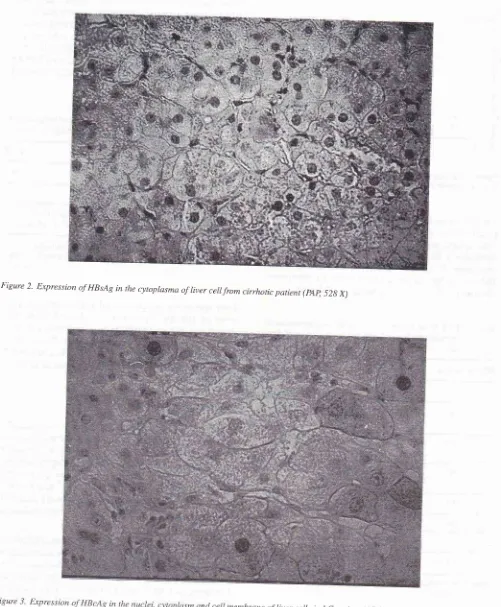
[image:4.595.28.523.79.488.2] [image:4.595.24.265.410.683.2]146 Tanurahnrdja et aL
Figure 2' Expression o.f HBsAg in the cytoprasma of liver ceil
from cirrhotic patient
(pAp
528x)
Med J Indones
Vol 5, No 3, July - Septernber 1996
CPH patients
who
hadseroconversion
topositive
anti-HBe.
TheHBcAg
expressionin
these cases werenega-tive.
It
is not known
whether the
casesin
this
study
have
anti-HBe in their
sera, since theclinical
datawere
incomplete. HBsAg
expression
in CpH in
this study
were
found
in
oneor
two liver
cells
andKupffer
cells,
which
was
similar to
the
solitary
pattern reported by
Akeyama
etal
17in mild
hepatitis. Ten KatË
18founâ
that
same expression
is
frequently seen
in
acute
hepatitis. The
basicpathological
processof CpH
wasdifferent from
that
of CAH
t3.In
CpH
patients
with
positive HBeAg
and
in
the healthy carriers, the
im-munologic
responsewas
inadequateor totally deficit
so that the virus replicate freely. Chu and
Liaw
lecalled
it
ashigh
replicative
immunetolerancephase.ln
CAH
,
the
patient's immunologic
response suppress anddestroy
viral replication irregularly. Irregular
im-munological
response and regenerationof liver
cells,aswell
asintegration of
HBV-DNA in liver
cells, causedHBsAg and
HBcAg
expression patterns
to
becomeirregular-non uniform.
Chu
andLiaw
called
jt
aslow
replicative immune
clearance
phase.
In
CpH
with
seroconversion
to
positive anti-HBe, patient.s
im-munologic
response increased and causeddistortion
of
viral
replication,
so that the patterns becameirregular.
This
phase.i^scalled
non-replicative recidual
integra-tiott
phase.r9Application
of
Knodell's
HAI
on
histopathological
specimens
,
showed
that
HAI
in
the group
of
CpH,
CAH
andLC differed significantly. Knodell,s
results
were that the
HAI
tend
to
be
low
in
mild
chronic
hepatitis (CPH)
,andhigh
in
severechronic
hepatiris
(CAH
andLC).13 In
thii
study
wetried
to compare thehistopathological diagnosis and
HAI
without
the
fourth
component.
The
results showed
that this
wassimilar with
previous results using complete
HAI.
It
meant
that to determine
histological activity
wecould
useeither
3or
4 componentsof HAI.
The key feature to determine piecemeal necrosis or
focal
necrosis was
infiltration of
mononuclearinflammatory
cells
in
that region.
Insteadof
piecemeal necrosis andfocal
necrosis one used
the term:
necroinflammatory
activity
which
wasusually
appliedin
chronic
hepatitis, becausenecrosis was almost always
u""o.purri"d
by
inflammatory
reaction. In this study we tried to correlatethe
first
and second component
(periportal
and focal
necrosis)
of HAI
with
the
third
component
@ortal in_flammation).
The results show apositive correlation.
It
meant
that
in
chronic
B
hepatitis, necrosis
is
almost
always accompanied
by inflammation.
All
specimensstained
by HBsAg
immunoperoxidase
staining
showedpositive
results in thecytoplasm
of theHBsAg - HBcAg - HAI in Liver
Biopsy
147liver
cells,
andnegative
in
thecell
membrane and thenuclei. The expression
of HBsAg
were not correlated
with necroinflammatory
activity
featured
by
HAI
assee
by
semiquantitative
score. Besides,
the
regular-uniform
expressionof
HBsAg in
theliver
tissueswere
obtained
in only 3 cases. The other twenty-six
(26)
casesshowed
irregular-
non
uniform
pattem.
HAI of
the
two
groups showed
no
differences statistically,
which
meant that the
distribution
patterns
of
HBsAg
and the number
of
HBsAg positive liver
cells
werenot
correlated
with
necroinflammatory
activity.
All
specimensstained
by HBcAg
immunoperoxidase
staining
were
positive
in
the
cytoplasm and cell
membrane. Semiquantitatively
thepositive liver
cells
were
1+in
20 cases(69%),2+in3
cases(10%),
and 3+in
6
cases
(21%).
HAI
from
each
groups were
not
statistically different. HBcAg
expression
in
thenuclei
of
liver
cells were found
in
14
cases
(4g%), with
ive
result
of
l+.
There
ence between
HAI
in
theive group. Regular-unifo
HBcAg
expression werefound in
4 cases, theother
25cases
showed
irregular-non uniform pattems.
Therewere
no
significant differences between
these
two
groups
in
their
HAI.
So,
HBcAg
expression
in
liver
tissue
wasnot
correlated
with necroinflammatory
ac-tivity.
The
absenceof
correlation was also found
betweenHBsAg
e
rylatory
activity.
The
same
Chu andLiaw.le
The
work
contrary
results.HBcAg
expression
in
this study was not
correlated
with necroinflammatory activity,
and
this
was
not in
agreement.with the-r^esults
of
many authors such asChu
and
Liaw
tn,Ruy
20,Suzuki et
ul
13,voo
ti
una trr"
others. Other studies are
needed
to clarify
this
dis_crepancy.
We concluded that
HBsAg
expression
in liver
tissueshad no
association
with
necroinflammatory
activity in
the
liver
tissues.The
absenceof
this
association
wasalso found
for
HBcAg. Whether necroinflammatory
activity
also
depend
on
other factors, such as
hostimmune
responses,
ther
studies. The
possibil
hadits role
in
this
procesREFERENCES
L
BianchiL,
nic hepatitis. In: À[acSweenRN
eds.pathologyofthe148
Tanurahardja et aI.2. Millward-Sadler GH. Cirrhosis. In: MacSween RNM,
An-thony PP, Scheuer Pf. eds. Pathology ofthe liver. Edinburg: Churchill Livingstone; 1987 : 342-63.
3. Millward-Sadler GH, Hahn EG, Wright R. Cirrhosis : An appraisal.
In: Wright
R,
Millward-SadlerGH,
AlbertiKGMM,
Kiran
S.
eds.
Liver
and
biliary
disease.Pathophysiology, diagnosis, management. [.ondon: Bail-liere Tindall Saunders ;1985: 821-6O.
4. Sherlock S, Dooley J, eds. Diseases ofthe liver and biliary
system. Oxford
:
Blackwell Scientific Publications, 1993:293- 313,357-369.
5. Anthony PP, Ishak KG, Nayak NC, Poulsen HE, Scheuer Pf, Sobin LH. The morphology of cirrhosis. Recommendations
on dehnition, nomenclature, and classifrcation by a working group sponsored by the World Health Organization. J Clin
Pathol 1978;31:395414
6. Scheuer PJ. Classification of chronic viral hepatitis: a need
for reassessment.
I
Hepatol 19911'13:372-4.7
.
Czaja AJ. Chronic active hepatitis : the challenge for a newnomenclature. Ann Intem Med 1993;l l9:510-7
8. Ludwig J. The nomenclature of chronic active hepatitis : an
obituary. Gastroenterolo gy 1993;lo5z21 4-8,
9. Desmet VJ, Gerber M, Hoofnagle JH, Manns M, Scheuer PI.
Classification
of
cfuonic hepatitis: diagnosis, grading andstaging. Hepatology L99 4;19 :l 5 13-20.
10. Ishak KG. Chronic hepatitis : morphology and nomencla-ture. Modern Pathology 1994;7 :69O-7 13.
tl.
Czaja AJ, CarpenterHA.
Diagnostic standardfor
chronic hepatitis. Gastroenterolo gy 1994;lO6:17 23-27 .12. Knodell RG, Ishak
KG,
BlackWC,
Chen TS, Craig R,Kaplowitz N, et al. Formulation and application of
numeri-cal
scoring systemfor
assesing histological âctivityin
Med J Indones
asymptomatic
chronic active
hepatitis.
Hepatology1981;5:431-5.
13. Suzuki K, Uchida T, Shikata T. Histopathological analysis
of
chronic hepatitisB
virus(HBV) infectionin
relation toHBV replication. Liver 1987 : 260-7 O.
14. International Group. Acute and chronic hepatitis revisited. Lancet 1977;lL:914-9.
15. De Groote
f,
Desmet VJ, Gedigk P, Korb G, Popper H,Poulsen H et al. A classification ofchronic hepatitis. Lancet
1968;1 1:626-8.
16. IIsu HC, Lin YH, Chang MH, Su IJ, Chen DS. Pathology
of
chronic hepatitis B virus infection in children: with special refetence to the intrahepatic expression of hepatitis B virus
antigens. Hepatology 1988;8 :378-82.
17. Akeyama T, Kamada T, Koyama
M,
Abe H. Distributionpatterns of hepatitis B antigen in the liver. Arch Pathol 1974;
98:252-6.
18. Ten Kate FfW. Hepatitis B.
A
light microscopical andim-muno histochemical
study
(
Dissertation). Rotterdam, Netherland University of Rotterdam, 1989.28 lp19. Chu CH, Liaw YF . Intrahepatic distribution of hepatitis B sutface and core antigens in chronic hepatitis B virus infec-tion. Gastroenterology 1987 : 22O-5.
20. Ray MB, Desmet
VI,
Bradburne AF, Desmyter I, Fevery J,De Groote J, Differential distribution of hepatitis B surface
antigen and hepatitis B core antigen in the liver of hepatitis B patients. Gastroenterolo
gy
197 6; 7 I:462-7.21. Yoo
fY,
Howard R, Waggoner IG, Hoofnagle IH. HepatitisB core antigen in the cytoplasma and nuclei of hepatocytes