p53 expression in synovial sarcoma and its association with prognostic
factors
Muhammad I. Ilmiawan,1 Puspita E. Wuyung,2 Nurjati C. Siregar2
1 Master Program in Biomedical Sciences, Faculty of Medicine, Universitas Indonesia, Jakarta, Indonesia 2 Departement of Pathological Anatomy, Faculty of Medicine, Universitas Indonesia, Jakarta, Indonesia
Abstrak
Latar belakang: Sinoviosarkoma adalah tumor agresif dan memiliki dua tipe histologi yang sering dijumpai yaitu bifasik dan monofasik. Tumor ini mengalami fusi gen SYT-SSX yang berefek menurunkan ekspresi supresor tumor p53. Prognosisnya berhubungan dengan mitosis dan diameter tumor. Penelitian ini bertujuan mempelajari bagaimana ekspresi p53 dan hubungannya dengan mitosis, diameter tumor, tipe histologi maupun faktor lainnya terkait prognosis sinoviosarkoma. Metode: Dua puluh kasus sinoviosarkoma yang terdiri 4 monofasik dan 16 bifasik di FKUI-RSCM tahun 2005-2011dianalisis kaitan ekspresi p53 dengan mitosis sebagai faktor prognostik. Sediaan hematoksilin-eosin digunakan untuk menghitung mitosis. Sediaan blok parain digunakan untuk menganalisis ekspresi p53 melalui imunohistokimia dan untuk mengetahui translokasi gen SYT melalui FISH (Fluorescein in situ Hybridization).
Hasil: Uji Fisher’s exact menunjukkan ekspresi positif p53 berhubungan dengan diameter tumor <5 cm meskipun tidak berhubungan dengan jumlah mitosis. Tipe histologi sinoviosarkoma tidak berhubungan dengan ekspresi p53 maupun mitosis. Melalui FISH diperoleh hanya 7/19 kasus mengalami translokasi gen SYT.
Kesimpulan: Pada sinoviosarkoma ekspresi p53 berhubungan dengan ukuran tumor. (Med J Indones. 2012;21:196-202)
Abstract
Background: Synovial sarcoma is an aggressive tumor and has two common histological subtype, biphasic and monophasic. It has SYT-SSX gene fusion that decreases expression of p53 tumor suppressor. The prognosis is associated with mitosis and tumor diameter. Therefore this study conducted to know the pattern of p53 expresion and its association with mitosis, histological subtype, and other prognosis factors.
Methods: Twenty synovial sarcoma cases consisted of 4 monophasic and 16 biphasic cases from Cipto Mangunkusumo Hospital – Faculty of Medicine, Universitas Indonesia (CMHospital-FMUI) 2005-2011 were analyzed for association of
p53 expression and mitosis as prognostic factor. Haematoxylin-eosin slides were used to count mitosis. Parafin block
materials were used to analyze p53 expression by immunohistochemistry and to detect SYT gene translocation by FISH (Fluorescein in situ Hybridization).
Results: The Fisher’s exact test showed that positive p53 expression was associated with tumor diameter <5 cm although it was not associated with mitosis. The histological subtype has no association with p53 expression and mitosis. Unfortunately, only 7/19 cases were positive for FISH-SYT gene translocation.
Conclusion: In synovial sarcoma, p53 expression is associated with tumor diameter. (Med J Indones. 2012;21:196-202)
Keywords: Mitosis, p53, synovial sarcoma, SYT-SSX fusion gene
Correspondence email to: [email protected]
chromosome. Three of nine SSX gene family involve in SYT-SSX translocation are SSX1, SSX2, or SSX4. The presence of SYT-SSX transcript in almost all synovial sarcoma cases indicated that the fusion protein play role as tumor etiology.3 D’Arcy et al reported that
SYT-SSX1 fusion protein can enhance stability of HDM2. HDM2 incresases p53 ubiquitinization and p53 degradation. Moreover, SYT-SSX1 shows pro-survival activity and increases cell growth.4
p53 plays important role to maintain genome integrity. Upon DNA damage, the p53 will be activated. This can leads to cell cycle arrest and induction of DNA repair. If DNA repair fails, p53 triggers apoptosis. In cells with loss or mutation of p53, DNA damage does not induce DNA repair and cell cycle arrest. The genetically Synovial sarcoma is an aggressive tumor than can recur
and have metastasis with low surviral rate.1 The tumor
is commonly found in extremity and surgery represents the primary therapy modality. Thus the therapy can render disability to the patient.2
Synovial sarcoma has two cellular type, spindle cell and epiteloid cell. The proportion of both cell are varied and can be classiied as several histological subtypes: biphasic subtype, monophasic subtype (monophasic ibrous or rarely monophasic epithelial), and poor differentiated subtype.3
damaged cells proliferate and may leads to malignant neoplasms.5,6
It has been reported that the prognosis associated with mitosis <10/10 HPF (high power ield), tumor diameter <5 cm, no necrosis, patients age ≤ 20 years and if tumor can be eradicated by surgery.1 To date,
factors that associated with the tumor patogenesis and prognosis still investigated. Therefore the objective of this study was to know the pattern of p53 expresion and to evaluate its association with mitosis, histological subtype, and other prognosis factors.
METHODS
Diagnosis
Twenty cases were retrieved from the archives of Ana-tomical Pathology Department of Cipto Mangunkusumo Hospital – Faculty of Medicine Universitas Indonesia (CMHospital-FMUI) 2005-2011. The hematoxylin-eo-sin slides were re-examined by a pathologist of soft tis-sue and Bone Division of Anatomical Pathology CM-Hospital-FMUI. Patients age, sex, tumor site and tumor
diameter were noted. One representative parafin block
per tumour was selected for immunostaining and FISH (Fluorescence in situ hybridization).
p53 expressions
The tumor p53 expression were observed through im-munohistochemistry. The cases were grouped as nega-tive p53 expression if the stain intensity 0-weak or if the tumor cell stained less than 10% and as possitive p53 expression if the nuclei stained moderate-strong and the tumor cell stained 10% or more. The examina-tion performed through digital camera Olympus DP25 from 10 HPF (40x objective lens) Olympus BX51 mi-croscop and then counted with ImageJ 1.46n software.
Parafin block materials were cut 2-5 µm thick, mount -ed on glass slides coat-ed by poly-l-lysine. The sections were deparainized in xylene and rehydrated in ethanol, and endogenous peroxidase was blocked by methanol containing 0,5% H2O2 for 30 min. For antigen retrieval, sections were pretreated with citrate buffer, at power level 8 and 1 each for 5 min in a microwave oven. For blocking unspesiic staining, the sections were treated with bacground sniper (Biocare) for 30 min. The sec-tions were incubated with primary antibody (1:750, monoclonal mouse anti-human p53 protein, M7001, DAKO Cytomation) at room temperature for 60 min. Then followed by treating with a Starr Trek universal HRP detection system (Biocare). The sections were i -nally reacted with Betazoid DAB chromogen solution (Biocare) and counterstained with haematoxylin.
Mitosis
The haematoxylin-eosin slides (which were coded) were used to count mitosis (metaphase or anaphase) microscopically on 10 HPF (40x objective lens). The cases were grouped as mitosis <10/10 HPF group and
≥10/10 HPF group. The counting was done three times,
one time by the author and twice by co-author. The re-sults then summed and averaged.
Translocation t(X;18) (p11.2;q11.2)
Translocation t(X;18) (p11.2;q11.2) detected by FISH method using ZytoLight ® SPEC SYT Dual Color Break Apart Probe (ZytoVision). This reagent
con-tains two probes, the irst probe labeled with orange luorophore hybridized with sequences distal to the
SYT gene and the second probe labeled with green
luorophore hybridized with sequences proximal to
the SYT gene. In cell without translocation involving SYT gene, two pairs of green/orange fusion signals appeared. Otherwise if the translocation involving one of SYT gene, one separate green signal and or-ange signal by a distance more than diameter of one signal appeared (Figure 1). The cases were grouped
as FISH positive if the translocations were counted ≥
20% of 50 nucleus.
Parafin block materials were cut 2-5 µm thick, mount -ed on glass slides coat-ed by poly-l-lysine. Sections were incubated on a hot plate at 60oC for 2 hours and at 70oC for 10 min. Sections were deparainized in xylene (2x
for 10 min) and rehydrated in ethanol (100%, 100%, 90%, and 70%, each for 5 min) followed by washing in distilled water (2x for 2 min). Sections were then incu-bated in pre-warmed heat pretreatment solution cytric (PT1, ZytoVision) at 98oC for 15 min and washed in
distilled water (2x for 2 min). For proteolysis, sections were reacted with pepsin solution (ES1, ZytoVision) and incubated at 37oC in a humidity chamber for 10
min. Then followed by washing with wash buffer ssc (WB1, ZytoVision) for 5 min and with distilled water for 1 min. Sections were dehydrated in ethanol (70%, 90%, and 100% each for 1 min) and air dried. The probe were applied to sections, covered with a coverslip and sealed with rubber cement. Sections were denatured at 75°C for 10 min on a hot plate. Followed by hybridiza-tion overnight at 37°C in humidity chamber. Sechybridiza-tions were washed with pre-warmed wash buffer A (WB2, ZytoVision) at 37oC for 3 min and again twice for 5
min. Sections were air dried in the dark then reacted with DAPI/Antifade (MT1, ZytoVision). Sections were covered with coverslip, sealed with nail varnish and in-cubated in room temperature for 15 min. The samples
were analyzed under a luorescent microscope (100x
SYT gene, chromosome 18
SYT SSX translocation
Figure 1. SYT gene translocation in chromosome 18. At the top left, 2 probes were labeled with 2 different luoro -chromes (green and red), they hybridize with the proximal and distal parts of SYT gene. In the absence of SYT gene translocation showed two pairs of red and green signals close to each other (bottom left). While if there was a SYT gene translocation (top right), then showed a sparated red and green signal (bottom right)
and digitally photographed (Olympus DP25 camera). The pictures were then edited and counted with ImageJ 1.46n software.7
Statistical analysis
Fisher’s exact tests were used to analyze differences between independent variables. Differences were
con-sidered signiicant if p <0.05. The software MYSTAT
12 Version 12.02.00 was used for statistical analysis.
RESULTS
The cases number were equal in female and male, most tumor cases were located at extremity. In one case, the tumor location was unknown because the medical record was incomplete. Majority of the cases were histologically biphasic type. Tumor diameter was classiied as <5 cm (9 cases) and ≥5 cm (10 cases) while in one case, the tumor diameter was unknown (Table 1).
The results of tumor p53 expresion, mitosis and FISH can be seen in table 2. The p53 positive cases were counted more than negative cases (Figure 2). Number of cases with mitosis ≥ 10/10 HPF was higher than cases with mitosis <10/10 HPF. The results of FISH revealed, 7/19 cases positive for SYT gene translocation (Figure 3). One case can not be analyzed because of pepsin overdigestion and was not restained because the probe was insuficient.
Clinical data
Age 15-70 year (mean 41.3 year)
Gender
Female 10
Male 10
Tumor location
Upper extremity 6
Lower extremity 8
Head and neck 2
Trunk 3
Unknown 1
Tumor diameter
< 5 cm 9
≥ 5 cm 10
Unknown 1
Histological subtype
Monophasic 4
Biphasic 16
Table 1. Clinical datas and histological subtype
DISCUSSION
In this study, synovial sarcoma cases occurred in the age ranged from young to old and their average age was adult, while the proportion of cases in both sexes were equal. Characteristics of the subjects were similar to those reported by Oda et al.8 In this
study, the tumor was found at the extremity, head
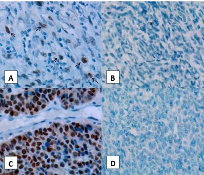
A
B
D
C
Figure 2. p53 expression in synovial sarcoma. (A) p53 positive expression case, several nucleus were brown stained by DAB (arrows), (B) Case with negative p53 expression, (C) positive control (breast tumors), (D) negative control. (Immunohistochemistry staining , 40x objective lens)
Examination Cases
p53 expression
Positive 11
Negative 9
Mitosis
< 10/HPF 12
≥ 10/HPF 8
SYT gene translocation
Positive 7
Negative 12
Can not be analyzed 1
Table 2. p53 expression, mitosis and SYT gene translocation and neck region and trunk. Most tumors were located in the extremity. These results were similar to those reported by Amary et al9 and Fang et al.10
Some cases expressing p53. This result was similar to that reported by Oda et al8 and Schneider-Stock
et al.11 In synovial sarcoma, p53 should decreases
as reported by D’Arcy et al4, though the soft tissue
tumors may have increased expression of p53 and this is associated with poor prognosis.12 Several
processes can increase p53 expression such as telomere shortening, DNA damage, oxidative stress, oncogenic stress and Ras activation.13 Therefore
in this study possible causes need to be further investigated. Moreover Oda et al8 and
Schneider-Stock et al11 reported in some cases with p53
expression have no p53 mutated.
A
B
Figure 3. SYT gene translocation. (A) SYT gene translocation in the nuclei, white arrow pointing a nuclei with a green signal (representing proximal segment of SYT gene) and a red signal (representing distal segment of SYT gene) separated by a distance exceeding diameter of one signal. (B) nuclei without SYT gene translocation, white arrow pointing nuclei with two pairs of green signal and red signal located adjacent (FISH, 100x objective lens with immersion oil).
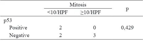
p53
p Mitosis p
Positive Negative < 10/HPF ≥ 10/HPF p53
Positive 8 3 0,362
Negative 4 5
Tumor diameter
< 5 cm 8 1 0,005* 6 3 0,650
≥ 5 cm 2 8 5 5
Tumor Location
Upper extremity 4 2 0,592 3 3 1,000
Lower extremity 3 5 5 3
Trunk 1 2 2 1
Head and neck 2 0 1 1
Unknown 1 0 1 0
Histological subtype
Monophasic 1 3 0,285 1 3 0,255
Biphasic 10 6 11 5
Gender
Female 5 5 1,000 5 5 0,650
Male 6 4 7 3
SYT gene translocation
Positive 2 5 3 4
Negative 8 4 5 7
Table 3. Statistical analysis of p53 expression and mitosis
p: Fisher’s Exact test two-tail, *: signiicant
al8 and Schneider-Stock et al11 reported p53 expression
in synovial sarcoma some are mutants. In addition to functioning as a tumor suppressor p53 can serve as an oncoprotein if it is mutated.15 Although Oda et al8 also
reported no association between p53 expression with p53
mutations, in these study whether p53 mutations were involved in the tumor pathogenesis was not known.
Mitosis
Table 4. p53 expression and mitosis of the cases with SYT gene translocation
there was no association between p53 expression with mitosis, this is similar as reported by Oda et al.8 This
may indicated that p53 signaling pathway was not the only major factor affecting tumor mitosis. Haldar et al3 explain there are other signaling pathways that
inluence synovial sarcoma growth. SYT-SSX fusion proteins can increase expression of factors that promote cell proliferation, such as cyclin D1, β-catenin, IGF2, IGF-1R, ERK1, ERK2, p-ERK and can decreased expression of factors that suppress cell proliferation, such as EGR1 and COM1.17
Because this study was conducted in population different from previous studies, there was possibility p53 and HDM2 polymorphism inluence tumor mitosis. Although this is not the cause of malignancy, polymorphism may affect both protein activity and may inluence tumor pathogenesis as well.18
In this study, tumor diameter was not associated with mitosis. Tumor diameter should relate to the number of mitosis, the higher mitotic number the greater tumor size. Therefore in this study tumor proliferation should be further conirmed by Ki-67 examination.
Almost all synovial sarcoma presents translocation between SYT gene on chromosome 18 with SSX gene on chromosome X.2 This translocation can be detected
by FISH or RT-PCR.19 FISH method as described in
igure 1 has been proved by Amary et al9 and Motoi
et al.20 Unfortunately, in this study a positive FISH
obtained only in 7/19 cases. The FISH results was different from the number of histopathologic diagnosis. Amary et al9 reported that not all cases showed SYT
gene translocation by FISH. This is because the FISH method may occur dificulty if one signal partner disappears or if there are SYT gene ampliication, we also experienced the same thing. Amary et al9 reported
in some of FISH negative cases, SYT-SSX fusion transcripts can be detected by PCR or RT-PCR. There was a possibility that the FISH negative cases were not synovial sarcoma as tumor histologically may resemble malignant peripheral nerve sheath tumor.2 Amary et
al9 reported the cases with negative SYT-SSX fusion
gene based on other tests obtained diagnosis other
p: Fisher’s Exact test two-tail
than synovial sarcoma. Therefore, in this study further investigations are needed to conirm the results of negative FISH.
Of seven positive FISH cases showed no association between p53 expression with mitosis. Mitosis can be inluenced by SYT-SSX fusion gene subtype.16 Inagaki
et al16 reported SYT-SSX1 fusion gene associated with
high expression of Ki-67 and high mitotis compared with SYT-SSX2 fusion gene. Although there was no association between SYT-SSX fusion gene subtype with p53 expression,16 in this study to investigate the
role of SYT-SSX fusion gene subtype, PCR or qRT-PCR is needed.
SYT-SSX fusion gene subtype associated with synovial sarcoma histological subtype, mitosis and Ki67.16 In
this study showed no correlation between histological type with p53 expression and with mitosis. These results were consistent with reports of Schneider-Stock et al11 and Oda et al.8 SYT-SSX1 translocation subtype
associated with glandular epithelial differentiation than SYT-SSX2. Expression of E-cadherin reported to be associated with the phenotype of SYT-SSX1 and glandular biphasic morphology.12 Furthermore Derksen
et al21 reported that the loss of expression of E-cadherin
and p53 synergize to form breast tumors that grow faster and invasive in mice. Therefore role of both factors in histological subtypes of synovial sarcoma may be important and need to be further studied.
In this study, there was an association between tumor diameter <5 cm with positive p53 expression. The result was in contrast to reports of Oda et al.8 This indicated
that positive p53 expression suppresses tumor growth and conversely negative p53 expression promotes tumor growth. Wild-type p53 has function as a tumor suppressor, therefore it was possible that p53 expressed in this study were wild-type. This inding should be further clariied by performing p53 mutation examination.
In this study showed no signiicant difference p53
studied revealed no expression of estrogen receptor and progesterone receptor found only in one case.
Acknowledgments
We would like to thank ZytoVision GmbH Bremerhaven Germany and assisted by PT. Biozatix Indonesia who have given the FISH reagents (ZytoLight SPEC SYT Dual Color Break Apart FISH Probe and ZytoLight-Tissue Implementation Kit) for SYT gene translocation examination in this study.
REFERENCES
1. World Health Organization. International agency for research on cancer. Fletcher C. Pathology and genetics of
tumours of soft tissue and bone. Lyon: IARC Press; 2002.
2. Eilber FC, Dry SM. Diagnosis and management of synovial
sarcoma. J Surg Oncol. 2008;97(4):314–20.
3. Haldar M, Randall RL, Capecchi MR. Synovial sarcoma:
from genetics to genetic-based animal modeling. Clin
Orthop Relat Res. 2008;466(9):2156–67.
4. D’Arcy P, Maruwge W, Ryan BA, Brodin B. The oncoprotein SS18-SSX1 promotes p53 ubiquitination and degradation by enhancing HDM2 stability. Mol Cancer
Res. 2008;6(1):127–38.
5. Robbins S. Robbins and Cotran pathologic basis of disease.
8th ed. Philadelphia PA: Saunders/Elsevier; 2010. 6. Ozaki T, Nakagawara A. p53: the attractive tumor suppressor
in the cancer research ield. J Biomed Biotechnol [Internet]. 2011;2011:[about 13 p]. Available from: http://www.
hindawi.com/journals/biomed/2011/603925/.
7. Abramoff M, Magelhaes P, Ram S. Image processing with
ImageJ. Biophotonics International. 2004;11(7):36–42.
8. Oda Y, Sakamoto A, Satio T, Kawauchi S, Iwamoto Y, Tsuneyoshi M. Molecular abnormalities of p53, MDM2, and H-ras in synovial sarcoma. Mod Pathol.
2000;13(9):994–1004.
9. Amary MFC, Berisha F, Bernardi FDC, Herbert A, James M, Reis-Filho JS, et al. Detection of SS18-SSX fusion
transcripts in formalin-ixed parafin-embedded neoplasms:
analysis of conventional RT-PCR, qRT-PCR and dual color FISH as diagnostic tools for synovial sarcoma. Mod Pathol.
2007;20(4):482–96.
10. Fang Z, Chen J, Teng S, Chen Y, Xue R. Analysis of soft tissue
sarcomas in 1118 cases. Chin Med J. 2009;122(1):51–3.
11. Schneider-Stock R, Onnasch D, Haeckel C, Mellin W,
Franke D-S, Roessner A. Prognostic signiicance of p53
gene mutations and p53 protein expression in synovial
sarcomas. Virchows Archiv. 1999;435(4):407–12.
12. Oda Y, Tsuneyoshi M. Recent advances in the molecular
pathology of soft tissue sarcoma: implications for diagnosis,
patient prognosis, and molecular target therapy in the future.
Cancer Sci. 2009;100(2):200–8.
13. Shangary S, Wang S. Targeting the MDM2-p53 interaction
for cancer therapy. Clin Cancer Res. 2008;14(17):5318–24.
14. Monoclonal mouse anti-human p53 protein, clone DO-7
[Internet]. [cited 2012 Jun 1]. Available from: http://
www.dako.com/dist/ar38/p105510/prod_products. h t m ? s e t C o u n t r y = t r u e & p u r l = a r 3 8 / p 1 0 5 5 1 0 / p r o d _ products.htm.
15. Suzuki K, Matsubara H. Recent advances in p53 research
and cancer treatment. J Biomed Biotechnol [Internet]. 2011;2011:[about 7 p]. Available from: http://www.hindawi.
com/journals/biomed/2011/978312/.
16. Inagaki H, Nagasaka T, Otsuka T, Sugiura E, Nakashima N, Eimoto T. Association of SYT-SSX fusion types with proliferative activity and prognosis in synovial sarcoma.
Mod Pathol. 2000;13(5):482–8.
17. Przybyl J, Jurkowska M, Rutkowski P, Debiec-Rychter M, Siedlecki JA. Downstream and intermediate interactions of synovial sarcoma-associated fusion oncoproteins and
their implication for targeted therapy. Sarcoma [Internet]. 2012;2012:[about 13 p]. Available from: http://www.
hindawi.com/journals/srcm/2012/249219/.
18. Whibley C, Pharoah PDP, Hollstein M. p53 polymorphisms: cancer implications. Nat Rev Cancer. 2009;9(2):95–107.
19. Bruce R P. Recent advances in the molecular diagnosis of paediatric soft tissue sarcomas. Diagnostic Histopathology.
2011;17(1):25–35.
20. Motoi T, Kumagai A, Tsuji K, Imamura T, Fukusato T. Diagnostic utility of dual-color break-apart chromogenic in situ hybridization for the detection of rearranged SS18 in
formalin-ixed, parafin-embedded synovial sarcoma. Hum Pathol. 2010;41(10):1397–404.
21. Derksen PWB, Liu X, Saridin F, van der Gulden H, Zevenhoven J, Evers B, et al. Somatic inactivation of E-cadherin and p53 in mice leads to metastatic lobular mammary carcinoma through induction of anoikis resistance and angiogenesis. Cancer Cell.
2006;10(5):437–49.
22. Bhargava R, Shia J, Hummer AJ, Thaler HT, Tornos C, Soslow RA. Distinction of endometrial stromal sarcomas from “hemangiopericytomatous” tumors using a panel of immunohistochemical stains. Mod Pathol.