Vol 7, No 3, July - September 1998 Isolation of hantavirus from Bandicota indica 115
Isolation
and presumptive
serological char acterization
of hantayirus
from
wild
rat
(Bandicota
indica)
Praseno*,
Suwarso**
Abstrak
Hantavirus termasuk dalam kelompok
famiti
Bunyaviridae dan ditemukandi
banyak bagian dunia. Secara klasik, infeksi Hantavirus dihubungkan dengan Hemorrhagic fever with Renal Syndrome (HFRS), meskipung"j;b
Wirik sangat beragam. Kàrena pembawa utama dari Hantavirus adalah tikus, maka survei secara serologik dilakukanpoao
titw
liar
(Banàicota indica)di
kota Yogyakarta. Isolasi virus dilakukan untuk memastikan keberadaan hantaviris. Hasil yang didapat menunjukkan bahwa, dengan teknik imunoJluoresensi antibodi, tingkat seropositif adalah 30Vo. Hantavirus telah diisolaiidiri iorirgo,
paru
tikus. S"roro ,"roîogik virus tersebut berhubungan dekat dengan virus Hantaan atau Seoul sampai saat ini maknakliiik
iiyeksi hantavirusdi
yogyakarta belum dikctahui dan memerlukan penelitian lebih tanjut. Keberadaan Haniavirus ini kemungkinan dapat menjadi masalahffiksi
virat yong akan muncul di Indonesia.Abstract
Hantavirus is a newly established
genus
d in many parts with hantavirus is associated withhemorrhagi
althoughilinica
the main reservoir of hantavirus is rodents,
s
wild urban ratsity'
virus. Results showed that, by immunofluorescence antibodyi:
ical significance of hantavirus Iung tissue of the animal. Serologically, hantavirus infectingin Indonesia is not yet known and warrants further investigations. I1 is likely that the presence of the virus could be an emerging viral infection problem in Indonesia.Keywords: hantavirus, Bandicota indica, hemorhagic fever with renal syndrome
INTRODUCTION
Hantavirus
is
anewly
established genusof
thefamily
Bunyaviridae
which is found
mainly in Asia
and east-ern partsof Europe.
At
least
sevendifferent
strains ofhantavirus have
beenrecognized,
including
Hantaan,Seoul, Puumula, Belgrade, Prospect
Hill,
andLeaky
virus. IPrototype
of hantavirus,
Hantaanvirus,
wasknown to
be the etiologic
agentof
Korean Hemorrhagic Fever
(KHF).
In
1982
The
WHO
recommended
ro use
"Hemorrhagic Fever with
Renal
Syndrome" (HFRS)
instead
of
KHF
and KHF-like
diseasesfor infection
causedby Hantaan
andHantaan-related virus.2
*
Department of Microbiology Faculty of Medicine, Gadjah Mada U niv ers ity, Yo gyalcarta, Indone s ia**
Department of Clinical Pathobgy Faculty of Medicine, Gadj ah M ada U niv e rs i ty, y o gy akar t a, Indone s iaSpecifically, the disease
is
characterized.
by
abrupt
onset
of
high fever, various hemorrhagic
manifesta-tions
and transient renal and hepatic disfunctions.
However,
theclinical
spectrum ofHantavirus
infection
varies greatly,
including'flu-like
illness,,
acuterenal
failure,
acute abdomen, acute respiratory
distress
synd2rome,and disseminated intravascular
coagula-tion.r
More
recenty,
anovel
hantavirus
called Sin Nombre
virus
has beenisolated
in United
States.This virus
isassociated
with
acute respiratory -distress syndrome
with mortality
rateof
70 percent.a')All
are spreadby
rodents where themajor
rou
ssion
is
via
aerosol
of
rodent urine,
sali
s.6116 Praseno and Suwarso
because of unawareness of
the clinicians about
thevirus and
the lack
of
diagnostic
kit for
laboratory
diagnosis
of
infection
with
hantavirus
in
Indonesia. Since casesof HFRS
have beenreported
in
Thailand,
Phillipine,
andMalaysia
it
is very
likely
thatinfection
with
thevirus
have alsooccured
in
Indonesia.So
far,
there
is no
dataon
seroepidemiological
studyof
infection with
hantavirus
in
urban ratsof
Bandicota
indica
in Indonesia.
This study is very important in
providing information
about the extent of thereservoir
in
these rodents.Isolation
of thevirus
will
establish the existenceof
hantavirus
in
Indonesia.
MATERIALS
AND METHODS
Preparation
of
sera
from wild rats
(Bandicota
indica)
Wild
rats were captured
by
trapping the animal
along ariver-side in
the center
of
Yogyakarta
municipality.
Blood
was taken
from
these
rats and sera
were
separatedfrom
theclot
andstored
at-20
Cuntil
used.Lungs
of
the animal were collected
andkept frozen
at -70oC
for further
viral isolation
experiments.
Preparation of viral
antigen
Vero E6 cell line
infected
with
Hantaan,
Seoul,
Puumula, and Sin Nombre virus, respectively
werecultured in
EagleMinimal
EssentialMedium (EMEM)
supplemented
with
lÙVo
fetal calf
serum
(FCS)
andantibiotic.T After
12days
of incubation at3'lo Cina
humidified CO2 incubator cultures were
trypsinized
and harvested.Cells
were washedwith
phosphate-buf-fered saline (PBS)
and resuspendedin EMEM
supple-mented
with 5Vo
FCS. The suspension was
then
distributed
on towells
ofteflon-coated
glass slides andfixed
with
aceton.After fixation
the
slides was stored at -70oC
until
used.Detection
of specifïc antibody
to
hantaviruses
by
indirect immunofluorescence antibody technique
Sera
of
Bandicota
indica
were
diluted
1:32with
PBS. Eachof
25pl of
thesediluted
serawere
dropped onto
wells
of
slides on which
viral
antigens
were fixed
(Hantaan, Seoul, Puumula, and
Sin
Nombre
virus,
respectively). Slides were incubated
at
37o
C in
ahumid
chamberfor
30minutes.
After
incubation
slides were washedwith
PBSfor
5minutes
and dried atroom
temperature.25
1tlof
FlTC-labeled rabbit
anti-rat
IgG
Med J Indones
was added on to
wells
and the slidesreincubated
for
30minutes. After
washing and
drying, small drops
of
mounting
fluid
applied
onto
wells,
the
slidescovered
with
coverslips and examined under
fluorescence
microscope. The
presence
of
cytoplasmic
granuleswith
greenish-yellow color
was recorded as apositive
result.
Titration
of the animals
sera
Positive
sera werefurther diluted
1:128,1:512,1:2048,
and
l:8I92.
These
serawere examined
aspreviously
described.
Determination
of
titer
of
the antibody
to
thesefour different
strains
of
hantaviruses
is
usedto
presumptively
identify
thenewly
isolated hantavirus.
Viral
isolation
Lungs
from
the animal showing antibody
titer
greaterthan
512were
used asstarting
material for
the
isola-tion. The lungs
were
minced
and
suspended as 20Vow/v in MEM
suplemented
with
lOVoFCS and
an-tibiotic. The
suspensionwas
allowed to
standfor
10minutes
to
settle larger tissue
fragments.
1ml of
thesuspension
was then inoculated
into
confluent
monolayer growth
of
Vero E6 cell culture.
After
10 daysincubation
thecells
was harvested and suspendedin
freshgrowth medium
(MEM with
lÙEo FCS).While
suspended,
slide
was prepared
andexamined
for
thepresence
of viral
antigen
by
immunofluorescence
assay as
previously
described
using known
positive
serumto Hantaan
virus.
Suspendedcells
were
further
cultured and examination
of viral
antigen was
per-formed after additional
5 daysincubation.
RESULTS
Two
hundredswild
rats were captured and usedin this
study. Seropositive
to
Hantaan, Seoul, Puumula,
andSin Nombre
virus were found
in
rats with
different
rates
(Table
l).
Table
l.
Seropositive rates of antibody to four different strainsof hantaviruses in Bandicota indica
Number of animals seropositive to strains
of
HANTAAN SEOUL
PUUMULA
SIN NOMBRE60
(3OVo)
50
(25Vo)
27
(135%)
2l
Vol 7, No 3, July - September l99B
It should be
noted that
a serum sample may react
tothree
or all of
theseviruses.
Titers of
the antibody to
Hantaan, Seoul,
Puumula,
and SinNombre
werein
therange
of
32-8912, 32-8912, 32-2048, and 32-512,
respectively (Table
2)."lable 2. Antibody titers of Bandicota indica
to
strains ofhantaviruses
Strains of hantavirus Titer of antibody* (range)
Isolation of hantavirus from Bandicota indica
tt7
DISCUSSION
Isolation
of
Hantaan
virus
and
development
of
serodiagnostic
test hasled
to
therecognition
thathan-taviruses
are
widely
spread
throughout the world.
Urban
ratsinfected
with
hantavirus
have beenreported
in
10 Asian countries
(Japan,
Korea, China,
Hong-kong, India,
Sri
Lanka,
The Philippines.
Fiji,
Singa-pore, and
Malaysia).' Using
immunofluorescence
antibody technique, seropositive
rateofhantavirus
in-fection in
rodents
in
Singapore
was 26Vo.8Our
studyshow that
overall seropositive
ratein
Bandicota
indica
was
30Vo.The
animals
was
seropositive
to
four
dif-ferent
strainsof
thevirus
andthis
isnot uncommon
asthere
is
serological cross
reactivity
among strains
of
hantaviruses.
From
titration
of
the
serait
seemsthat
the
virus is
more
closely
related
to
Hantaan
or
Seoulvirus. Further investigation
isstill
needed to determineidentity
of
thevirus in
these animalsusing monoclonal
antibody or nucleotide
sequence analysis.Our
study
obviously
showed that hantavirus exists
in
Yogyakarta. However,
clinical
significance
of
thevirus is
not yet
known.
It
is very
likely
that
infection
with
thevirus
doresult
in clinical
signs and symptoms resemble to thoseof other
viral
orbacterial infections.
HantaanSeoul Puumula Sin Nombre
32 - 8192 32 - 8192
32-
5t2
32 - 2048
x reciprocal titer
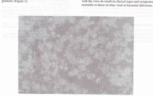
Viral
antigen expression was observed
after
l0
daysincubation
of
infected
Vero
E6
cell
culture.
Im-munofluorescence staining
of
infected cells
will
showcharacteristics greenish-yellow
intracytoplasmic
[image:3.595.39.536.384.697.2]granules
(Figure
l).
118 Praseno and Suwarso Med J Indones
REFERENCES
l.
Lee HW, Lee PW, Baek LJ, Chu YK. GeographicalDistribu-tion of Hemorrhagic Fever and Renal Syndrome and Han-taviruses. Arch
Virol
1990 (Suppl. 1):5-18.2. Hemorrhagic Fever
with
Renal Syndrome: memorandum from a WHO meeting. Bull WHO 1982;61:269-75. 3. LeeHW
and van deer Groen G. Hemonhagic Fever withRenal Syndrome. Prog Med
Virol
1989;36:62-102. 4. HjelleB,
Jenison S, MartinezNT,
YamadaT,
Nolte K,Zumwalt R, Innes
K,
Myers G.A
Novel Hantavirus As-sociated with an Outbreak of Fatal Respiratory Disease in The Southwestern United States. JVirol
1994;68(2):592-6.5. Schmal John AL, Li D, Negley DL, Bressler DS, Tunel MJ.
Isolation and Initial Characterization of a Newfound Han-tavirus from Califomia.
Virol
I 995 ;20 6:9 63 -7 2.6. Niklasson BS. Hemorrhagic Fever with Renal Syndrome,
Virological and Epidemiological Aspects. Peditr Nephrol, 1992;6(2):2Ol-4.
7. Lee PW, Meegan JM, Tkachenko EA, Kitamura T. Serologic
Techniques for Detection of Hantavirus Infection, Related Antigens and Antibodies,
in
LeeHW
and Dalrymple JM(eds). Manual of Hemorrhagic Fever with Renal Syndrome.
WHO Collaborating Center
for
Virus Reference andRe-search. Seoul, Korea University 1989:75-l 10.
8.
Wong
Tlùy', ChanYC,
Joo
YG, Lee HW,
Lee
PW, Yanagihara R. Hantavirus Infectionin
Humans andCom-mensal Rodents in Singapore. Trans R Soc Trop Med Hyg.
1989;83(2):248-51.
9. Chun CH, Laehdevirta J, Lee HW. Clinical Manifestations
of HFRS, in Lee HW and Dalrymple JM (eds). Manual of
Hemorrhagic Fever
with
Renal Syndrome.WHO
Col-laborating Center for Virus Reference and Research. Seoul,Korea University I 989; 19-38.
10. Imam Supardi, Kosasih. A Retrospective Study on Dengue Hemorrhagic Fever in St. Bartolomeus Hospital of Bandung.
Mikrobiologi Klinik Indonesia. 1989;7:88-91.
ll.
Wuryadi S. Isolasi Virus Dengue dari Pasien Demam Ber-darah Dengue Pada Waktu Wabah di Jakarta Tahun 1988.Cermin Dunia Kedokterer;' 1990;60:27 -30.
Different
strainof hantavirus
may causeinfection
with
different
signs andsymptoms.
Infection with
Puumula
virus
has been associated
with
nephritis
without
hemorrhagic manifestations, whereas Seoul virus
causes
mild
forms
of
HFRS.eIt
wasreported
in
Singapore that
infections with
han-tavirus were
found
in
lOVoof
casesof
suspectedden-gue
fever,
5Vo cas^es suspected leptospirosis,
and
l7ocases
of
hepatitis.8Previàus
studyby
^Supardiin
Ban-dung showed
that more
than
50Vo casesof
clinically
dengue
fever were
seronegative
when confirmed
by
hemagglutination inhibitio"n t.st.10 In
asimilar
studyconducted
in
Jakarta
by
Wuryadi
showed that
seronegative werefound
in46.3Voof
suspected denguefever."
It
is
possible that infection
with hantavirus
might
haveoccured
in
these cases.Differential
diagnosis
of infection
with
hantavirus
in-cludes
hepatitis, nephritis,
leptospirosis,
dengue
hemorrhagic
fever,
pneumonia, and
septicaemia.r
Thus,
diseaseswith clinical
features resemble tothem
should be
examined
for
possible hantavirus
infection.
Since the
main reservoir of hantavirus
iswild
rats, andour environment, especially
in
slum
areas,is
still
den-selypopulated
with
the animals,
the existenceof
han-tavirus could be
anemerging
viral infection
problem
in
Indonesia.
Acknowledgement
Parts
of this work
werecarried out
at TheDepartment
of
Virology
AsanInstitute
for
Life
ScienceKorea. We