1
ISOLATION AND CHARACTERIZATION OF ACTINOMYCETES FROM PINEAPPLE PLANTATION
POTENTIAL AS ANTAGONIST OF
DICKEYA
SP., THE PINEAPPLE SOFT ROT PATHOGEN
Titik Nur Aeny
1a, Joko Prasetyo
1b, Radix Suharjo
1c, Suskandini R. Dirmawati
1d, Efri
1f, Meri D. Saputri
1e& Wulandari
1f1Plant Protection Department, Faculty of Agriculture, University of Lampung,
Jl. Prof. Soemantri Brojonegoro No.1 Bandar Lampung 35145, Indonesia
aE-mail: [email protected] bE-mail: [email protected] cE-mail: [email protected] dE-mail: [email protected]
eE-mail: [email protected] fE-mail: [email protected]
gE-mail: [email protected]
Abstract
:
This study was aimed to isolate and characterize actinomycetes having antagonist activity to Dickeya sp., the pathogen of pineapple soft rot. Soil samples were collected from four different pineapple plantation in Lampung. Samples were air-dried, serially diluted, and plated on actinomycetes isolation agar media; and potential colonies were selected and purified in malt agar medium. Isolates were grouped in different color series based on their aerial-mycelia color, and then morphologically characterized. Antagonist activity of the actinomycetes isolates on Dickeya sp. were evaluated using the same medium. From totally 51 actinomycetes isolates, only 34 isolates that grew well, then screened and tested for antagonistic activity. There were 35 tested isolates (including the control) exhibited a clear zone around the colonies indicating antagonistic activity; only 14 isolates showed clear zone higher than that of the positive control (chloramphenicol). From the smallholder pineapple plantation, isolate A-12 exihibited the highest antagonist activity against Dickeya sp. (diameter of clear zone = 13,69 mm) and from the private pineapple plantation isolate GGP-9 showed the best (diameter of clear zone = 19,44 mm). Based on their cultural and morphological charactyeristics, the isolated actinomyecetes were suggested as genus of Streptomyces. Since many isolates showed inhibitory activity in vitro against Dickeya sp., one of important pineapple soft rot pathogens, it is suggestive that pineapple plantation could be an interesting source to explore for actinomycetes potentially used as antagonist.Keywords: actinomycetes; antagonist activity; Dickeya sp.; pineapple soft rot
I. INTRODUCTION
Actinomycetes are a group of gram-positive bacteria, aerobic, grow and have aerial hypha like fungi. They are easily found in the soil, especially in dry alkaline soil. Actinomycetes has been reported as the common producers of antibiotics, especially the genus Streptomyces [1], [2], [3]. So far, actinomycetes have been widely developed and utilized in many diferent fields such as in the pharmaceutical industry,
2
from soil in Malaysia capable of inhibiting the growth of some types of plant pathogens [8], [9]. Actinomycetes has been used as biocontrol agents of onion bacterial rot diseases [10]. Sme actinomycetes showed antagonistic effect on Xanthomonas pv.
oryzae, the causal agent of bacterial leaf blight on rice [11]. There are still vey limited information, however, concerning the use of actinomycetes as biocontrol agents of bacterial soft rot diseases in Indonesia.
This study was aimed to characterize actinomycetes isolates capable of inhibit the in vitro growth of Dickeya sp., the bacterial pathogen of pineapple soft rot disease in Lampung.
II. THE MATERIAL AND METHODS
2.1 Collection of Soil Samples and Isolation of Actinomycetes
The soil samples were taken from predetermined pineapple plantation sites located in Lampung during July – December 2016. The source of soil samples collection consisted of four pineapple plantations in different locations, two came from private companies: PT Great Giant Farm (GGF) located in Terbanggi Besar (Central Lampung) and PT Nusantara Tropical Farm (NTF) located in Way Jepara (East Lampung) and another two came from smallholder plantations located in Astomulyo (A) located in Punggur - Central Lampung and Mulyajaya (M) located in Tulang Bawang Tengah - West Tulang Bawang. From each of the plantation, soil samples were collected from five diagonal points in a depth of 15 cm below the soil surface, composited, kept in a plastic bag, and stored in a cooler during transportation.
Prior to isolation, the soil was air-dried for a week to reduce the growth of gram-negative bacteria. Furthermore, the isolation process followed a procedure described in [7]. Colonies of suspected actinomycetes having different appearance were subcultured in malt agar medium and incubated at room temperature for one week to obtain pure culture of each isolate.
2.2 Antagonistic Test
An antagonistic test was performed to investigate the potential of actinomycetes isolates as antagonistic agents or growth inhibitors of Dickeya sp. Antagonistic test was performed by agar diffusion method developed by [12] to determine the antagonistic effect of actinomycetes toward
Dickeya sp. The tested pathogen, 24 hour Dickeya sp., was suspended in sterile water and mixed into culture medium. The actinomycetes isolates were taken from a 3-day culture using a 5 mm diameter sterile cork and placed on the surface of the medium mixed with the tested pathogen. Positive reactions are indicated by the formation of clear zones around the
actinomycetes. Observations and measurements of clear zones were conducted daily for 5 days.
2.3 Characterization
Collected isolates were characterized based on their cultural and morphological features of colonies grown on malt agar medium (colour of the aerial mycelia, substate mycelia, and diffusible pigment). Visual observation of microscopic characteristics was done directly and indirectly under a compound light microscope and scanning electron microscope. SEM analysis was condacted at the Unit Pelaksana Teknis Laboratorium Terpadu dan Sentra Inovasi Teknologi (UPT LTSIT) University of Lampung. Important criteria observed included characteristics of the structures of aerial and substrate hyphae, spore chains, and ornamentation of spore surface .
III. RESULTS AND DISCUSSION
3.1 Isolates of Actinomycetes
The initial in vitro growth of actinomycetes colonies was relatively slow, it took about 3 days after incubation. The colonies that grew in malt agar media varied in their surface colors, surface structures, and color of diffusible pigment. Color series of colony surface included white, beige, gray, brown, yellow, and pink. Reference [13] described that the color of the aerial and substrate mycelia produced actinomycetes varied with different media. The colonies of actinomycete isolates were round-shaped with starchy surfaces, having clear edges and were firmly attached to the surface of the media. Those characters were in line with the description by
Reference [14] showed that colonies of actinomycetes show powdery consistency and stick firmly to agar surface; in culture media they produced hyphae and spora-like fungi. Any isolates suspected as actinomycetes were isolated and re-purified on the same medium for further characterization. From this step was obtained totally 51 isolates suspected as actinomycetes; however, only 45 isolates that grew well.
3
Figure 1. Some variations of shapes and colours of actinomycetes colonies
3.2 Antagonistic test
In general, the result of antagonistic test showed clear zone formed around actinomycetes colonies started one day after incubation and increased up to following five days. The clear zone suggested that the growth of Dickeya sp. in agar medium was inhibited by actinomycetes (Figure 2). The clear zone mplied that the pathogen did not grow well when exposed to actinomycetes, thus indicated that the actinomycetes may produced substance that acted as antibacterials [7]. References [16] and [17] showed that the antibacterial activity of actinomycetes inhibited the growth of E. carotovora subsp. carotovora and E. chrysanthemi, respectively. The presence of a growing clear zone also indicated that the more antibacterial compounds were produced, the stronger the antibacterial effect of the actinomycetes is.
Figure 2. Formation of a clear zone around the actinomycete colonies
Some actinomycetes isolates did not form any clear zone, or in other words they were not be able to inhibit the growth of
Dickeya sp. Based on samples source, out of 25 isolates from smalholder pineapple plantation tested for antagonistic only 19 isolates showed antagonistic effect (8 Astomulyo isolates and 11 Mulyajati isolates (Table 1). And out of 20 isolates from private pineapple plantation, only 10 GGF isolates and 5 NTF isolates that showed inhibition effect (Table 2). Various publications stated that the growth of actinomycetes is influenced by soil type and soil acidity. The population of actinomycetes will be especially high in dry and alkaline soils [18], [19]. However, it turned out that on pineapple plantations which is the soils tend to be wet and acid (pH < 6.0) still can be obtained various actinomycete isolates.
Table 1. Inhibition of in vitro growth of Dickeya sp. by actinomycetes isolates from smallholder pineapple plantation.
Table 2. Inhibition of in vitro growth of Dickeya sp. by
actinomycetes isolates from private company’s pineapple plantation
3.3 Characterization of Selected Actinomycetes
4
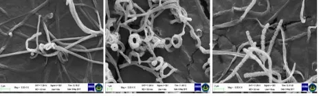
Streptomyces. Among actinomycetes genera, Streptomyces represented the largest genus [21]. The characters including the color of aerial and substrate mycelia, formation of diffusible pigments, structure of the substrate and aerial mycelium, branching of hyphae containing spores, and the spore surface. Observation under scanning electron microscope showed spore surface morphology that was very much alike Streptomyces [22].
Figure 3. SEM of actinomycetes of two different isolates indicated as Streptomyces sp.
IV. CONCLUSIONS
Actinomycetes were abundantly found in pineapple plantations. From several locations of pineapple plantations in Lampung can be collected as many as 34 isolates that potential as bacterial antagonist of Dickeya sp. Out of those 34 isolates, six isolates were considered as the best antagonist. From the smallholder pineapple plantation, isolate A-12 exihibited the highest antagonist activity against Dickeya sp. (diameter of clear zone = 13,69 mm) and from the private pineapple plantation isolate GGP-9 showed the best (diameter of clear zone = 19,44 mm). Based on the cultural and morphogical characteristics, the actinomycete isolates were grouped as the genus of Streptomyces. Since many isolates showed inhibitory activity against Dickeya sp., one of the important pineapple soft rot pathogens, it is suggestive that pineapple plantation could be an interesting source to explore for actinomycetes potentially used as antagonist.
ACKNOWLEDGMENT
The authors would like to thank Directorate of Research and Community Service - Director General for Research and Development at the Ministry of Research, Technology and Higher Education of the Republic of Indonesia for the funding given. Special thanks to the Great Giant Farm Co., Nusantara Tropical Farm Co. and Mr. Wahyu for allowing us to collect the soil samples from their pineapple plantation. Thanks is also extended to Mr. Efri and Ms. Widyaningrum A. Sari for their technical assistance.
REFERENCES
[1] Saadoun I, Gharaibeh R. 2003. The Streptomyces flora of Badia region of Jordan and its potential as a source of
antibiotics active against antibiotic-resistant bacteria. J Arid Environ. 53:365–371.
[2] Miyadoh, S., and M. Otoguro. 2004. Workshop on Isolation Methods and Classification of Actinomycetes. Bogor (ID): Biotechnology Centre, LIPI.
[3] Kumar, V., Alpana Bharti, Omprakash Gusain, and Gajraj Singh Bisht. 2011. Scanning Electron Microscopy Of Streptomyces Without Use of Any Chemical Fixatives. Scanning Vol. 33, 446–449. DOI 10.1002/sca.20261
[4] Singh, L.S., I. Baruah, and T.C. Bora. 2006.
Actinomycetes of loktak habitat : isolation and screening for antimicrobial activities. Biotchnology. 5 (2) : 217-221.
[5] Arifuzzaman.M, Khatun.M.R and Rahman.H. 2010. Isolation and screening of actinomycetes from Sundarbans soil for antibacterial activity , African Journal of Biotechnology Vol. 9(29), pp. 4615-4619.
[6] Dhananjeyan V., N. Selvan and K. Dhanapal. 2010. Isolation, Characterization, Screening and Antibiotic Sensitivity of Acrinomycetes from Locally (Near MCAS) Collected Soil Samples. Journal of Biological Science, 10: 514-519. DOI: 10.3923/jbs.2010.514.519.
[7] Oskay, M., Tamer, A.U., and Azeri, C. 2004. Antibacterial activity of some actinomycetes isolated from forming soils of Turkey. African Journal of Biotechnology, 3: 441-446.
[8] Jeffrey LSH, Sahilah AM, Son R, Tosiah S. 2007. Isolation and screening of actinomycetes from Malaysian soil for their enzymatic and antimicrobial activities JTAFS. 35: 159-164
[9] Jeffrey, L. S. H. 2008. Isolation, characterization and identification of actinomycetes from agriculture soils at Semongok, Sarawak African Journal of Biotechnology Vol. 7 (20), pp. 3697-3702.
[10] Abdallah ME, S.A. Haroun , A.A. Gomah , N.E. El-Naggar & H.H. Badr. 2013. Application of actinomycetes as biocontrol agents in the management of onion bacterial rot diseases, Archives of Phytopathology
and Plant Protection.
DOI:10.1080/03235408.2013.778451
[11] Giyanto, C.P. 2014. Compatilbility of Bacillus spp. and Actinomycetes as Biocontrol Agent for Xantomonas oryzae pv. oryzae and Growth Promoter for Rice. Jurnal Fitopatologi Indonesia Vol. 10 No. 5 Oktober 2014 (160-169). DOI: 10.14692/jfi.10.5.160
5
[13] Sowndhararajan K. and S. Kang. 2012. In vitro antagonistic potential of Streptomyces sp. AM-S1 against plant and human pathogens, Journal of Agricultural Chemistry and Environment, Vol. 1 No. 1, pp. 41-47. doi: 10.4236/jacen.2012.11007.
[14] Qinyuan Li, Xiu Chen, Yi Jiang and Chenglin Jiang. 2016. Morphological Identification of Actinobacteria. In Actinobacteria – Basic and Biotechnological Application. edited by Dharumadurai Dhanasekaran and Yi Jiang, ISBN 978-953-51-2248-7
[15] Powers E.M. 1995. Efficacy of the Ryu Nonstaining KOH Technique for Rapidly Determining Gram Reactions of Food-Borne and Waterborne Bacteria and Yeasts. Applied And Environmental Microbiology, Oct. 1995, p. 3756–3758. Vol. 61, No. 10. 0099-2240/95/$04.0010
[16] Zamanian S, Bonjar GH, Saadoun I. 2005. First report of antibacterial properties of a new strain of Streptomyces plicatus (strain 101) against Erwinia carotovora subsp. carotovora from Iran. Biotechnology. 4:114–120.
[17] El Karkouri A, El Hassani F, El Mzibri M, Benlemlih M, El Hassouni M. 2010. Isolation and identification of an actinomycete strain with a biocontrol effect on the phytopathogenic Erwinia chrysanthemi 3937VIII responsible for soft rot disease. Ann Microbiol. 60:263– 268.
[18] Athalye M, Lacey J, Goodfellow M. 1981. Selective isolation and enumeration of actinomycetes using rifampicin. J. Appl. Bac. 51: 289297.
[19] Goodfellow, M. and Williams, S.T. 1983. Ecology of ac-tinomycetes. Annual Review of Microbiology, 37, 189- 216. doi:10.1146/annurev.mi.37.100183.001201
[20] Kumar V., Alpana Bharti, Omprakash Gusain, and Gajraj Singh Bisht. 2011. Scanning Electron Microscopy Of Streptomyces Without Use Of Any Chemical Fixatives. Scanning Vol. 33, 446–449. DOI 10.1002/sca.20261
[21] Taddei A, Rodriguez MJ, Vilchez EM, Castelli C. 2006. Isolation and identification of Streptomyces spp. from Venezuelan soils: Morphological and biochemical studies
I. Microbiol Res. 161: 222-231.
doi:10.1016/j.micres.2005.08.004.
[22] Kumar,S.V, Sahu, M.K and Kathiresan K. 2005. Isolation and characterization of streptomycetes producing antibiotics from a mangrove environment, Asian Jr.of Microbial. Biotech Env. Sc.Vol. 7 No. (3); 457-464.