Tommy Heryantho
1,2, Andi Wijaya
1,2, and Teguh Santoso
3R E S E A R C H A R T I C L E
1 Prodia Clinical Laboratory, Jakarta
2 Post Graduate Program in Clinical Biochemistry, Hasanuddin University, Makassar 3 Faculty of Medicine – University of Indonesia, Jakarta
Biochemical Markers for Determining Vulnerable
Atherosclerotic Plaque in Stenotic Patient
A Biochemical Markers Study of Myeloperoxidase (MPO), Matrix Metallo-Proteinase-9
(MMP-9), Secretory Phospholipase A2 (SPLA2) and CD40 Ligand
B
ACKGROUND: Thrombus is a main causeof cardiac death. Therefore identifying which coronary artery plaque is vulnerable to rupture is a critical step for cardiac intervention to prevent future cardiac events. Systemic biochemical markers are used for predicting rupture of coronary plaque or identifying stenotic coronary artery plaque(s) vulnerable to rupture.
METHODS: Blood samples of 2 x 24 locations (2 x
10 controls, 2 x 12 stable plaques and 2 x 2 unstable plaques) of 13 patients to undergo stent placement were taken from an artery which showed no stenosis (control), 70% or more stenosis of stable plaques and unstable plaques, respectively. The blood samples were taken by using micro-catheter distally and proximally. Concentrations of MPO, MMP-9, SPLA2 and CD40L of each sample were assayed.
RESULTS: Concentration of MMP-9 in unstable
coronary artery plaque (94.7 + 14.4 ng/ml) significantly increased compared with that of stable coronary artery plaque (71.0 + 67.8 ng/ml, p=0.024). SPLA2 concentration significantly decreased in unstable coronary artery plaque (45.9 + 14.0 pg/ml) compared with that of stable coronary artery plaque (80.9 + 39.3 pg/ml, p=0.015). Nine of ten studied subjects showed an average of 14.5% (range: 0.0 – 28.8%) decrease of the SPLA2 concentration in stable plaques compared with that of the non-stenotic coronary artery.
CONCLUSION: MMP-9 increased in unstable
coronary artery plaque compared with that of stable coronary plaque. Unstable coronary artery plaques
absorbed SPLA2 from the vasculars more than the stable plaques and control plaques. MMP-9 and SPLA2 may be used as markers of stability of a plaque in coronary artery in relation to its rupture potential.
KEYWORDS: stable and unstable plaque,
Myeloperoxidase, Matrix Metallo-Proteinase-9, Secretory Phospholipase A2, CD40 Ligand.
Introduction
Of 241 cardiac deaths, 125 or 52% are caused by acute thrombi related to ruptures, erosions, and calcified nodules (1). Vulnerable plaques have large lipid cores and thin fibrous caps, and are infiltrated by macrophages (2). Angiographic definition of unstable plaque underlines the existence of 50% or more stenosis accompanied by at least 2 of the followings: intra luminal filling defect, ulcer in the plaque, irregular surface, and impaired blood flow (3).
The association between Myeloperoxidase (MPO) and vulnerable plaque has been investigated recently (4). Libby et al. showed co-localization of macrophages that expressed MPO and HOCl modified protein in plaque of a person who died of cardiac event (5). MPO can be used as a predictor of plaque rupture (6), even in Troponin T negative patients (7).
higher than that of controls (10). Plasma level of MMP-9 increases in infarction and unstable angina (11).
Inwald et al. explain the role of CD40 mediator in activating platelet in thrombosis, .
Monocytes/macrophages in plaque stimulated by CD40L or T-cell membrane of CD4+ express pro-coagulant tissue factor and matrix degradation proteinase (13). Using MRI,
CD40L in blood reflects platelet activation and instability of plaque (15).
Circulating sPLA2-II in blood has been demonstrated to be able to predict coronary events in initially healthy subjects and in patients with frank coronary heart disease including acute coronary syndromes (16). Lipoprotein that contains Apo-B and SPLA2-IIA can facilitate enzymatic hydrolysis of phospholipid lipoproteins through interaction with proteoglycan in the artery. Further modification of lipoprotein causes aggregation and lipoprotein fusion within the plaque (17).
Methods
PATIENTS
Thirteen patients scheduled to undergo stent placement in the coronary artery were involved in the study. Blood samples were taken from 12 x 2 (distal and proximal of the plaque) coronary artery stable plaques from 11 patients, and 2 x 2 (distal and proximal of the plaque) coronary unstable plaques from another two patients. Control blood samples were taken from each of the same patients from coronary artery that showed no stenosis (0% stenosis).
METHODS
Blood samples from coronary artery were taken using a micro-catheter. Unstable plaque is defined according to the criteria of Goldstein. The level of CD40L and MMP-9 in the blood .
MPO and SPLA2 levels were measured using ELISA kits from Oxis Research and Cayman Chemicals, respectively.
STATISTICAL ANAL SIS
ruskal- allis and Mann- hitney tests were used to compare means between groups.
Results
Table 1. Characteristics of the study subjects
T
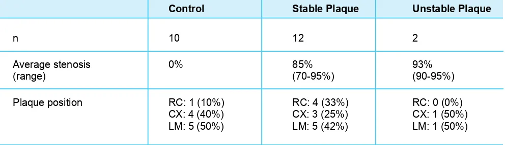
able 2. Characteristics of the coronary artery plaques
e u q a l P e l b a t s n U e
u q a l P e l b a t S l
o r t n o C
2 2
1 0
1 n
% 3
9
% 5 8 %
0
s
i
s o
n e t
s
e
g
a r e v A
)
% 5
9
-0
9 ( )
% 5
9
-0 7
( )
e
g
n a r
(
Plaque position RC: 1 (10%) RC: 4 (33%) RC: 0 (0%)
)
% 0 5
(
1 : X C
)
% 5 2
(
3 : X C
)
% 0 4
(
4 : X C
)
% 0 5
(
1 : M L
)
% 2 4
(
5 : M L
)
% 0 5
(
5 : M L
RC = right coronary artery, CX = circumflex, LM = left main.
Characteristics of t he s tudy subjects are shown i n Table 1. Characteristics of the coronary artery p laques a re shown in Table 2.
Levels of MMP-9, MPO, SPLA2 and CD40L of the distal and proximal coronary artery plaques are shown in Table 3.
Table 3. MMP-9, MPO, SPLA2 and CD40L levels (mean + SD) in the distal and proximal
coronary artery plaques
Biomarker Control S table Plaque U nstable Plaque p 1)
MMP-9 (ng/ml) 77.5 ± 74.4 71.0 ± 67.8 94.7 ± 14.4 0.024*
MPO (ng/ml) 16.6 ± 6.7 15.2 ± 6.3 21.0 ± 4.7 0.082
SPLA2 (pg/ml) 99.7 ± 48.4 80.9 ± 39.3 45.9 ± 14.0 0.015*
CD40L (ng/ml) 1 .07 ± 0.69 1 .11 ± 1.09 0 .79 ± 0.38 0 .681
1) between stable and unstable plaque * significance (p<0.05)
Among t he b iomarkers used, MMP-9 showed a significant i ncrease in unstable coronary artery p laque as compared with stable coronary artery plaque (p=0.024). On the other hand, SPLA2 showed a significant decrease in unstable coronary artery plaque compared with that in stable coronary artery plaque (p=0.015).
Discussion
MMP-9 level increased, but SPLA2 level decreased in unstable coronary artery plaques. MMP-9 is a proteinase . e r u t p u r o t e n o r p s u h t d n a r e n n i h t e m o c e b p a c s u o r b fi e h t e k a m o t x i r t a m r a l u l l e c a r t x e e d a r g e d o t e l b a s i t a h t
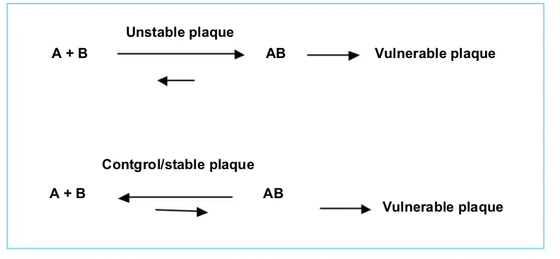
SPLA2 becomes atherogenic when associated with Apo-B. In other words, decrease levels of soluble free SPLA2 indicate increase of associated forms, as illustrated below:
Unstable plaque
Vulnerable plaque AB
A + B
Contgrol/stable plaque
Vulnerable plaque AB
A + B
Figure 1. Schematic model of SPLA2 in associated and dissociated forms.
In vulnerable to rupture plaque SPLA2 is associated with LDL-cholesterol, but in stable or control plaques SPLA2 are in free form. A=SPLA2, B=LDL-cholesterol.
MPO did not increase significantly in unstable plaque as compared with that in stable plaque. In plaque, MPO causes apoptosis of endothelial cells and erosion. According to Virmani, et al. (1), erosion occurs rarely in men, but the male subjects involved in this study comprised 92% of all study subjects.
CD40L di not increase or decrease significantly in unstable plaques as compared with that in stable plaques. It is possible that CD40L is a platelet activation marker, which plays role in thrombus generation after rupture of the plaque.
MMP-9 increased in unstable coronary artery plaques . s e u q a l p y r a n o r o c e l b a t s n i t a h t h t i w d e r a p m o c s a
nstable coronary artery plaques absorbed SPLA2 from the vasculars in a greater amount than in stable plaques and non- plaques (control). In conclusion, MMP-9 and SPLA2 can be used as markers of stability of a plaque in coronary artery towards rupture vulnerability.
Conclusion
Acknowledgement:
The authors thank the Prodia Foundation for Research and Training for the invaluable support in this research.
References:
2.Naghavi, M., Libby, P., Falk, E., Casscells, S.W., Litovsky, S. From vulnerable plaque to vulnerable patient - a call for new definitions and risk assessment strategies: part I. Circulation. 2005;. 108:1664-1672.
5.Libby, P., Theroux, P. Pathophysiology of Coronary Artery Disease.
Circulation. 2005; 111:3481-3488.
6.Baldus, S., Heeschen, C., Meinertz, T., Zeiher, A.M., Eiserich, J.P.
Myeloperoxidase serum levels predict risk i n patients with
acute coronary syndromes. Circulation. 2003;
108:1440-1445.
7.Brennan, M.L., Penn, M.S., Van Lente F., Nambi V., Shishehbor, M.H., et al. Prognostic value of myeloperoxidase in patients with chest pain. N Engl J Med. 2003; 349:1595-1603.
8.Blankenberg, S., Rupprecht, H.J., Poirier, O., Bickel, C., Smieja,
M., et a l. Plasma concentrations and genetic variation of
matrix metalloproteinase 9 and prognosis of patients with
cardiovascular disease. Circulation. 2003;107:1579-1585.
9.Sundstrom, J., Evans, J .C., Benjamin, E .J., Levy, D., Larson,
M.G., et al. Relations of plasma matrix metalloproteinase-9
to clinical cardiovascular risk factors and echocardiographic left ventricular m easures: the Framingham heart study.
Circulation. 2004; 109:2850-2856.
10.Kalela, A ., Koivu, T.A., Sisto, T., Kanervisto, J ., Hoyhtya, M ., et a l. Serum matrix m etalloproteinase-9 concentration in angiographically assessed coronary artery disease. Scand J Clin Lab Invest. 2002; 62(5):337-342.
11.Szmitko, P.E., Wang, C.H., Weisel, R.D., Jeffries, G.A., Anderson, T.J., Verma, S . Biomarkers of vascular disease linking
inflammation to endothelial activation, part II, Circulation,
2003; 108:2041-2048.
12.Inwald, D.P., McDowall, A ., Peters, M .J., C allard, R .E., Klein, N.J., CD40 is constitutively expressed on platelets and
provides a novel mechanism for platelet activation, C irc
Res. 2003; 92:1041-1048.
13.Mach, F ., Schonbeck, U., Bonnefoy, J.-Y., Pober, J.S., Libby,
P. Activation of m onocyte/macrophage functions related
to acute atheroma complications by ligation of CD40.
Circulation. 1997;. 96:396-399.
14.Blake, G.J., Ostfeld, R.J., Yucel, E.K., Varo, N., Schonbeck, U.
Soluble CD40 ligand levels indicate lipid accumulation in
carotid atheroma: an in vivo study with high-resolution MRI.
Arterioscler Thromb Vasc Biol. 2003; 23:e11-e14.
15.Freedman, J.E., Loscalzo, J . Platelet-Monocyte aggregates:
bridging thrombosis and inflammation. Circulation.
2002;105:2130-2132.
16.Koenig, W . Khuseyinova, N . Biomarkers of Atherosclerotic
Plaque Instability and Rupture. Arterioscler Thromb Vasc
Biol. 2007; 27: 15-26.
17.Hurt-Camejo, Camejo, G., Peilot, H ., Oorni, K., K ovanen, P .,
Phospholipase A2 i n Vascular D isease. C irc Res. 2001;