Mutations of the
p53 Gene in Nasal
NK/T-Cell Lymphoma
Ting Li, Tadashi Hongyo, Mukh Syaifudin, Taisei Nomura, Zhiming Dong,
Norihisa Shingu, Shizuo Kojya, Shin-ichi Nakatsuka, and Katsuyuki Aozasa
Departments of Pathology (TL, ZD, NS, SN, KA) and Radiation Biology (TH, MS, TN), Osaka University Medical School, Osaka; and Department of Otorhinolaryngology (SK), University of the Ryukyus, Faculty of Medicine, Okinawa, Japan
SUMMARY: Mutations of thep53tumor suppressor gene are reported in various kinds of malignancies including lymphomas. However,p53gene mutations in nasal NK/T-cell lymphoma have not been reported because most parts of tumors are necrotic and a small amount of living tumor tissues is available for the molecular study. Expression and mutations of thep53gene were examined in the paraffin-embedded specimens of the nasal lesions from 42 Chinese (Beijing and Chengdu) and Japanese (Okinawa and Osaka) patients with nasal NK/T-cell lymphoma by the immunohistochemistry and single strand conformation polymorphism (SSCP) analysis of polymerase chain reaction (PCR) amplified products followed by direct sequencing. Thirty single-nucleotide substitution mutations were observed in 20 of 42 cases (47.6%). Among the 30 mutations, 18 were missense (mainly G:C to A:T transitions), 9 were silent, and 1 was a nonsense mutation. The remaining 2 mutations involved intron 5 and exon 5 terminal points. Abnormal expression of thep53protein was also observed in 19 of 42 (45.2%) cases. The incidence was significantly (4-fold) higher in the cases of Osaka than those in other areas, although the incidence ofp53mutations in the cases of Osaka was one-half to one-third of those in the other three areas. The results may suggest some racial, environmental, or lifestyle differences in the cause of nasal tumorigenesis. (Lab Invest 2000, 80:493– 499).
N
asal lymphoma frequently shows a polymorphous pattern of proliferation, consisting of large atypical cells, small lymphocytes, plasma cells, benign looking macrophages occasionally showing with phagocytic fig-ures, neutrophils, and eosinophils much less frequently. Thus the term polymorphic reticulosis (PR) was pro-posed for this condition (Eichel et al, 1966). PR consti-tutes lethal midline granuloma (LMG), which is charac-terized by necrotic and granulomatous lesions mainly affecting the nasal cavity. Malignant lymphoma with monomorphous proliferation and Wegener’s granuloma-tosis also show clinical features of LMG (Kassel et al, 1969). Majority of nasal lymphoma, especially PR, was recently categorized as NK/T cell lymphoma (Jaffe et al, 1996). Through immunophenotypic and genotypic stud-ies, we have shown that PR is an NK cell lymphoma (Ohsawa et al, 1999). Epidemiologic studies of PR re-vealed the clustering of patients in the east Asian coun-tries (Aozasa et al, 1989) and Epstein-Barr virus (EBV) association (Harabuchi et al, 1990; Tomita et al, 1995), however little is known about mechanisms for develop-ment of PR.p53 is a well-known tumor suppressor gene that causes cells with damaged DNA to arrest at the G1 phase of cell cycle or stimulating expression of thebax
gene, the protein that promotes apoptosis (Levine et al, 1991). In a wide variety of human cancers, p53
gene mutations have been detected mainly in exons 5 though 8 (Hollstein et al, 1991). High incidence of malignant lymphoma in the p53 knockout mice has been reported (Donehower et al, 1992), suggesting an
important role of p53 gene mutations in
lym-phomagenesis. The mutatedp53gene encodes mu-tant p53 protein, which has a much longer half-life time than that of wild typep53protein, thus accumu-lating in the cytoplasm in an amount sufficient for immunohistochemical detection. Previous immuno-histochemical study showed that p53 is overex-pressed in a high percentage of cases with nasal NK/T-cell lymphoma (Quintanilla-Martinez et al, 1998). These findings suggest that thep53gene mutations might be frequent in nasal NK/T-cell lymphoma. In this study, we analyzed the mutations of thep53gene in a series of 42 cases of nasal lymphomas of PR type collected from China and Japan by polymerase chain reaction-single strand conformation polymorphism (PCR-SSCP) and direct sequencing.
Results
Histological and Immunohistochemical Findings
Histologically, varying degrees of necrosis were found in the upper respiratory lesions. Diffuse proliferation of large atypical mono- or multinucleated cells was
ob-Received November 2, 1999.
Supported by a grant from the Vehicle Racing Commemorative Founda-tion, grants from the Ministry of EducaFounda-tion, Science and Culture, Japan (09670184, 09770148, 10042005, 11470353, 11670212, 11680546), and Research for the Future.
Address reprint requests to: Dr. K. Aozasa, Department of Pathology(C3), Osaka University Medical School, 2–2 Yamadaoka, Suita, Osaka 565-0871, Japan. Fax: 81 6 6879 3713; E-mail: [email protected]
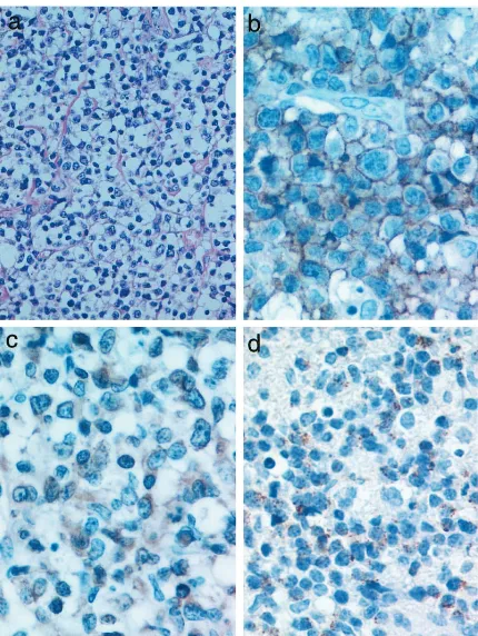
served with various numbers of lymphocytes, plasma cells, and macrophages, giving a polymorphous ap-pearance. An angiocentric pattern of proliferation was occasionally observed. Histologic grade based on the criteria described by Lipford et al (1988) was Grade II in 17 cases and Grade III in 25 cases. Percentage of large atypical cells among infiltrating cells was more than 30% and 80% in Grade II and III lesions, respec-tively. Immunohistochemically, tumor cells were CD202, TIA-11, CD561, CD31, or CD431(Fig. 1).
Expression and Mutation of the p53 Gene
Positive immuno-reactivity for DO-7, suggestive of abnormal expression or stabilization of p53 protein, was found in 19 of 42 (45.2%) cases (Table 1). The incidence of abnormal p53 expression was 4-fold higher in the cases in Osaka than those in Okinawa and 2-fold higher than those in China (Table 2).
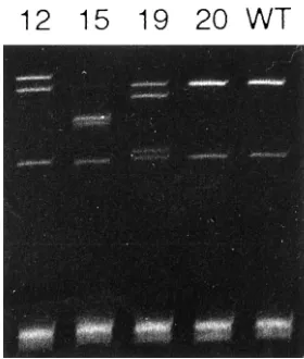
By the direct sequencing of SSCP products, 30 single-nucleotide substitution mutations were de-tected in 20 of 42 cases: 13 cases had a single mutation, 5 had two mutations, 1 had three mutations, and 1 had four mutations (Fig. 2) (Table 1). With the exception of 2 mutations involving the intron 5 and exon 5 terminal point (codon 187), 18 were missense mutations leading to amino acid substitutions, 1 was a nonsense mutation, and 9 were silent mutations
re-sulting in no amino acid changes. No specific muta-tions were observed, although G:C to A:T transition was predominant (19 lesions), followed by A:T to G:C (3 lesions) transition. For mutation frequency by histo-logic grade, 10 of 17 cases (58.8%) in Grade II and 10 of 25 cases (40%) in Grade III showedp53mutations. There were no significant differences in the inci-dence ofp53mutation among the cases in the four areas. However, the incidence in the cases in Osaka was one-half to one-third of those in other areas, although the incidence of abnormal p53 expression was very high in the cases in Osaka (Table 2). The results suggest the difference in the cause of nasal lymphomagenesis between the cases in Osaka and the three other areas, Beijing, Chengdu, and Okinawa. Histologic grade of the cases that stained positive for
p53, but were negative by SSCP, was Grade II in 3 cases and Grade III in 8.
EBV
The proliferating cells in 8 of 10 cases showed positive signals in the nucleus. Percentage of positive cells per 1000 cells counted under high power fields ranged from 11% to 64% (mean, 35%).
Discussion
Pathological characteristics of 42 nasal lymphomas, ie, varying degrees of necrotic change in the upper respiratory tract, and the polymorphous pattern of proliferation were identical with those in LMG of PR type (Eichel et al, 1966; Kassel et al, 1969), which is now classified as an NK/T- or NK-cell lymphoma (Jaffe et al, 1996). Indeed immunohistochemical findings of the present 42 cases were in agreement with those of NK/T- or NK-cell lymphoma, ie, TIA-11, CD561,
CD31, or CD431.
In the non-nasal and ordinary lymphomas, p53
mutations were rare; none of 43 cases in the United States (Gaidano et al, 1991) and 8 of 48 (17%) in Japan (Ichikawa et al, 1992) had mutations. Mean-while it was reported that aggressive high-grade B-cell non–Hodgkin’s lymphoma (NHL) had approximately 30% incidence ofp53mutations, whereas its indolent counterpart rarely had incidence (Lo Coco et al, 1993). Among T-cell lymphomas, frequency of p53 gene mutations was reported to be less than 10%. Con-versely frequency of p53 mutations was high in the specific category of lymphomas; greater than 50% in Burkitt’s lymphoma (Villuendas et al, 1993) and ap-proximately 70% in cases with pyothorax-associated lymphoma (Hongyo et al, 1998). Both types of lym-phoma are known to be EBV-associated. The present study on NK/T-cell lymphoma revealed a high fre-quency (47.6%) of p53 gene mutations. NK/T-cell lymphoma is also EBV-associated (Harabuchi et al, 1990; Tomita et al, 1995). There have been no reports on p53 gene mutations in NK/T-cell lymphoma by using the PCR-SSCP method followed by direct se-quencing. This must be due to availability of a small Figure 1.
Table 1. Mutations and Overexpression ofp53 gene in 42 Cases of Nasal NK/T-Cell Lymphoma from China and Japan
Regions Cases No Age Sex Site
Histologic grade
p53 Expression
p53 mutation
Exon Codon Nucleotide Amino acid
China
Beijing 1 53 M paranasal III 2 nonea
2 47 M nose III 2 7 246 ATG3GTG Met3Val 3 22 M nasopharynx II 1 7 245 GGC3AGC Gly3Ser 8 276 GCC3ACC Ala3Thr
4 34 M tonsilla II 2 none
5 57 M paranasal II 1 none
6 67 M nose III 2 none
7 18 M trachea II 2 5 144 CAG3CTG Gln3Leu
8 31 M nose III 2 5 NDb
9 28 M nose II 1 7 251 ATC3AGC Ile3Ser 10 39 M nose II 1 7 251 ATC3AGC Ile3Ser 11 40 M nose III 2 6 204 GAG3GAA Glu3Glu
intron 5 third position G3A
12 42 M nose II 2 5 142 CCT3CTT Pro3Leu 13 32 M nose II 1 6 193 CAT3TAT His3Leu 14 33 M nasopharynx II 1 none
Chengdu 15 24 M paranasal III 1 5 151 CCC3CTC Pro3Leu 16 79 M nose III 2 5 151 CCC3TCC Pro3Ser 7 245 GGC3AGC Gly3Ser 8 273 CGT3CAT Arg3His
17 60 M nose III 1 none
18 48 M nose III 2 none
19 38 M nose III 2 5 162 ATC3GTC Ile3Val 8 269 AGC3AAC Ser3Asp Japan
Okinawa 20 45 M nose II 2 none
21 64 F nose II 2 5 39terminal G3A
22 39 M nose III 2 6 203 GTG3GAG Val3Term 7 244 GGC3GAC Gly3Asp
23 60 M nose III 2 none
24 53 M nose II 2 5 159 GCC3GCT Ala3Ala 25 69 F nose II 2 8 273 CGT3CAT Arg3His
26 34 M nose III 2 none
27 51 M oral II 2 none
28 71 F nose II 2 none
29 62 M nose III 1 none
30 64 M nose III 2 8 285 GAG3AAG Glu3Lys 31 44 F nose II 2 6 193 ATG3ACG Met3Thr 8 294 GAG3GAA Glu3Glu 32 37 M nose III 1 5 149 TCC3TCT Ser3Ser 5 154 GGC3GGT Gly3Gly 5 161 GCC3GCT Ala3Ala 8 276 GCC3GCT Ala3Ala
33 57 M nose III 1 none
Osaka 34 78 F nose III 1 6 206 TTG TTA Leu3Leu
35 71 M nose II 1 none
36 60 M nose III 1 6 193 CAT GAT His3Asp
37 71 M nose III 1 none
38 48 M nose III 1 none
39 57 M nose III 1 none
40 52 M nose III 2 none
41 50 F oral III 1 none
42 49 M nose III 1 none
amount of samples with necrotic change from PR lesions.
The ability to find a mutation is partially dependent upon the percentage of tumor cells. Based on the criteria proposed by Lipford et al (1988), our cases had Grade II or III histology, composing more than 30% of large atypical cells. Nasal NK/T-cell lymphoma is one of the EBV-associated lymphomas, and the percent-age of EBV genome-containing cells was more than 10% (mean, 35%) in our cases. We previously re-ported that the PCR-SSCP method used in this study could detect the alterations of the p53 gene in 3% cells of population (Hongyo et al, 1993). Thereforep53
mutations could be detected in all of our cases if they occurred.
Transitions at the CpG dinucleotides site were the predominant pattern of substitutions in NHL cases, and G:C to T:A transversion is uncommon (Hollstein et al, 1991). In our series of NK/T-cell lymphoma, more than 70% of substitutions were transitions, however the CpG site was involved in only three lesions. The predominance of transition mutations (G:C to A:T) in our series of NK/T-cell lymphoma suggests that some “endogenous” mutagens act in lymphomagenesis. The transition pattern of thep53mutation is known to be more susceptible to spontaneous genetic instabil-ity than transversion. Meanwhile 75% of the cases in the current series had at least one mutation that
changed an amino acid, which might have provided the selection pressure for expansion.
Mutational analysis of thep53gene has been con-fined principally to exons 5 through 8, because 90% of the mutations in human tumors occurred in this region (Hollstein et al, 1991). In NHL, the predominant site for mutations did not present in previous reports (Adam-son et al, 1995; Ichikawa et al, 1992). In our cases with NK/T-cell lymphoma, exon 5 is the most common site for mutations, ie, 13 of 30 (43%) mutations occurred in exon 5. Some carcinogens might cause mutation in specific codons, as was observed in the mutation of codon 249 in lung and liver cancers induced by irradiation (Anderson et al, 1995; Taylor et al, 1994). Relatively restricted distribution of mutational spots were found in our cases; codon 273, one of the so-called mutational “hot spots” (Hollstein et al, 1991), was involved in 2 cases, and codons 151, 193, and 251 were involved in 2 or 3 cases. Distribution of hot spots inp53 mutations might reflect the function of specific mutant alleles being selected for promoting cell proliferation or the nature and activity of mutagens in different tissue fashion.
Alteration of thep53gene results in accumulation of thep53mutant protein due to its longer half-life time than that of wild type. Thus immunohistochemical detection of the p53 protein could be performed in various kinds of malignancies under supposition that
p53gene mutations might occur frequently in these
tumors. On the nasal lymphomas,
Quintanilla-Martines et al (1998) reported that thep53protein was overexpressed in 22 of 29 cases (76%) examined. In our cases with nasal NK/T-cell lymphomas, 19 of 42 cases (45%) overexpressed thep53protein. Our study showed a discordance betweenp53protein overex-pression and the presence of mutations: 11 of 19 cases (58%) overexpressing the p53protein had no mutations. This kind of discordance had been re-ported in many types of tumors including lymphomas (Adamson et al, 1995; Martinez et al, 1997), and reasons for this were variously speculated. Recently a link between accumulation of a wild typep53protein and EBV has been postulated in the nasopharyngeal carcinoma (Effert et al, 1992). Nasal NK/T-cell lym-phoma is also an EBV-associated disease (Harabuchi et al, 1990; Tomita et al, 1995). Accumulation of a wild type p53 protein was also reported in the acquired immunodeficiency syndrome-related lymphomas (Na-kamura et al, 1993). EBV was studied only in 10 of the current cases, therefore we could not evaluate the relationship between abnormal p53 expression and EBV.
Functional inactivation ofp53through interaction of the wild type gene product with various viral products such as SV 40 large T antigen, adenovirus E1B, human papilloma virus (HPV) E6 protein, or cellular protein MDM2 was reported (Capoulade et al, 1998). In HPV-positive tumors, binding of the E6 protein obviates the need forp53mutations in the genesis of such tumors, but this association was not noted in EBV infection and Hodgkin’s disease (Niedobitek et al, 1993). How-ever, EBV immediate-early protein, BZLF1, which is Table 2. Overexpression and Mutations ofp53 Gene in
the 4 Different Areas
Regions No. of Cases p53 overexpression p53 mutation
Beijing 14 6 (42.9%) 8 (57.1%) Chengdu 5 2 (40.0%) 3 (60.0%) Okinawa 14 3 (21.4%) 7 (50%) Osaka 9 8 (88.9%)a 2 (22.2%)
Total 42 19 (45.2) 20 (47.6)
ap,0.01 versus Okinawa by Fisher’s exact test.
Figure 2.
highly expressed in immunodeficiency syndrome-related lymphoma, can interact withp53and inhibit its function (Zhang et al, 1994). EBNA-5, another EBV encoded protein necessary for transformation of in-fected B cells, can also form complexes with both wild type and mutant p53 protein (Szekely et al, 1993). Taken together, it is possible that EBV gene products could indirectly suppressp53function, thus resulting in overproduction and accumulation of wild typep53
as a compensating function. The lowest incidence of
p53gene mutations and the highest incidence ofp53
protein expression in the Osaka cases in our series suggest the different causes for nasal lymphomagen-esis including racial, environmental, or lifestyle causes.
In conclusion, mutations of the p53 gene are fquent in nasal NK/T-cell lymphoma with rather re-stricted sites for mutations. These findings give an insight on the lymphomagenesis of nasal NK/T-cell lymphoma and also give some suggestions for its treatment.
Materials and Methods
Forty-two cases with PR, 19 from China and 23 from Japan, were selected for the current study: they were admitted to hospitals during the period of 1986 to 1997 (Table 1). All patients presented with the necrotic and granulomatous lesions in the upper respiratory tract, which were biopsied for histologic diagnosis before treatment. Age of patients ranged from 18 to 79 years (median, 49.5) with a male to female ratio (M/F) of 6:1. There were differences in the distribution of age
and sex between Chinese and Japanese patients: in China, a median age of 42 years and all men, and in Japan, a median age of 53 years and M/F ratio of 2.8:1. The nasal cavity and paranasal sinuses were the most common sites (37 cases) for involvement fol-lowed by naso-/oropharynx (3 cases), tonsil (1 case), and trachea (1 case). Histologic specimens were fixed in 10% formalin and routinely processed for paraffin-embedding. All of the paraffin blocks were gathered from Osaka University, and 3 mm sections were cut and stored at 4° C before staining with hematoxylin-eosin and immunohistochemical procedures at the same time. Histologic slides were reviewed by two of the authors (LT, KA) for diagnosis. To show the num-ber of the proliferating cells in the lesions, histologic grade was determined based on the criteria described by Lipford et al (1988).
Immunohistochemistry
Immunohistochemical study of the paraffin sections was carried out using the ABC method. The primary antibodies used in the study, their suppliers, and dilutions were as follows: CD3 (1:100; Dakopatts, Glostrup, Denmark), MT-1(CD43) (1:50; Bioscience, Emmenbrucke, Switzerland), Mx-PanB (CD20) (1:200; Kyowa Medex, Tokyo, Japan), 123C3 (CD56) (1:40; Zymed, South San Francisco, California), ZH7 (CD16) (1:200; Novocastra, Newcastle, United Kingdom), and TIA-1(1:500; Coulter, Hialeah, Florida). The alka-line phosphatase-anti-alkaalka-line-phosphatase (APAAP) method was used inp53protein detection with use of monoclonal anti-human p53 protein (DO-7) (Dako-Table 3. p53 Mutations in Malignant Lymphomas
Lymphomas Histology
Burkitt Lymphoma Burkitt type 2/4 (50) 3/4 (75) 1/3 (33) 1/3 (33) 1/3 (33)
Burkitt type NDd 9/27 (33) 5/11 (45) 4/11 (36) 2/11 (18) Gaidano et al,
1991 AIDS-Related
Lymphoma
Diffuse large cell 22/34 (65) 1/34 (3) 1/1 (100) 0/1 (0) 1/1 (100)
Small non-cleaved cell
10/24 (42) 10/27 (37) 8/10 (80) 5/10 (50) 5/10 (50)
Mucosa-Associated B-cell type ND 21/75 (28) 6/21 (29) 4/21 (19) 3/21 (14) Lymphoid Tissue
Lymphoma
aone deletion.
bp50.05 by 2 test with Yates’ correction and/or Fisher’s exact tests versus other kinds of lymphomas or cancers except squamous cell carcinoma of skin and
radon-associated lung cancer.
cp50.05 except basal cell carcinoma and squamous cell carcinoma of the skin and AIDS-related lymphoma. Statistical analysis were not done for the samples
with less than four cases.
patts) diluted at 1:10 as the primary antibody. When CD3, CD56, and DO-7 were used as primary antibody, sections were treated in a microwave oven for 15 minutes in 10 mM citrate buffer (10 mM citrate
mono-hydrate in distilled water, pH 6.0) for antigen retrieval. Cases with more than 10% of tumor cells positive for DO-7 were regarded as positive.
DNA Extraction and PCR for p53 Gene
DNA for PCR amplification was extracted using chela-tion resin. Three 10-mm thick paraffin sections were cut, transferred into sterile distilled water containing 20% chelating resin iminodiacetic acid (Sigma, St. Louis, Missouri), and boiled for 30 minutes. After centrifugation, the supernatant was transferred to a sterile 500ml tube and stored at 220° C. The PCR primer pairs for the amplification of thep53gene exons 5 through 8 were: (a) 59-GTACTCCCCTGCCCTCAACA-39and 59 -CTCACC-ATCGCTATCTGAGCA-39 for exon 5; (b) 59
-TTGCT-CTTAGGTCTGGCCCC-39 and 59
-CAGACCTCAGGC-GGCTCATA-39for exon 6; (c) 59
-TAGGTTGGCTCTG-ACTGTACC-39 and 59
-TGACCTGGAGTCTTCCAGT-GT-39 for exon 7; and (d) 59
-AGTGGTAATCTACTG-GGACGG-39 and 59-ACCTCGCTTAGTGCTCCCTG-39
for exon 8.
Hot start PCR was performed as follows: 45 cycles of denaturation at 95° C for 30 seconds; annealing at 58° C, 62° C, 60° C, and 60° C for 30 seconds for exon 5, 6, 7, and 8, respectively; extension at 72° C for 1 minute, and final extension at 72° C for 7 minutes. Paraffin blocks containing no sample were cut and used as negative controls throughout the procedures. The amplified products were subjected to electro-phoresis in 1.5% agarose gel containing 2 mg/ml ethidium bromide in TBE buffer. After electrophoresis, the gels were examined under an ultraviolet light transilluminator.
SSCP
Nonradioactive SSCP was performed as previously reported (Hongyo et al, 1995). Twenty microliters of reaction mixtures containing 5 ml of PCR product (20 –200 ng of DNA), 0.2 ml of 1M methylmercury hydroxide, 3ml of loading buffer (15% Ficoll, 0.25% bromphenol blue, 0.25% xylene cyanol), and TBE buffer were heated to 90° C for 4 minutes and put on ice and then electrophoresed in 18% polyacrylamide TBE gel at 300 volts, while maintaining the tempera-ture at 35° C for exon 5, 5° C for exon 6, and 25° C for exons 7 and 8. The gels were stained with 0.5mg/ml ethidium bromide in TBE buffer for 20 minutes at room temperature. The bands migrated apart from that of wild type were determined as SSCP positive. The bands possibly mutated by SSCP were extracted from the gels and amplified by 25 cycles of PCR to enrich the mutated alleles.
Direct Sequencing
Sequencing was carried out on PCR products of SSCP positive cases. To purify single or double
stranded PCR products with a range of 100 bp, the PCR products were processed using the QIAquick PCR purification kit (QIAGEN Inc., Valencia, California) according to the manufacturer’s protocol. Sequencing was performed by the dideoxy chain termination
method using the Big Dye Terminator
cycle-sequencing kit (Perkin-Elmer Corporation, Foster City, California). The same primers were used as for PCR. Cycle sequencing was performed following the proto-col, ie, 30 cycles of denaturation (95° C, 20 seconds), annealing (54° C, 30 seconds), and extension (72° C, 3 minutes). After ethanol precipitation, the samples were analyzed by the Genetic Analyzer (ABI Prism 310, Perkin-Elmer Corporation). The PCR-SSCP analysis and sequencing of the possible positive cases were repeated three times to rule out the contamination and artifacts.
In Situ Hybridization for EBV
EBV RNA in situ hybridization (ISH) was performed in 10 cases as previously described (Weiss et al, 1991). As a positive control, the Raji cell line was used. Hodgkin’s disease of mixed cellularity type with the EBV genome was also included as positive control. As negative controls, the hybridizing mixture was used with (a) sense probe and (b) antisense probe after RNase (Sigma) treatment.
Acknowledgements
The authors thank Dr. Wei-ping Liu (Department of Pathology, West-China University of Medical Science, Chengdu, China) for providing histologic materials.
References
Adamson DJA, Thompson WD, Dawson AA, Bennett B, and Haites NE (1995).p53mutation and expression in lymphoma. Br J Cancer 72:150 –154.
Anderson M, Jonsson M, Nielsen LL, Vyberg M, Visfeldt J, Storm HH, and Wallin H (1995). Mutations in the tumor suppressor gene p53 in human liver cancer induced by alpha-particles. Cancer Epidemiol Biomarkers Prev 4:765– 770.
Aozasa K, Ohsawa M, Tajima K, Sasaki R, Maede H, Matsu-naga T, and Friedmann I (1989). Nation-wide study of lethal midline granuloma in Japan: frequencies of Wegener’s gran-ulomatosis, polymorphic reticulosis, malignant lymphoma and other related conditions. Int J Cancer 44:63– 66.
Capoulade C, Bressac PB, Lefrere I, Ronsin M, Feunteun J Tursz T, and Wiels J (1998). Overexpression of MDM2, due to enhanced translation, results in inactivation of wild-typep53 in Burkitt’s lymphoma cells. Oncogene 16:1603–1610.
Donehower LA, Harrey M, Slagle BL, McArthur MJ, Mont-gomery CA Jr, Butel J, and Bradley A (1992). Mice deficient forp53are developmentally normal but susceptible to spon-taneous tumors. Nature 356:215–221.
Eichel BS, Harrison EGJ, Devine KD, Scanlon PW, and Brown HA (1966). Primary lymphomas of the nose including rela-tionship to lethal midline granuloma. Am J Surg 112:597– 605.
Gaidano G, Ballerini P, Gong JZ, Inghirami G, Neri A, New-comb EW, Magrath IT, Knowles DM, and Dalla-Favera R (1991). p53 mutations in human lymphoid malignancies: association with Burkitt lymphoma and chronic lymphocytic leukemia. Proc Natl Acad Sci USA 88:5413–5417.
Harabuchi Y, Yamanaka N, Kataura A, Imai S, Kinoshita T, Mizuno A, and Osato T (1990). Epstein-Barr virus in nasal T-cell lymphomas in patients with lethal midline granuloma. Lancet 335:128 –130.
Hollstein M, Sidransky D, Vogelstein B, and Harris SR (1991). p53mutations in human cancers. Science 253:49 –53.
Hongyo T, Buzard GS, Calvert RJ, and Weghorst CM (1993). ‘Cold SSCP’: a simple, rapid and non-radioactive method for optimized single strand conformation polymorphism analy-ses. Nucl Acid Res 21:3637–3642.
Hongyo T, Buzard GS, Palli D, Weghorst CM, Amorosi A, Galli M, Caporaso NE, Fraumeni JF, and Rice JM (1995). Mutations of the k-ras andp53gene in gastric adenocarci-noma from a high incidence region around Florence, Italy. Cancer Res 55:2665–2672.
Hongyo T, Kurooka M, Taniguchi E, Iuchi K, Nakajima Y, Aozasa K, and Nomura T (1998). Frequentp53mutations at dipyrimidine sites in patients with pyothorax-associated lym-phoma. Cancer Res 58:1105–1107.
Ichikawa A, Hotta T, Takagi N, Tsushita K, Kinoshita T, Nagai H, Murakami Y, Hayashi K, and Saito H (1992). Mutations of p53gene and their relation to disease progression in B-cell lymphoma. Blood 79:2701–2707.
Jaffe ES, Chan JKC, Su I-J, Frizzera G, Mori S, Feller AC, and Ho FCS (1996). Report of the workshop on nasal and related extranodal angiocentric T/natural killer cell lymphomas. Am J Surg Pathol 20:103–111.
Kassel S, Echevaria RA, and Guzzo FP (1969). Midline malignant reticulosis (so-called lethal midline granuloma). Cancer 23:920 –935.
Levine AJ, Momand J, and Finley CA (1991). Thep53tumor suppressor gene. Nature 351:453– 456.
Lipford EH Jr, Margolick JB, Longo DL, Fauci AS, Jaffe ES (1988). Angiocentric lymphoproliferative lesions: A clinico-pathologic spectrum of post-thymic T-cell proliferations. Blood 72:1674 –1681.
Lo Coco F, Gaidano G, Louie DC, Offit K, Chaganti RSK, and Dalla-Favera R (1993).p53 mutations are associated with histologic transformation of follicular lymphoma. Blood 82: 2289 –2295.
Martinez DB, Robledo M, Arranz E, Infantes F, Echezarreta G, Marcos B, Sanz C, Rivas C, and Benitez J (1997). Correlation between mutations inp53 gene and protein expression in human lymphomas. Am J Hematol 55:1– 8.
Nakamura H, Said JW, Miller CW, and Koeffer HP (1993). Mutation and expression of p53 in acquired immunodefi-ciency syndrome-related lymphoma. Blood 82:920 –926.
Niedobitek G, Rowlands DC, Yong LS, Herbst H, Williams A, Hall P, Padfield J, Rooney N, and Jones EL (1993). Overex-pression ofp53in Hodgkin’s disease: lack of correlation with Epstein-Barr virus infection. J Pathol 169:207–212.
Ohsawa M, Nakatsuka S, Kanno H, Miwa H, Kojya S, Harabachi Y, Yang WI, and Aozasa K (1999). Immunopheno-typic and genoImmunopheno-typic characterization of nasal lymphoma with polymorphic reticulosis morphology. Int J Cancer 81:865– 870.
Quintanilla-Martinez L, Guerrero I, Franklin JL, Naresh KN, Krenacs L, Rama-Rao C, Bhatia K, Magrath IT, and Raffeld M (1998). Nasal NK/T-cell lymphoma from Peru. High preva-lence ofp53overexpression. Mod Pathol 11:139A.
Szekely L, Selivanova G, Magnusson KP, Klein G, and Wiman KG (1993). EBNA-5, an Epstein-Barr virus-encoded nuclear antigen, binds to the retinoblastoma andp53proteins. Proc Natl Acad Sci USA 90:5455–5459.
Taylor JA, Watson MA, Devereux TR, Michels RY, Saccom-anno G, and Anderson M (1994).p53mutation hotspot in radon-associated lung cancer. Lancet 8:86 – 87.
Tomita Y, Ohsawa M, Mishiro Y, Kubo T, Maeshiro N, Kojya S, Noda Y, and Aozasa K (1995). The presence and sub-type of Epstein-Barr virus in B- and T-cell lymphomas of the sino-nasal region from the Osaka and Okinawa districts of Japan. Lab Invest 73:190 –196.
Villuendas R, Piris MA, Algara P, Sanchez-Beato M, Sanchez-Verde L, Martinez JC, Orradre JL, Garcia P, Lopez C, and Martinez P (1993). The expression ofp53protein in non-Hodgkin’s lymphoma is not always dependent onp53 gene mutations. Blood 82:3151–3156.
Weiss LM, Chen YY, Liu XF, and Shibata D (1991). Epstein-Barr virus Hodgkin’s disease: a correlative in situ hybridiza-tion and polymerase-chain-reachybridiza-tion study. Am J Pathol 139: 1259 –1265.