Shin-ichi Nakatsuka,1
Tadashi Hongyo,2
Mukh Syaifudin,2
Taisei Nomura,2
Norihisa Shingu1 and Katsuyuki Aozasa1, 3
Departments of 1Pathology and 2Radiation Biology, Osaka University Graduate School of Medicine, 2-2
Yamadaoka, Suita, Osaka 565-0871
Malignant lymphoma of the adrenal gland is a rare disease, usually with diffuse large cell morphol-ogy and B-cell immunophenotype, and often associated with Epstein-Barr virus infection. In this study, mutations of p53, c-kit, K-ras, and ββββ-catenin gene were analyzed in 17 cases (13 males and four females with ages ranging from 25 to 84 years) of such lymphomas by polymerase chain reac-tion-single strand conformation polymorphism followed by direct sequencing. Selected exons in each gene, representing hot spots, were analyzed. All 44 mutations detected were single-nucleotide substitutions and 33 were missense mutations. Nineteen mutations were detected in exon 5 and/or 7 of the p53 gene in nine of 17 cases (52.9%) and 21 in exon 11 and/or 17 of the c-kit gene in 10 of 14 cases (71.4%). Bilateral adrenal lesions in one case who had not received any adjuvant therapy showed different mutational patterns of the p53 and c-kit genes, suggesting different clonal evolu-tion of lymphoma between the left and right sides. Mutaevolu-tion at codon 13 of the K-ras gene was detected in one of 14 cases (7.1%), and in exon 3 of the ββββ-catenin gene in three of 12 cases (25%). All but one mutation were transition mutations, indicating that some endogenous mutagens act in lymphomagenesis in the adrenal gland. Our results suggest that p53 and c-kit gene mutations might play a role in adrenal lymphomagenesis .
Key words: Adrenal lymphoma — p53 — c-kit — K-ras — β-Catenin
The adrenal gland is involved during the course of non-Hodgkin’s lymphomas (NHL) in approximately one-fourth of cases.1) However, initial manifestation of NHL in the adrenal gland is extremely rare.2)
In such cases, patients usually present with nonspecific signs and symptoms, such as fever, fatigue, and weight loss. Physical and roentgeno-graphic examinations usually reveal huge and bilateral adrenal masses without lymphadenopathy, accompanied by adrenal insufficiency in some cases.2) Prognosis is very poor compared to other types of extranodal NHL. Histo-logic and immunohistochemical studies show that most of cases are diffuse large B-cell lymphomas.2)
Adrenal lymphoma is one of the Epstein-Barr virus (EBV)-asso-ciated lymphomas: EBV genome was detected in 45% of the cases with occasional expression of latent membrane protein-1.3)
Accumulation of gene mutations results in development of malignant tumors, including NHL. In this study, the mutations of the p53, c-kit, K-ras, and β-catenin genes were examined in 17 cases of NHL with initial manifesta-tion in the adrenal gland.
The p53 gene is a well-known tumor suppressor gene that causes cell cycle arrest at the G1 phase or stimulates expression of the bax gene, the protein that promotes
apo-ptosis in cells with damaged DNA.4) In a wide variety of human cancers, p53 gene mutations have been detected mainly in exons 5 through 8.5) High incidence of malig-nant lymphoma in p53 knockout mice has been reported,6) suggesting an important role of p53 gene mutations in lymphomagenesis.
The c-kit gene encodes a receptor tyrosine kinase, and the signal transduction mediated by c-kit receptor tyrosine kinase (KIT) plays a crucial role in proliferation and dif-ferentiation of hematopoietic stem cells, mast cells, and interstitial cells of Cajal.7, 8) Activating mutation of KIT (Asp816→Val) in the kinase domain of the c-kit gene was described in mast cell disorders.9) The development of acute leukemia or malignant lymphoma was also reported in transgenic mice expressing a KIT mutant (Asp816→ Val).10)
Recently, a relative high frequency of c-kit gene muta-tions in human nasal NK/T-cell lymphoma was reported.11) The K-ras gene encodes a 21-kD ras protein, GTP- and GDP-binding protein, which plays a role in signal trans-duction through transmembrane signaling systems.12)
K-ras mutations are frequently observed in pancreatic, colo-rectal, and lung adenocarcinomas,12) but rarely in the ordi-nary type of NHL.13) Thyroid lymphoma, a lymphoma which develops in autoimmune thyroiditis, showed rela-tively frequent mutations of the K-ras gene (25% of cases).14) The mutations tended to accumulate in high-grade B-cell lymphoma with replication error phenotype, 3To whom requests for reprints should be addressed.
suggesting a possible association of alterations of the K-ras gene with a subset of high-grade B-cell lymphomas. Because the majority of adrenal lymphoma was high-grade B-cell lymphoma, we examined K-ras mutations in the present study.
β-Catenin is associated with E-cadherin-mediated cell-cell adhesion and acts downstream of the Wnt signaling pathway.15, 16) Activating mutations of the β-catenin gene at a crucial regulatory site in exon 3 result in accumulation of β-catenin protein in the cytoplasm.17) The activated β-catenin complex leads to overexpression of c-myc,18) which is related to cell proliferation, thus causing tumori-genesis. In fact, mutations of the β-catenin gene were reported in cancers of the colon,19) endometrium,20) and liver.21) Although there have been no reports describing mutations of the β-catenin gene in malignant lymphomas, Knowles et al. reported a crucial role of overexpression of the c-myc gene in the development of EBV-associated lymphoproliferative disorders.22) Because adrenal lym-phoma is occasionally EBV-associated, we examined β-catenin gene mutations.
MATERIALSAND METHODS
Case selection Seventeen cases of NHL with initial man-ifestation in the adrenal gland were selected for this study through a review of the “Annual of the Pathological Autopsy Cases in Japan (1992–1996)” (11 cases) and the Japanese medical journals (six cases). Clinical findings of these cases are summarized in Table I. There were 13 males and four females with ages ranging from 25 to 84 (median 68) years. They were admitted to hospitals during 1985 to 1996. The histologic diagnosis of adrenal NHL was made by biopsy (eight cases), surgery (two cases), or autopsy (seven cases). Bilateral adrenal glands were involved at presentation in all but two cases (cases 6, 16). In all cases, the main masses were located in the adrenal region, and the clinical stage was I in one case, II in 11, and III in two. Mediastinal mass was not noted in any patient during the clinical course. Endocrinological abnor-malities such as serum adrenocorticotropic hormone, serum renin, urine noradrenaline, dopamine, low serum aldosterone, and/or lack of responsiveness to rapid adrenocorticotropic hormone test were found in eight of the 11 cases evaluated. In two cases (cases 8 and 10), sam-ples from adrenal glands on both sides were examined (Table II). Metastatic lesions of lymph nodes and liver were also examined in cases 5 and 12, respectively. Histo-logic specimens were fixed in 10% buffered or neutral for-malin, and routinely processed for paraffin-embedding. Histologic sections, cut at 3 µm, were stained with hema-toxylin-eosin and by means of immunohistochemical pro-cedures. Sources of samples for molecular study are shown in Table II. Biopsy and surgery were performed
prior to chemotherapy and/or radiotherapy. Among 12 cases in which autopsy materials were used for analysis, seven received chemotherapy or radiation, but five did not receive any adjuvant therapy.
Immunohistochemistry Immunohistochemical study on the paraffin-embedded sections was carried out using the avidin-biotin-peroxidase complex method. Primary anti-bodies used in the study, suppliers and dilutions were as follows: CD3 (Dakopatts, Glostrup, Denmark; 1:100), CD43 (Bioscience, Emmenbrucke, Switzerland; 1:50), CD20 (Kyowa Medex, Tokyo; 1:200), CD45RO (Dako-patts; 1:100), and MB-1 (Bioscience; 1:50). Sections were treated with 0.1% trypsin solution (Sigma, St. Louis, MO) at 37°C for 30 min before reaction with anti-CD3. Histo-logic sections were reviewed by two of the authors (S. N. and K. A.), and lymphomas were classified based on the Revised European-American classification of lymphoid neoplasms.23) The alkaline phosphatase-anti-alkaline-phos-phatase method was used in p53 protein detection with monoclonal anti-human p53 protein (DO-7) (Dakopatts) diluted at 1:10 as the primary antibody. When DO-7 was used as the primary antibody, sections in 10 mM citrate buffer (10 mM citrate monohydrate in distilled water, pH 6.0) were treated with a microwave oven for 15 min for antigen retrieval. Cases with more than 10% of tumor cells positive for DO-7 were regarded as positive.
Detection of p53, K-ras, c-kit, and ββββ-catenin gene muta-tions DNA for PCR amplification was extracted from paraffin sections using chelating resin. Selected exons in each gene, representing hot spots, were analyzed in this study. Sequences of the PCR primer pairs for the amplifi-cation of exon 5 of p53 gene were 5′-TCTGTCTCCTTC-CTCTTCCTA-3′ and 5′-CATGTGCTGTGACTGCTTGT-3′ for the upper half region, and 5′-TGTGCAGCT-GTGGGTTGATTC-3′ and 5′-CAGCCCTGTCGTCTCTC-CAG-3′ for the lower half. Primer sequences for exons 6 through 8 of the p53 gene, exon 1 of the K-ras gene, exons 11 and 17 of the c-kit gene, and exon 3 of the β-catenin gene were described previously.11, 14, 24) DNA amplification and non-radioactive single strand conforma-tion polymorphism (Cold SSCP) analyses were carried out to detect mutations as described previously.25) The mutated bands at SSCP were extracted from the gel and reampli-fied by polymerase chain reaction (PCR) for 25 cycles to enrich mutated alleles.
RESULTS
Histological and immunohistochemical findings Histo-logically all cases showed a diffuse proliferation of large lymphoid cells of predominantly noncleaved cell morphol-ogy, with highly pleomorphic cells in one case (case 5). Immunohistochemically, 15 cases were CD20+, MB-1+/ −, CD43−/+, CD45RO−/+, CD3−; they were judged as diffuse large B-cell lymphomas. Two cases (cases 6 and 12) were CD20−, MB-1−, CD43+/−, CD45RO+, CD3+, and thus were judged as peripheral T-cell lymphomas, not specified (Table II). Tumor cells invaded the surrounding fat tissue, frequently yielding necrosis and fibrosis, often with hyalinization. Prominent intravascular proliferation of tumor cells in the adrenal gland and other organs was found in nine cases (Fig. 1). Positive immunoreactivity for DO-7 was found in seven of 21 (33.3%) lesions examined.
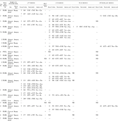
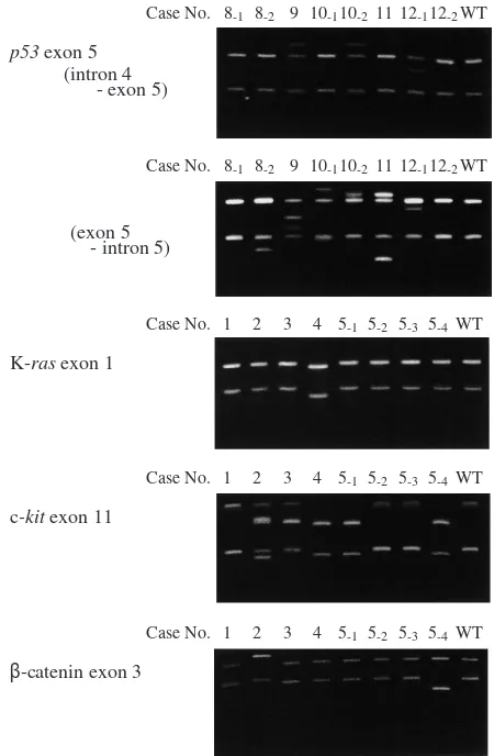
Mutations of p53, c-kit, K-ras, and ββββ-catenin gene Results of gene mutation analyses are summarized in Table II, and the electrophoretic patterns of representative cases are illustrated in Fig. 2. Not all genes/exons could be studied due to failure of PCR in some target fragments of genes, which might be caused by artifacts from sample processing. By the direct sequencing of SSCP products, 19 single-nucleotide substitution mutations of p53 gene were detected in nine of 17 cases (52.9%); 17 mutations (eight cases) at exon 5 and two mutations (two cases) at exon 7. In cases 3, 9, 10 (left gland), and 11, two or more muta-tions in exon 5 were detected in the extracts from aberrant bands on SSCP electrophoresis. In that case, direct sequencing of the full length of the products was per-formed in the same experiment. This indicated that these multiple mutations were present in the same allele. Cases 8 and 10 showed different patterns of mutations between
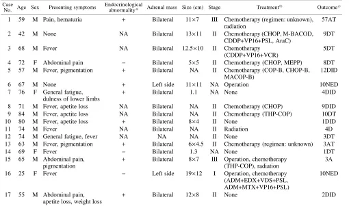
Table I. Clinical Findings in 17 Cases of Adrenal Lymphoma
Case
No. Age Sex Presenting symptoms
Endocrinological
abnormalitya) Adrenal mass Size (cm) Stage Treatmentb) Outcomec)
1 59 M Pain, hematuria + Bilateral 11×7 III Chemotherapy (regimen: unknown), radiation
57AT
2 42 M None NA Bilateral 13×11 II Chemotherapy (CHOP, M-BACOD,
CDDP+VP16+PSL, AraC)
9DT
3 68 M Fever NA Bilateral 12.5×10 II Chemotherapy
(CDDP+VP16+VCR)
5DT
4 72 F Abdominal pain − Bilateral 5×5 II Chemotherapy (CHOP, MEPP) 8DT 5 57 M Fever, pigmentation + Bilateral NA II Chemotherapy (COP-B, CHOP-B,
MACOP-B)
12DID
6 67 M None + Left side 11×11 NA Operation 10NED
7 76 F General fatigue, dulness of lower limbs
+ Bilateral 1.1 NA None 4DID
8 71 M Fever, apetite loss NA Bilateral NA II Chemotherapy (CHOP) 9DID
9 84 M Fever, apetite loss NA Bilateral NA II Chemotherapy (THP-COP) 10DT
10 80 M Fever, apetite loss + Bilateral 8×4 II None 1DID
11 74 M Fever NA Bilateral NA II Radiation 4D
12 74 M General fatigue, fever NA NA NA II None 3DT
13 63 M Fever, pigmentation + Bilateral 6×4.5 II Chemotherapy (regimen: unknown) 3AT
14 69 F Fever − Bilateral 1.3 NA None 1DT
15 65 M Abdominal pain, pigmentation
+ Bilateral 8×7 III Operation, chemotherapy (THP-COP), radiation
3A
16 25 F Fever − Left side 19×12 I Operation, chemotherapy
(ADM+EDX+VDS+PSL, ADM+MTX+VP16+PSL)
10NED
17 55 M Abdominal pain, apetite loss, weight loss
+ Bilateral 12×8 II None 2DID
a) High in serum adrenocorticotroptic hormone, serum renin, urine noradrenaline, dopamine, low in serum aldosterone, and no respon-sive to rapid adrenocorticotropic hormone test.
b) ADM, adriamycin; AraC, cytarabine; BLE, bleomycin; CDDP, cisplatinum; EDX, cyclophosphamide; MTX, methotrexate; PSL, prednisolone; VCR, vincristine; VDS, vindesine; VP16, etposide. CHOP: EDX, ADM, VCR, PSL. CHOP-B: CHOP+BLE. COP-B: CTX, VCR, PSL, BLE. MACOP-B: MTX, doxorubicin, CTX, VCR, PSL, BLE. M-BACOD: MTX, BLE, ADM, EDX, VCR, dexa-methasone. MEPP: mitoxantrone, VP16, CDDP, PSL. THP-COP: therarubicin, CTX, VCR, PSL.
the left and right lesions. In case 10, a single mutation was found in the right adrenal gland and four in the left. Four-teen mutations were missense mutations leading to amino acid substitutions, and five were silent mutations resulting
in no amino acid changes. G:C to A:T transitions were the predominant pattern of mutations (14 of 19 mutations, 73.7%), and the others were A:T to G:C transitions. Trans-version mutations were never observed. Expression of p53 Table II. Mutations of p53, c-kit, K-ras, and β-Catenin Gene in 17 Cases of Adrenal Lymphoma
Case No.
Histo-logic
diagno-sis
Samples for
molecular study p53 mutation DO-7 expres-sion
c-kit mutation K-ras mutation β-Catenin gene mutation
Site
Proce-dure Exon Codon Nucleotide Amino acid Exon Codon Nucleotide Amino acid Exon Codon Nucleotide Amino acid Exon Codon Nucleotide Amino acid
1 DLBL Adrenal gland
Biopsy 5 168 CAC→TAC His→Tyr − — — — 7 247 AAC→AAT Asn→Asn −
2 DLBL Adrenal
gland Autopsy — 11 566 AAC→AAT Asn→Asn — 32 GAC→CAC Asp→His 17 825 GTT→GCT Val→Ala
3 DLBL Adrenal gland
Autopsy 5 142 CCT→TCT Pro→Ser + 11 566 AAC→AAT Asn→Asn — ND 5 151 CCC→CTC Pro→Leu 11 585 CCC→CCT Pro→Pro
4 DLBL Adrenal
gland Autopsy — − 11 557 TGG→CGG Trp→Arg 1 13 GGC→GAC Gly→Asp — 17 825 GTT→GCT Val→Ala
5-1 DLBL Adrenal
gland Autopsy — − 11 557 TGG→CGG Trp→Arg — — 17 822 AAT→AAC Asn→Asn
17 825 GTT→GCT Val→Ala -2 DLBL Adrenal
gland Autopsy — − — — —
-3 DLBL Virchow
LN Biopsy — + — — —
-4 DLBL Cervical
LN Autopsy —
− 11 557 TGG→CGG Trp→Arg — 40 ACT→ACC Thr→Thr
17 825 GTT→GCT Val→Ala 6 PTCL Adrenal
gland Surgery — + ND — ND
7 DLBL Adrenal
gland Autopsy — − 17 825 GTT→GCT Val→Ala — ND 8-1 DLBL Adrenal
gland (lt)Autopsy — ND 17 825 GTT→GCT Val→Ala — ND -2 DLBL Adrenal
gland (rt)Autopsy 5 172 GTT
→GCT Val→Ala + — — —
9 DLBL Adrenal
gland Autopsy 5 133 ATG
→ACG Met→Thr − 17 825 GTT→GCT Val→Ala — —
5 144 CAG→CGG Gln→Arg 5 177 CCC→CTC Pro→Leu 10-1 DLBL Adrenal
gland (rt)
Autopsy 5 168 CAC→CAT His→His − 11 554 GAA→GGA Glu→Gly ND ND 11 559 GTT→ATT Val→Ile
17 825 GTT→GCT Val→Ala -2 DLBL Adrenal
gland (lt)
Autopsy 5 133 ATG→ACG Met→Thr − 11 566 AAC→GAC Asn→Asp ND — 5 144 CAG→CGG Gln→Arg
5 170a) ACG→ACA Thr→Thr
5 171 GAG→GAA Glu→Glu 11 DLBL Adrenal
gland
Autopsy 5 166 TCA→TTA Ser→Leu − — — — 5 177 CCC→CCT Pro→Pro
12-1 PTCL Adrenal
gland Autopsy 5 172 GTT→ATT Val→Ile + 11 574 ACA→ATA Thr→Ile — — 7 248a) CGG→TGG Arg→Trp
-2 PTCL Liver Autopsy — + — — —
13 DLBL Adrenal
gland Biopsy — ND ND ND ND
14 DLBL Adrenal gland
Autopsy 5 172 GTT→ATT Val→Ile − 11 551 CCC→TCC Pro→Thr — 40 ACT→ACC Thr→Thr 11 557 TGG→CGG Trp→Arg
15 DLBL Cervical
LN Biopsy — + — — —
16 DLBL Adrenal
gland Surgery 5 177 CCC→CTC Pro→Leu − ND ND ND 17 DLBL Adrenal
gland Autopsy —
− — — —
a) G:C to A:T transition at CpG dinucleotides site.
protein was found in four of 11 lesions without gene muta-tions and three of 10 with mutamuta-tions.
As for the c-kit gene, 21 single-nucleotide substitution mutations were detected in 10 of 14 cases (71.4%); 12 mutations (seven cases) at exon 11 and nine mutations (seven cases) at exon 17. As found in the p53 mutations, two mutations in exon 11 of cases 3, 10, and 14, and in exon 17 of case 5 (sample 5-1) were considered to occur in the same allele. Bilateral lesions in cases 8 and 10 showed different patterns of c-kit mutations, as observed in the case of p53 mutations. Case 10 showed three muta-tions in the right adrenal gland and one in the left. Codon 825 was involved in seven cases, and both codons 557 and 566 were involved in three. Seventeen mutations were missense and four were silent. Six (28.6%) of 21 muta-tions were G:C to A:T transimuta-tions and the others were A:T to G:C transitions.
Mutation of the K-ras gene was detected only in one case (case 4) (7.1%). It was a G:C to A:T transition muta-tion at codon 13 of exon 1, and was a missense mutamuta-tion (Gly→Asp). Three mutations of the β-catenin gene were detected in three of 12 cases (25.0%).
The frequencies of p53, c-kit, K-ras, and β-catenin gene mutations found in samples before chemotherapy or radio-therapy were 60%, 80%, 0%, and 25%, respectively, and those after therapies was 50%, 67%, 10%, and 25%. The frequencies before and after therapies were not signifi-cantly different.
DISCUSSION
Recent reports have shown that p53 mutations are asso-ciated with poor chemoresponsiveness and unfavorable
prognosis in patients with aggressive B-cell lymphoma.26) Döhner et al. reported that 17p deletion, possibly includ-ing the p53 gene, was involved in the disease progression and affected the survival in chronic lymphocytic leuke-mia.27) As for NHL, none of 43 cases in the United States28) and eight of 48 (17%) in Japan29) were reported to have p53 mutations. In contrast, Lo Coco et al. reported that p53 mutations were rather frequent (approximately 30%) in the aggressive type of B-cell NHL.30) p53 muta-tion is frequent in EBV-associated lymphomas; more than 50% in Burkitt’s lymphoma,31) 67% in pyothorax-associ-ated lymphoma(PAL),32)
the lymphoma developing in the pleural cavity of patients with long-standing pyothorax, and 48% in nasal NK/T-cell lymphoma.33) Adrenal lym-phoma is also associated with EBV infection,3) and in the
Fig. 1. Lymphoma cells show an intravascular proliferation in the connective tissue surrounding the adrenal gland (hematoxylin and eosin ×200).
Case No.
p53 exon 5 (intron 4
- exon 5)
8-1 8-2 9 10-110-211 12-112-2WT
Case No.
K-ras exon 1
1 2 3 4 5-1 5-2 5-3 5-4WT
Case No.
c-kit exon 11
1 2 3 4 5-1 5-2 5-3 5-4WT
Case No.
β-catenin exon 3
1 2 3 4 5-1 5-2 5-3 5-4WT
Case No.
(exon 5 - intron 5)
8-1 8-2 9 10-110-211 12-112-2WT
present study, we found a high frequency of p53 mutations in adrenal lymphomas (nine of 17 cases, 52.9%), as in other EBV-associated lymphomas.
Predominant sites of p53 mutations were not found in the previous studies on NHL,34) but most of the mutations in adrenal lymphoma were observed in exon 5 (17 of 19, 89.5%). This was also observed in EBV-associated lym-phomas, PAL32)
and nasal NK/T-cell lymphoma.33) In case 12, mutation was found at codon 248, one of the so-called mutational “hot spots,” 5) which involves an amino acid residue directly binding to DNA. Thus, this mutation might induce impairment of transcriptional activity of p53 protein. Mutations at codons 133, 142, 172, and 177 found in our cases were located at highly conserved regions in the core domain of the p53 protein, although whether these mutations alter the function of p53 protein is unclear. Restricted distribution of the mutations affecting codons 133, 144, 168, 172, and 177 might be a consequence of the selection of tumor clones with specific mutant alleles or an effect of specific mutagens.
As for the c-kit gene, the current study revealed a high frequency of mutations (10 of 14, 71.4%) in adrenal lym-phoma. Previous reports showed that mutations at codon 816 in the kinase domain of the human the c-kit gene were frequently found in myelodysplastic disorders with masto-cytosis9) and most gastrointestinal stromal tumors had mutations within an 11 amino acid stretch (codon 550– 560) in the juxtamembrane domain.35) All these muta-tions proved to be gain-of-function mutamuta-tions, because the cells transfected with these c-kit gene mutants show phos-pholyration of tyrosine of KIT and activation of KIT in the absence of stem cell factor, a ligand for KIT.35, 36) In this study, 12 of 21 (57.1%) c-kit mutations were detected in exon 11, i.e., in the juxtamembrane domain. Eight of nine mutations in exon 17 were located at codon 825. Mutation at codon 825 was observed frequently in nasal NK/T-cell lymphoma,11) but this c-kit mutant could induce neither phosphorylation of tyrosine nor acti-vation of KIT in the absence of stem cell factor, and thus proved not to be a gain-of-function mutation.11)
Analyses for functional activity of other kinds of c-kit gene muta-tions in our cases were not performed.
Six cases in our series had both p53 and c-kit gene mutations. Four of 10 cases (40.0%) with c-kit gene muta-tions did not have p53 gene mutations. It was reported that c-kit signals inhibit p53-dependent apoptosis,37) and thus c-kit mutations might play a role in oncogenesis in tumors without p53 gene mutation. Bilateral adrenal lesions were examined in two cases (cases 8 and 10), showing different mutational patterns of p53 and c-kit genes. Samples were obtained at autopsy in both cases. These were chosen because case 8 had received chemotherapy, which might induce a different mutational pattern, whereas case 10 did not receive any adjuvant therapy. The results suggest
dif-ferent clonal evolution of lymphoma between the left and right sides.
Except for thyroid lymphoma, mutation of ras genes in NHL is reported to be rare; none of 88 cases13)
and only three of 123 cases38) were reported. Also in our cases, only one point mutation of the K-ras gene was observed in 14 cases examined.
There have been no comprehensive investigations on β-catenin gene mutations in malignant lymphomas. In this study three mutations of the β-catenin gene were found at exon 3, and two of these were silent mutations, indicating that involvement of the β-catenin gene in the development of adrenal lymphoma might be limited.
All of the mutations in p53, c-kit, K-ras, and β-catenin gene found in this study were transition mutations, with the exception of one mutation (β-catenin gene in case 2). As for p53 gene mutations, G:C to A:T transitions were pre-dominant (14 of 19 mutations, 73.7%), which is consistent with previous reports on lymphoid malignancies.5) G:C to A:T transition is associated with endogenous oxidative or spontaneous deamination of 5-methylcytosine leading to replacement of cytosine by thymine. This replacement is not readily recognized by repair enzymes, thus resulting in G:C to A:T transition. These findings suggest that some endogenous mutagens act in adrenal lymphomagenesis.
Is the mutation frequency associated with any kind of treatment or not? Four of six cases without adjuvant ther-apy showed mutations in a gene, whereas one case receiv-ing combined chemotherapy and radiation did not show any genetic abnormalities. In case 5, no genetic abnormal-ities were found in the lymph node lesion biopsied before treatment, whereas three transition mutations (T→C) were detected in the adrenal and lymph node lesions obtained from autopsy after adjuvant therapy. Myelodysplastic syn-dromes and acute myelogenous leukemia developing in patients treated with cyclophosphamide, an alkylating agent, preferentially showed single-base substitutions at A:T pairs in the p53 gene.39)
In conclusion, the p53 and c-kit genes were frequently mutated in cases with adrenal lymphoma, suggesting that these genetic alterations might play an important role in lymphomagenesis. Adrenal lymphomas frequently show bilateral involvement, which might be a bifocal and biclonal development of lymphoma, but not a homing of lymphoma cells from one side to the other. Because selected exons in each gene, representing hot spots, were analyzed, it is not possible to draw any conclusions as to the mutation frequency of the genes investigated here.
ACKNOWLEDGMENTS
Uni-versity), Masayuki Fukase (Tsuruoka Municipal Shonai Hospital), Keiichi Nemoto (Niigata Cancer Center Hospital), Teruo Watanabe, Etsu Suzuki (University of Tsukuba), Noriko Yamamoto (Cancer Institute, Japanese Foundation for Cancer Research), Toshiyuki Mitsuya (Showa University Fujioka Hospi-tal), Isao Okayasu (Kitasato University), Masahide Watanabe (Nagano Red Cross Hospital), Nobuo Itoh (Shinshu University), Hideharu Miyabayashi (Nagano Cancer Detection Center), Kazuyoshi Katayanagi, Yasuni Nakanuma (Kanazawa Univer-sity), Takao Satou, Shigeo Hashimoto (Kinki UniverUniver-sity), Naoki Kanomata, Hirokazu Tanaka (Hyogo Prefectural Kakogawa
Hos-pital), Kenji Notohara, Shigeru Okada (Okayama University), Toshiaki Sano (University of Tokushima), and Ichiro Yamamoto, Toshimitsu Matsusaka (Matsuyama Red Cross Hospital). Thanks are also due to Mr. Y. Kabutomori for technical assistance. This work was supported in part by grants (10042005, 10151225, 11470353, 11670212, 11680546, 12576004, 12670159, 1277087) from the Ministry of Education, Culture, Sports, Science and Technology, Japan.
(Received October 19, 2001/Revised December 14, 2001/ Accepted December 21, 2001)
REFERENCES
1) Rosenberg, S. A., Diamond, H. D., Jaslowitz, B. and Craver, L. F. Lymphosarcoma: a review of 1269 cases.
Medicine, 40, 31–84 (1961).
2) Al-Fiar, F. Z., Pantalony, D. and Shepherd, F. Primary bilateral adrenal lymphoma. Leuk. Lymphoma, 27, 543–549 (1997).
3) Ohsawa, M., Tomita, Y., Hashimoto, M., Yasunaga, Y., Kanno, H. and Aozasa, K. Malignant lymphoma of the adrenal gland: its possible correlation with the Epstein-Barr virus. Mod. Pathol., 9, 534–543 (1996).
4) Levine, A. J., Momand, J. and Finley, C. A. The p53 tumor suppressor gene. Nature, 351, 453–456 (1991).
5) Hollstein, M., Sidransky, D., Vogelstein, B. and Harris, C. C. p53 mutations in human cancers. Science, 253, 49–53 (1991).
6) Donehower, L. A., Harvey, M., Slagle, B. L., McArthur, M. J., Montgomery, C. A., Jr., Butel, J. and Bradley, A. Mice deficient for p53 are developmentally normal but suscepti-ble to spontaneous tumors. Nature, 356, 215–221 (1992). 7) Reith, A. D. and Bernstein, A. Molecular biology of the W
and Steel loci. In “Genes and Phenotypes, Vol. 3,” ed. K. E. Davis and S. Tilghman, pp. 105 (1991). Cold Spring Harbor Laboratory, New York.
8) Huizinga, J. D., Thuneberg, L., Klffippel, M., Malysz, J., Mikkelsen, H. B. and Bernstein, A. W/kit gene required for interstitial cells of Cajar and for intestinal pacemaker activ-ity. Nature, 373, 347–349 (1995).
9) Nagata, H., Worobec, A. S., Oh, C. K., Chowdhury B. A., Tannenbaum, S., Suzuki, Y. and Metcalfe, D. D. Identifica-tion of a point mutaIdentifica-tion in the catalytic domain of the pro-tooncogene c-kit in peripheral blood mononuclear cells of patients who have mastocytosis with an associated hemato-logic disorder. Proc. Natl. Acad. Sci. USA, 92, 10560– 10564 (1995).
10) Kitayama, H., Tsujimura, T., Matsumura, I., Oritani, K., Ikeda, H., Ishikawa, J., Okabe, M., Suzuki, M., Yamamura, K., Matsuzawa, Y., Kitamura, Y. and Kanakura, Y. Neo-plastic transformation of normal hematopoietic cells by con-stitutively activating mutations of c-kit receptor tyrosine kinase. Blood, 88, 995–1004 (1996).
11) Hongyo, T., Li, T., Syaifudin, M., Baskar, R., Ikeda, H., Kanakura, Y., Aozasa, K. and Nomura, T. Specific c-kit
mutations in sinonasal natural killer/T-cell lymphoma in China and Japan. Cancer Res., 60, 2345–2347 (2000). 12) Barbacid, M. ras oncogenes: their role in neoplasia. Eur.
J. Clin. Invest., 20, 225–235 (1990).
13) Neri, A., Knowles, D. M., Greco, A., McCormick, F. and Dalla-Favera, R. Analysis of RAS oncogene mutations in human lymphoid malignancies. Proc. Natl. Acad. Sci. USA, 85, 9268–9272 (1988).
14) Takakuwa, T., Hongyo, T., Syaifudin, M., Kanno, H., Matsuzuka, F., Narabayashi, I., Nomura, T. and Aozasa, K. Microsatellite instability and k-ras, p53 mutations in thy-roid lymphoma. Jpn. J. Cancer Res., 91, 280–286 (2000). 15) Aberle, H., Butz, S., Stappert, J., Weissig, H., Kemler, R.
and Hoschuetzky, H. Assembly of the cadherin-catenin complex in vitro with recombinant proteins. J. Cell Sci., 12, 3655–3663 (1994).
16) Behrens, J., von Kries, J. P., Kuhl, M., Bruhn, L., Wedlich, D., Grosschedl, R. and Birchmeier, W. Functional interac-tions of β-catenin with the transcription factor LEF-1.
Nature, 382, 638–642 (1996).
17) Rubinfeld, B., Robbins, P., El-Gamil, M., Albert, I., Porfiri, E. and Polakis, P. Stabilization of β-catenin by genetic defects in melanoma cell lines. Science, 275, 1790–1792 (1997).
18) He, T. C., Sparks, A. B., Rago, C., Hermeking, H., Zawel, L., da Costa, L. T., Morin, P. J., Vogelstein, B. and Kinzler, K. W. Identification of C-Myc as a target of the APC path-way. Science, 281, 1509–1512 (1998).
19) Sparks, A. B., Morin, P. J., Vogelstein, B. and Kinzler, K. W. Mutational analysis of the APC/β-catenin/Tcf pathway in colorectal cancer. Cancer Res., 58, 1130–1134 (1998). 20) Fukuchi, T., Sakamoto, M., Tsuda, H., Maruyama, K.,
Nozawa, S. and Hirohashi, S. β-Catenin mutations in carci-noma of the uterine endometrium. Cancer Res., 58, 3526– 3528 (1998).
21) De La Coste, A., Romagnolo, B., Billuart, P., Renard, C. A., Buendia, M. A., Soubrane, O., Fabre, M., Chelly, J., Beldjord, C., Kahn, A. and Perret, C. Somatic mutations of the β-catenin gene are frequent in mouse and human hepa-tocellular carcinomas. Proc. Natl. Acad. Sci. USA, 95, 8847–8851 (1998).
Chen, J., Rose, E. A. and Michler R. E. Correlative mor-phologic and molecular genetic analysis demonstrates three distinct categories of posttransplantation lymphoprolifera-tive disorders. Blood, 85, 552–565 (1995).
23) Harris, N. L., Jaffe, E. S., Stein, H., Banks, P. M., Chan, J. K. C., Cleary, M. L., Delsol, G., De Wolf-Peeters, C., Falini, B., Gatter, K. C., Grogan, T. M., Isaacson, P. G., Knowles, D. M., Mason, D. Y., Muller-Hermelink, H. K., Pileri, S. A., Piris, M. A., Ralfkiaer, E. R. and Warnke, A. A revised European-American classification of lymphoid neoplasms: a proposal from the International Lymphoma Study Group. Blood, 84, 1361–1392 (1994).
24) Garcia-Rostan, G., Tallini, G., Herrero, A., D’Aquila, G. D., Carcangiu, M. L. and Rimm, D. L. Frequent mutation and nuclear localization of β-catenin in anaplastic thyroid carcinoma. Cancer Res., 59, 1811–1815 (1999).
25) Hongyo, T., Buzard, G. S., Calvert, R. J. and Weghorst, C. M. “Cold SSCP”: a simple, rapid and non-radioactive method for optimized single strand conformation polymor-phism analyses. Nucleic Acids Res., 21, 3637–3642 (1993). 26) Ichikawa, A., Kinoshita, T., Watanabe, T., Kato, H., Nagai, H., Tsushita, K., Saito, H. and Hotta, T. Mutations of the
p53 gene as a prognostic factor in aggressive B-cell lym-phoma. N. Engl. J. Med., 337, 529–534 (1997).
27) Döhner, H., Stilgenbauer, S., Benner, A., Leupolt, E., Kröber, A., Bullinger, L., Dohner, K., Bentz, M. and Lichter, P. Genomic aberrations and survival in chronic lymphocytic leukemia. N. Engl. J. Med., 343, 1910–1916 (2000).
28) Gaidano, G., Ballerini, P., Gong, J. Z., Inghirami, G., Neri, A., Newcomb, E. W., Magrath, I. T., Knowles, D. M. and Dalla-Favera, R. p53 mutations in human lymphoid malig-nancies: association with Burkitt lymphoma and chronic lymphocytic leukemia. Proc. Natl. Acad. Sci. USA, 88, 5413–5417 (1991).
29) Ichikawa, A., Hotta, T., Takagi, N., Tsushita, K., Kinoshita, T., Nagai, H., Murakami, Y., Hayashi, K. and Saito, H. Mutations of p53 gene and their relation to disease progres-sion in B-cell lymphoma. Blood, 79, 2701–2707 (1992). 30) Lo Coco, F., Gaidano, G., Louie, D. C., Offit, K., Chaganti,
R. S. K. and Dalla-Favera, R. p53 mutations are associated with histologic transformation of follicular lymphoma.
Blood, 82, 2289–2295 (1993).
31) Villuendas, R., Piris, M. A., Algara, P., Sanchez-Beato, M.,
Sanchez-Verde, L., Martinez, J. C., Orradre, J. L., Garcia, P., Lopez, C. and Martinez, P. The expression of p53 protein in non-Hodgkin’s lymphoma is not always dependent on
p53 gene mutations. Blood, 82, 3151–3156 (1993). 32) Hongyo, T., Kurooka, M., Taniguchi, E., Iuchi, K.,
Nakajima, Y., Aozasa, K. and Nomura, T. Frequent p53
mutations at dipyrimidine sites in patients with pyothorax-associated lymphoma. Cancer Res., 58, 1105–1107 (1998). 33) Li, T., Hongyo, T., Syaifudin, M., Nomura, T., Dong, Z., Shingu, N., Kojya, S., Nakatsuka, S. and Aozasa, K. Muta-tions of the p53 gene in nasal NK/T-cell lymphoma. Lab. Invest., 80, 493–499 (2000).
34) Adamson, D. J. A., Thompson, W. D., Dawson, A. A., Bennett, B. and Haites, N. E. p53 mutation and expression in lymphoma. Br. J. Cancer, 72, 150–154 (1995). 35) Hirota, S., Isozaki, K., Moriyama, Y., Hashimoto, K.,
Nishida, T., Ishiguro, S., Kawano, K., Hanada, M., Kurata, A., Takeda, M., Muhammad Tunio, G., Matsuzawa, Y., Kanakura, Y., Shinomura, Y. and Kitamura, Y. Gain-of-function mutations of c-kit in human gastrointestinal stro-mal tumors. Science, 279, 577–580 (1998).
36) Furitsu, T., Tsujimura, T., Tono, T., Ikeda, H., Kitayama, H., Koshimizu, U., Sugahara, H., Butterfield, J. H., Ashman, L. K., Kanayama, Y., Matsuzawa, Y., Kitamura, Y. and Kanakura, Y. Identification of mutations in the cod-ing sequence of the proto-oncogene c-kit in a human mast cell leukemia cell line causing ligand-independent activa-tion of c-kit product. J. Clin. Invest., 92, 1736–1744 (1993).
37) Abrahamson, J. L., Lee, J. M. and Bernstein, A. Regulation of p53-mediated apoptosis and cell cycle arrest by Steel fac-tor. Mol. Cell. Biol., 15, 6953–6960 (1995).
38) Nedergaard, T., Guldberg, P., Ralfkiær, E. and Zeuthen, J. A one-step DGGE scanning method for detection of muta-tions in the K-, N-, and H-ras oncogenes: mutations at codons 12, 13 and 61 are rare in B-cell non-Hodgkin’s lym-phoma. Int. J. Cancer, 71, 364–369 (1997).