STUDY OF ANTIMICROBIAL ACTIVITY FROM GUAVA (Psidium guajava L.) LEAF EXTRACT TOWARDS PATHOGENIC MICROBES
ELISA FRISKA ROMASI1*,ADOLF J. N. PARHUSIP1, YUNIWATY2
1)
Lecturer of Universitas Pelita Harapan, UPH Tower, Jl. MH Thamrin Boulevart 00-00, Lippo Karawaci, Tangerang 15811, Indonesia,
2)
Alumnus from Food Technology Department of Universitas Pelita Harapan, UPH Tower, Jl. MH Thamrin Boulevart 00-00, Lippo Karawaci, Tangerang 15811, Indonesia.
*
Coresponding author address :
UPHTower, Jl. MH Thamrin Boulevart 00-00, Lippo Karawaci, Tangerang15811, Indonesia, phone : (021) 5460901, Fax : (021) 5460910, e-mail : [email protected]
ABSTRACT
Guava leaves have been utilized traditionally as medicine and known as an antimicrobial agent as well. In this research, guava leaves were extracted using maceration method. The solvents used in this research were water, ethyl acetate, and hexane. Guava leaves extracts were tested towards Escherichia coli, Staphylococcus aureus, Bacillus cereus, and Penicillium sp. by the agar diffusion method. The objectives of this research were to (1) determine the MIC and MBC of guava leaves extracts towards tested microbes, (2) determine the active compound in guava leaves extract, (3) observe the influence of certain pH, sugar concentration, salt concentration, and heat treatment on the antimicrobial activity of guava leaves extract. The result showed that ethyl acetate extract could inhibit all the tested bacteria excluding
Penicillium sp. The MIC and MBC for Escherichia coli, Staphylococcus aureus, and
Bacillus cereuswas0.017% and 0.067%, 1.177% and 4.707%,0.126% and 0.504%, respectively. The active compounds found in guava leaves were alkaloid, saponin, tannin, phenol, flavonoid, triterpenoid, and steroid. The results indicate ethyl acetate extract was influenced by pH and effective at pH 4. Sugar addition could increase the antimicrobial activity. Furthermore, low concentration of salt could decrease the antimicrobial activity towards B. cereus as well as that by heat. Moreover, the results also indicate ethyl acetate extract could inhibit the growth of B. cereus spores.
Keywords : guava leaf, antimicrobial activity
INTRODUCTION
Guava plant (Psidium guajava L.) is a tropical plant that is easily found in
Indonesia. Many parts of this plant are utilized by human, especially its fruits and
leaves. Particularly, its fruit is commonly consumed as fresh fruit or processed food.
Guava fruit contains tryptophan lysine, pectin, calcium, phosphor, minerals and
vitamin. Currently, its fruit is also used to treat diabetes mellitus patient and people who
Besides its fruit prospective, other part of this plant is utilized for medicinal
purpose as well. Its root has potential utilizations, to stop dysentery, its young branch is
used to treat leucorrhea patient and its leaf is used to cure diarrhea, stomatitis, and
stomach-ache. Leaves of guava are reported to have antibacterial activity. Morton
(2006) reported about essential oil found in its leaves have, such as dendrene aromatic, -selinen, nerolidiol, caryophyllene oxide, triterpenoids and -sitosterol.
Hence, this research was carried out to observe the antibacterial activity of
guava leaves extract against pathogenic microbe and consequently would increase the
economical applications of guava leaves.
METHODE
The guava leaves used in this research were obtained from Muara Karang. All
the microbes were from PAU, Bogor Agriculture University and most of the chemicals
were purchased from Merck. The guava leaves were washed, freeze dried, then blended
to become powder. The powder was macerated with three kind of solvent, i.e. : water,
ethyl acetate, and hexane. The maceration process took 24 hour at room temperature.
The mixture then filtrated, condensed at 45oC with oven (for water as the solvent) or vacuum evaporator (for ethyl acetate and hexane as the solvent) to obtained the extracts.
The three kind of extracts then analyzed by Harborne method (Harborne, 1996) to
determine the active compound.
The antibacterial activities of all the extracts were tested by using agar diffusion
method. Four kinds of microbes, Escherichia coli, Staphylococcus aureus, Bacillus
cereus, and Penicillium sp. were used to test the antimicrobial activity of those extracts.
Every extract that were obtained from every solvent were tested in five concentrations
10%, 20%, 30%, 40%, and 50% and the solvent were used as control. The test was done
in 37oC. After 24 hour the diameter of inhibition zones were measured and the extract that gave the highest diametrical inhibition with minimal concentration were chosen to
be used in the next analysis. Bloomfield method (1991) was used to determine the MIC
and MBC of the extracts. To observe the influence of pH, the chosen extracts were
tested in five kinds of pH value, 4, 5, 6, 7, and 8. The extract also tested in four kind of
3, and 4%, and also in two kind of temperatures : 80oC and 100oC for 5, 10, and 15 minutes. The extract also tested against the Bacillus cereus spore for 24 hours in 37oC.
RESULT AND DISCUSSION
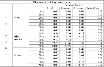
The water extracts did not inhibit all the microbes tested, in contrast the ethyl
acetate could inhibit all the bacteria tested but not Penicillium. The diameter of
inhibition of ethyl acetate extracts was between 6.17 mm – 12.95 mm. Furthermore,
hexane extract could only inhibit B. cereus and the diameter of inhibition was 0.00 mm
– 6.79 mm. (Table 1). For next analysis Pencillium was not used as tested microbes.
Table 1. Diameter of Inhibition Zone of Guava Leaves Extract
Diameter of Inhibition Zone (mm)
Kind of Bacteria
E.coli S. aureus B. cereus Penicillium
s
o
l
v
e
n
t
water
0% 0.00 0.00 0.00 0.00
10% 0.00 0.00 0.00 0.00
20% 0.00 0.00 0.00 0.00
30% 0.00 0.00 0.00 0.00
40% 0.00 0.00 0.00 0.00
50% 0.00 0.00 0.00 0.00
ethyl -
acetate
0% 0.00 0.00 0.00 0.00
10% 9.34 7.99 6.17 0.00
20% 9.28 8.58 6.32 0.00
30% 9.49 9.52 7.17 0.00
40% 9.73 11.81 7.25 0.00
50% 10.06 12.95 7.51 0.00
hexane
0% 0.00 0.00 0.00 0.00
10% 0.00 0.00 0.00 0.00
20% 0.00 0.00 5.04 0.00
30% 0.00 0.00 5.81 0.00
40% 0.00 0.00 5.79 0.00
50% 0.00 0.00 6.79 0.00
The MIC and MBC was determined for ethyl-acetate extract only. The
Bloomfield method was used and the result is in Table 2. The MIC and MBC for
Escherichia coli, Staphylococcus aureus, and Bacillus cereuswere0.017% and 0.067%,
Table 2. The MIC and MBC against tested Bacteria
Kind of Bacteria
E.coli S. aureus B. cereus
MIC MBC MIC MBC MIC MBC
0.017 % 0.067% 1.177% 4.707% 0.126% 0.504%
For ethyl – acetate 10% extract could inhibit the tested bacteria with no
significant differences with the next higher concentration; the inhibition test was done
with the lower concentration, i.e. 2, 4, 6, 8, and 10% and the result shown in Table 3.
Base on the result, ethyl – acetate 4% was chosen for next analysis to inhibit E. coli and
S. aureus, and ethyl acetate 6% was chosen to inhibit B. cereus.
Table 3. Diameter of Inhibition Zone of Ethyl – acetate extract
Diameter of Inhibition Zone (mm)
Kind of Bacteria
E.coli S. aureus B. cereus
Concentration
0 % 0.00 0.00 0.00
2 % 9.06 7.45 6.49
4 % 9.33 8.19 7.29
6 % 9.59 8.36 8.44
8 % 9.81 8.43 8.49
10 % 10.84 8.49 9.04
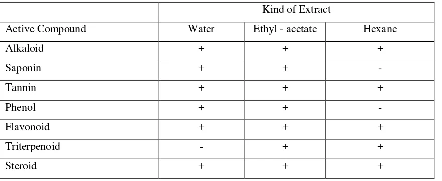
There were a lot of active compound in guava leaves. The active compounds in
guava leaves were alkaloid, saponin, tannin, phenol, flavonoid, triterpenoid, and steroid
(Table 4).
Table 4. The Active Compaound Found in Guava Leaves Extract
Kind of Extract
Active Compound Water Ethyl - acetate Hexane
Alkaloid + + +
Saponin + + -
Tannin + + +
Phenol + + -
Flavonoid + + +
Triterpenoid - + +
Influence of pH on Extract Activity
It was found that ethyl acetate extract was effective under acid condition. It
could inhibit all the tested bacteria at pH 4, but at pH 5 it could not inhibit S. aureus,
moreover it could not inhibit all the tested bacteria at pH 6, 7, and 8 (Figure 1).
0.00 0.00 1.62 0.00 0.00 0.00 0.00 0.00 0.00
4.17
0.00 6.16
0.00 0.00 0.00
0.00
4.73
1.68
0 2 4 6 8 10 12
Control 4 5 6 7 8
pH value
Diameter of Inhibition
Zone (mm)
E. coli S. aureus B. cereus
Figure 1. Diameter of Inhibition Zone of Guava Leaves Extract in Several pH Value
Most of the extract components were weak acid. At low pH, weak acids were
not dissociated. Non dissociated form weak acid would easy to diffuse inside the cell,
then the cell would react to maintain its pH. The cell reaction needs more energy, then
the energy to grow would decrease.
Influence of Sugar on Extract Activity
The result in Figure 2 shown that there was sugar concentration influence on the
antibacterial activity of the extract. The diameter inhibition range was 2.78 – 9.70 mm.
The higher the sugar concentration, the higher the diameter inhibition. The sugar
concentration influenced the Aw value (water activity). At sugar concentration 10 –
30%, the water activity was 0.978, and at sugar concentration 40%, the water activity
was 0.973. Not all water in the solution can be used by the bacterial for its growth. The
water that can be used by bacteria is stated as water activity, the water activity restrict
0.00
7.59
9.70 9.43
0.00
4.12 3.99
3.34 2.78
3.92
0.00
5.43 4.83
4.84 4.55
0 2 4 6 8 10 12
Control 10 20 30 40
sugar concentration (%)
Diameter of Inhibition
Zone (mm)
E. coli S. aureus B. cereus
Figure 2. Diameter of Inhibition Zone of Guava Leaves Extract in Several Sugar
Concentration
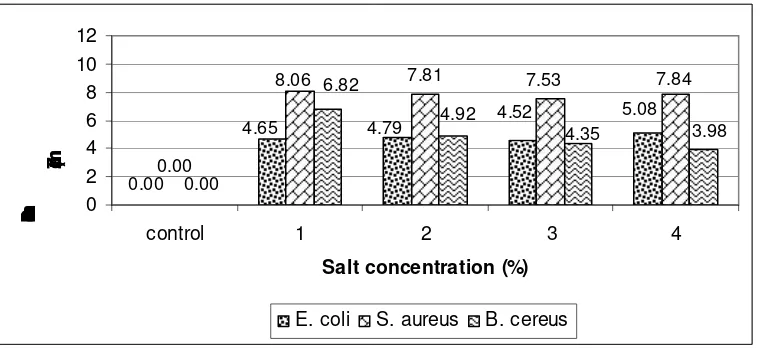
Influence of Salt on Extract Activity
The data in Figure 3 shown that different kind of bacteria showed different
result. The diameter of inhibition zone were 4.52 – 5.08 mm for E. coli 7.53 – 8.06 mm
for S. aureus, and 3.98 – 6.82 mm for B. cereus The extract activity could be influenced
in inhibiting B. cereus dissimilar with in inhibiting E. coli and S. aureus.
The salt will reduce the water activity value (Aw). Generally pathogen bacterial
can be inhibited at Aw (water activity) less than 0.92 that is the same with 13% (w/v)
salt concentration. The highest salt solution in this experiment was 4% (w/v). This
concentration was chosen for those were usually used for food. This salt concentration
was not sufficient to inhibit the bacterial growth. This data strengthen that the inhibition
0.00 0.00
4.65 4.79 4.52
5.08
0.00
8.06 7.81 7.53 7.84
4.35 6.82
3.98 4.92
0 2 4 6 8 10 12
control 1 2 3 4
Salt concentration (%)
Diameter of Inhibition
Zone (mm)
E. coli S. aureus B. cereus
Figure 3. Diameter of Inhibition Zone of Guava Leaves Extract in Several Salt Concentration
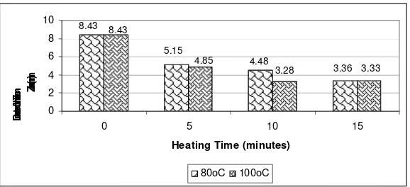
Influence of Heating on Extract Activity
The data in Figure 4 – 6 shown that the ability of the antibacterial to inhibit the
bacterial growth will decrease when the heating temperature and time increase. The
diameter of inhibition zones were 5.24 – 7.29 mm for E. coli (Fig. 4), 3.28 – 5.15 mm
for S. aureus (Fig. 5), and 5.89 – 8.04 (Fig. 6). The higher the heating temperature and
the longer the heating time, the less the active compound and the less the volatile
component of the extract (Ardiansyah, 2002).
7.29
5.58 6.80
10.09
5.24 6.03
6.83 10.09
0 2 4 6 8 10 12
0 5 10 15
Heating Time (minute)
Diam
eter
of In
hibit
ion Zone
(mm
)
80oC 100oC
3.28 3.36 4.48
5.15 8.43
3.33 4.85
8.43
0 2 4 6 8 10
0 5 10 15
Heating Time (minutes)
Diam
eter
of In
hibit
ion Zone
(mm
)
80oC 100oC
Figure 5. Diameter of Inhibition Zone of Guava Leaves Extract in Several Heating Time towards S. aureus
7.28
6.83 8.94
8.44
7.83 7.14
5.89 8.44
0 2 4 6 8 10
0 5 10 15
Heating Time (minutes)
Diam
eter
of In
hibit
ion Zone
(mm
)
80oC 100oC
Figure 6. Diameter of Inhibition Zone of Guava Leaves Extract in Several Heating Time towards B.cereus
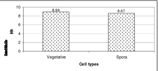
Extract Activity towards B. cereus Spore
Figure 7 shown that the inhibition zone of vegetative cell of B. cereus was 8.94
mm in diameter and the inhibition zone of B. cereus spore was 8.67 mm in diameter.
Bacterial spore is more complex in structure than vegetative cell (Madigan et al., 2006).
Bacterial spore is resistant to heat, drying, radiation, acid, and disinfectant. This result
showed that the extract could inhibit bacterial spore, even though the spore was more
8.94 8.67
0 2 4 6 8 10
Vegetative Spora
Cell types
Diam
eter
of I
nhib
ition
Zon
e
(mm
)
Figure 7. Diameter of Inhibition Zone of Guava Leaves Extract towards B. cereus Spore
CONCLUSION
From the entire experiment, it can be concluded that guava leaves have
antibacterial activity. The activity was influenced by pH, sugar, salt, and by heating
process. Moreover the antibacterial activity was strong enough to inhibit B. cereus
spores. This research indicates that guava leaves have potential natural antibacterial
compound and can be applied for certain food such as sour food, sugar added food, food
with no heating process or short heating process. Further research is suggested to study
the application of antibacterial activity of guava leaves.
REFERENCES
Ardiansyah. Kajian Aktivitas Antimikroba Ekstrak Daun Beluntas (Plucea indica L.). Program Pascasarjana IPB, 2002.
Bloomfield, S.F. Assesing Antimicrobial Activity. Oxford: Blackwell Scientific Publication, 1991.
Harborne, J.B. Metode Fitokimia: Penuntun Cara Modern Menganalisis Tumbuhan.
Bandung: Penerbit ITB, 1996.
Madigan, M.T., H.M. Martinko, dan J. Parker. Brock Biology of Microorganisms. Southern Illinois : Prentice Hall, 2006.
Morton, J. F. “Guava”. Home n-line. o Available from