www.elsevier.com/locate/jinsphys
Plasmatocytes from the moth Pseudoplusia includens induce
apoptosis of granular cells
Louis L. Pech
1, Michael R. Strand
*Department of Entomology, University of Wisconsin–Madison, Madison, WI 53706, USA
Received 31 January 2000; accepted 2 May 2000
Abstract
The primary immune response toward internal parasites and other large foreign objects that enter the insect hemocoel is encapsul-ation. Prior studies indicated that granular cells and plasmatocytes are the two hemocyte types required for capsule formation by the moth Pseudoplusia includens (Lepidoptera: Noctuidae). Capsules formed by P. includens also have a defined architecture with primarily granular cells attaching directly to the target, multiple layers of plasmatocytes adhering to this inner layer of granular cells, and a monolayer of granular cells attaching to the capsule periphery. Dye-exclusion assays indicated that granular cells die shortly after attaching to the capsule periphery, leaving a basal lamina-like layer around the capsule. In examining the mechanisms underlying granular cell death, we found that culture medium preconditioned by plasmatocytes induced apoptosis of granular cells. Characteristics of plasmatocyte-induced apoptosis included condensation of chromatin, cell surface blebbing and fragmentation of nuclear DNA. Plasmatocyte-conditioned medium did not induce apoptosis of other hemocyte types, and medium conditioned by other hemocyte types did not induce apoptosis of granular cells. The adhesive state of granular cells and plasmatocytes also affected levels of apoptosis. Conditioned medium from spread plasmatocytes induced higher levels of granular cell apoptosis than medium conditioned by unspread plasmatocytes. Reciprocally, spread granular cells underwent significantly higher rates of apoptosis than unspread granular cells in medium conditioned by spread plasmatocytes. In situ analysis indicated that granular cells on the periphery of capsules also undergo apoptosis. Collectively, our results suggest that spread plasmatocytes release one or more factors that induce apoptosis of granular cells, and that this response is important in the final phases of capsule formation. 2000 Elsevier Science Ltd. All rights reserved.
Keywords: Hemocytes; Apoptosis; Insect immunity; Pseudoplusia includens; Cell–cell cooperation
1. Introduction
The primary immune response toward internal para-sites and other foreign entities that enter the insect hemo-coel is encapsulation (Lackie, 1988; Strand and Pech, 1995a; Gillespie et al., 1997). Plasmatocytes and granu-lar cells are the two types of hemocytes most often observed in capsules produced by Lepidoptera. Both hemocyte types circulate freely in the hemocoel, but upon immune challenge these cells attach rapidly to the foreign target and one another to form an overlapping sheath. Pech and Strand (1996) used monoclonal
anti-* Corresponding author. Tel: +1-608-262-6902; fax: + 1-608-262-3322.
E-mail address: [email protected] (M.R. Strand).
1 Present address: Department of Biology, Carroll College,
Wauke-sha, WI, USA.
0022-1910/00/$ - see front matter2000 Elsevier Science Ltd. All rights reserved. PII: S 0 0 2 2 - 1 9 1 0 ( 0 0 ) 0 0 0 8 3 - 4
cells are no longer visible. Instead, a diffuse, basal lam-ina-like layer surrounds the capsule periphery. Immuno-cytochemical studies indicate that this layer is labeled by monoclonal antibodies, like 48F2D5, that also label inclusions in the cytoplasm of granular cells (Pech and Strand, 1996; Gardiner and Strand, 1999). While targets are encapsulated more rapidly in vivo, their architecture is virtually identical to capsules produced in vitro (Pech and Strand, 1996; Loret and Strand, 1998).
These observations suggest that, after attaching to the target, granular cells initiate capsule formation by releas-ing factors that induce plasmatocytes to adhere to the target. This is supported by experimental studies which indicate that neither granular cells nor plasmatocytes are capable of forming a capsule independently. However, plasmatocytes readily encapsulate targets if they are pre-incubated in medium conditioned by granular cells (Pech and Strand, 1996; Strand and Clark, 1999). That encap-sulation ceases after attachment of granular cells to the capsule periphery also suggests a role for this hemocyte type in terminating an encapsulation response. However, why these peripheral granular cells die is unclear. Her-ein, we report that these cells die by apoptosis, and that this response is induced by factors released by spread plasmatocytes.
2. Materials and methods
2.1. Insects and hemocyte culture
Pseudoplusia includens was reared and
physiologi-cally staged as described by Strand (1990). Hemocytes were collected from fifth stadium (36–48 h post-ecdysis)
P. includens larvae as outlined by Pech et al. (1994).
Briefly, 36–48 h fifth instar larvae were anesthetized with CO2 and bled from an incision across the last abdominal segment into anticoagulant buffer (98 mM NaOH, 186 mM NaCl, 17 mM Na2EDTA and 41 mM citric acid, pH adjusted to 4.5). After a 40 min incubation at 4°C, hemocytes were washed twice by centrifugation in Ex-cell 400 medium (JRH Biosciences). Four differ-ent types of hemocytes are found in P. includens hemo-lymph: granular cells, plasmatocytes, spherule cells and oenocytoids. Hemocytes collected directly from the lar-val hemocoel are referred to here as unseparated hemo-cytes. Hemocyte types were identified by using estab-lished morphological characters and the monoclonal antibody (mAb) markers 49B11C6, 42C3A3 and 48F2D5 (Gardiner and Strand, 1999). MAb 49B11C6 specifically recognizes plasmatocytes, 42C3A3 labels spherule cells, and 48F2D5 labels granular cells. Plasma-tocytes, granular cells and spherule cells account for ca. 35%, 60% and 4%, respectively, of circulating hemo-cytes in fifth stadium P. includens larvae (Gardiner and Strand, 1999). The different hemocyte types can also be
isolated on Percoll gradients (Pech et al., 1994). Plasma-tocytes, granular cells and spherule cells were isolated on 47.5 and 62% Percoll gradients made in Ex-cell 400 medium as described by Gardiner and Strand (1999). Plasmatocyte and granular cell fractions are ca. 92% and 94% pure, respectively, as determined by antibody labe-ling (Gardiner and Strand, 1999). The primary contami-nant of plasmatocyte fractions are granular cells, whereas granular cell fractions are contaminated by plas-matocytes. Spherule cells were ca. 98% pure with the contaminant being oenocytoids.
2.2. Preparation of conditioned medium
Gradient-purified granular cells, plasmatocytes or spherule cells (1×10⁵cells/well) were incubated in 70µl of Ex-Cell 400 (JRH Scientific) medium in 96-well cell-culture plates (Corning). After 24 h, the conditioned cul-ture medium was removed from each hemocyte type and filtered through 0.2µm filters (Acrodisc, Millipore). The resulting solutions were referred to as 100% conditioned media. Subsequent dilutions were made with Ex-Cell 400 medium.
2.3. Apoptosis of cultured hemocytes
was then added to a final concentration of 100µg/ml followed by a 1 h incubation at 50°C. Lysates were removed, placed into centrifuge tubes, and then extracted three times with chloroform/phenol (1:1) and once with chloroform. Nucleic acids were precipitated with 1/10 volume of 3 M sodium acetate and 2.5 volumes of ethanol. The nucleic acid pellet was resuspended in water. RNA was removed by adding 20µg/ml ribonu-clease A and incubating for 1 h at 37°C. DNA was ana-lyzed by agarose gel electrophoresis on a 1% agarose gel followed by staining with ethidium bromide.
2.4. In situ detection of apoptosis in capsules
In vitro encapsulation assays were conducted as described by Pech et al. (1995). Ninety-six-well culture plates were precoated with Matrigel (Collaborative Biomedical Products, Becton Dickinson Labware, Bedford, MA); a commercially available preparation of mammalian basement membrane. Matrigel was diluted 1:100 with ice-cold Ex-cell 400 medium and plates were coated with 70µl of diluted Matrigel for 1 h at 27°C, followed by five washes with medium. P. includens plas-matocytes and granular cells do not spread on the surface of Matrigel-coated plates (Pech et al., 1995). Wells were filled with 50µl of Ex-Cell 400 medium and then 4×10⁵ unseparated hemocytes plus 50–100 Dowex 1X2 beads were added. After 24 h, capsules were fixed in 10% for-malin in phosphate-buffered saline (PBS), washed three times in PBS, and transferred to 75% ethanol for 10 min. Capsules were then permeablized with three 10 min washes of PBS containing 0.1% Triton X-100, followed by a 5 min incubation with 50µg/ml Proteinase K. Cap-sules were washed with PBS and postfixed with 10% formaldehyde. DNA strand breaks diagnostic of apoptosis were detected by TdT-mediated dUTP–digoxi-genin nick end labeling (TUNEL) using a commercially available kit (ApoTag, Oncor). End-labeled DNA frag-ments were detected using an alkaline-phosphatase-con-jugated, anti-digoxigenin monoclonal antibody (Boeheringer-Mannheim). To identify granular cells and plasmatocytes, capsules were also labeled with the monoclonal antibodies 49B11C6 and 48F2D5. Capsules were double-labeled by first incubating with a given pri-mary and secondary antibody, rinsing, and then incubat-ing with the other primary and secondary antibody (Gardiner and Strand, 1999). The secondary antibodies used were fluorescein- or Texas-red-conjugated goat-antimouse IgG (Jackson Labs). Capsules were then examined by Hoffman and epifluorescent microscopy.
2.5. Image processing and analyses
All samples were examined with a Nikon Diaphot epi-fluorescent microscope fitted with Hoffman modulation contrast optics. Microscope images were captured as
electronic images by use of Metamorph software (Metamorph 1.0) interfaced with a Photometrics high-resolution camera. Files were printed from Adobe photo-shop using a Tektronix Phaser IISDX dye-sublimation printer. We determined treatment differences of ranked data by analysis of variance using the GLM procedure of JMP (2.04) for Macintosh (SAS Institute Inc.).
3. Results
3.1. Plasmatocyte-conditioned medium induces apoptosis of granular cells
We had observed P. includens hemocytes in primary culture on many previous occasions (see Pech et al., 1994; Pech and Strand, 1996; Strand and Clark, 1999 for examples). During these studies, we noticed that rela-tively few granular cells die after 24 h when maintained as a pure population, whereas large numbers of granular cells die by this time in cultures of unseparated hemo-cytes. We also noted that significant numbers of granular cells die after 24 h when co-cultured with plasmatocytes. We thus hypothesized that plasmatocytes release factors that kill granular cells, and that plasmatocytes may also be involved in the death of granular cells on the periph-ery of capsules.
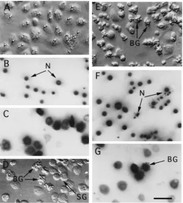
Fig. 1. Plasmatocyte-conditioned medium induces apoptosis of granular cells. (A) Hoffman modulation contrast image of spread granular cells in unconditioned Ex-Cell 400 medium. Granular cells were isolated on Percoll gradients and had been in primary culture for 24 h. Nuclei (N) of granular cells in unconditioned medium, visualized with the nuclear stain Hoechst 33342, are uncondensed (B) and contain intact mitochondria as evidenced by positive staining with rhodamine 123 (C). (D) Hoffman image of granular cells after 16 h in 100% plasmatocyte-conditioned Ex-Cell 400 medium. Most granular cells remain attached and spread on the surface of the culture plate (SG), whereas some granular cells are in the process of blebbing into membrane-bound bodies (BG). (E) A much larger percentage of granular cells are in the process of blebbing after 24 h in 100% plasmatocyte-conditioned medium. The nuclei (N) of blebbing granular cells (BG) are highly condensed or fragmented (F), but still contain intact mitochondria (G). Scale bar for (A)–(F) equals 75µm.
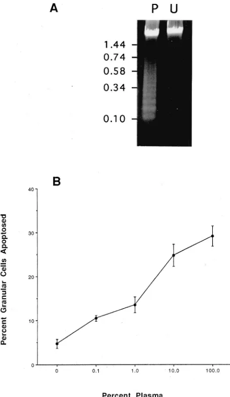
DNA by agarose gel electrophoresis revealed that samples from 100% plasmatocyte-conditioned medium formed a nucleosomal ladder, whereas samples from unconditioned medium remained in a high-molecular-mass form [Fig. 2(A)]. Lastly, serial dilution of plasma-tocyte-conditioned medium with Ex-Cell 400 resulted in a linear decrease in the percentage of granular cells undergoing a blebbing response after 24 h in culture [Fig. 2(B)]. Taken together, these results indicated that medium conditioned by plasmatocytes induces apoptosis of granular cells.
3.2. The effects of plasmatocyte-conditioned medium on granular cell apoptosis are specific
Fig. 2. Fragmentation of granular cell DNA and dose-dependent effects of plasmatocyte-conditioned medium. (A) Agarose gel showing granular cell DNA (5µg/lane) from cells cultured for 24 h in 100% plasmatocyte-conditioned medium (P) and unconditioned Ex-Cell 400 medium (U). Size markers are indicated to the left. (B) Effect of con-ditioned-medium concentration (v/v) on apoptosis of granular cells in vitro. Apoptosis was assayed by counting the number of blebbing and normally spread granular cells after 24 h in 10–100% plasmatocyte-conditioned medium. Unplasmatocyte-conditioned Ex-Cell 400 medium served as the negative control (0% plasmatocyte-conditioned medium on the graph). Each data point is the mean percentage±standard deviation (SD) of apoptosed granular cells from five independent collections of hemocytes.
percentage of granular cells to apoptose than plasmato-cytes or spherule cells (Table 1). We concluded from these experiments that apoptosis-inducing activity was produced specifically by plasmatocytes and acted only on granular cells.
3.3. Apoptosis-inducing activity is associated with hemocyte spreading on foreign surfaces
Capsule formation by P. includens requires that both granular cells and plasmatocytes change from
non-Table 1
Effects of conditioned medium from different hemocyte types on apoptosis of granular cells, and apoptosis of different hemocyte types in plasmatocyte-conditioned mediuma
Medium n %
Apoptosis±SD of granular cells
Granular cell 7 4.0±2.0
Plasmocyte 7 28.5±5.1
Spherule cell 7 6.5±2.6
Ex-cell 400 (unconditioned) 7 6.0±2.5 Test statistic: H=14.88, df=3, P,0.0019
Target cell n %
Apoptosis±SD in plasmatocyte-conditioned medium
Granular cells 6 28.7±3.5
Plasmatocytes 6 1.2±0.9
Spherule cells 6 1.6±1.1
Test statistic: H=11.96, df=2, P,0.0025
a Each replicate is cells or conditioned medium prepared from
dif-ferent gradients. Conditioned medium was prepared by culturing hem-ocytes in unmodified 96-well cell culture plates (see Materials and methods). Each hemocyte type was cultured in 100% conditioned medium. The percentage of cells undergoing apoptosis was scored 24 h later as the number of blebbing cells divided by the total number of hemocytes counted (see Materials and methods). Analyses were perfor-med on ranked data (Kruskal–Wallis test).
Signifi-Table 2
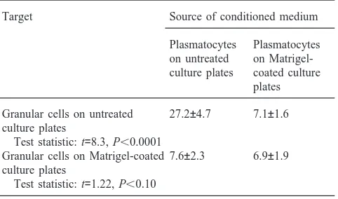
Percentage (±SD) of granular cells that undergo apoptosis in plasmato-cyte-conditioned mediuma
Target Source of conditioned medium
Plasmatocytes Plasmatocytes on untreated on Matrigel-culture plates coated culture
plates
Granular cells on untreated 27.2±4.7 7.1±1.6 culture plates
Test statistic: t=8.3, P,0.0001
Granular cells on Matrigel-coated 7.6±2.3 6.9±1.9 culture plates
Test statistic: t=1.22, P,0.10
aEach replicate represents cells or conditioned medium prepared
from five different gradients. Granular cells were cultured in 100% plasmatocyte-conditioned in medium. Each hemocyte type was cul-tured in 100% conditioned medium. The percentage of granular cells undergoing apoptosis was scored 24 h later as the number of blebbing cells divided by the total number of hemocytes counted (see Materials and methods). Analyses were performed on ranked data (Wilcoxon two-sample test).
cantly more granular cells underwent apoptosis on unco-ated wells than on Matrigel-counco-ated wells (Table 2).
3.4. Granular cells on the periphery of capsules die by apoptosis
We encapsulated targets in vitro and then used TUNEL assays to determine whether hemocytes in cap-sules undergo apoptosis. We examined more than 500 capsules at different stages of development. Fig. 3 sum-marizes the progression of events we observed. After 16 h, capsules were nearly fully formed as indicated by the presence of granular cells around the capsule periph-ery (labeled by mAb 48F2D5) and the overlapping layers of plasmatocytes (labeled by mAb 49B11C6) that form the capsule’s interior [Fig. 3(A)]. In situ labeling by TUNEL assay revealed that almost no hemocytes were labeled by dUTP–digoxigenin at this time (data not presented). By 24 h, however, many of the peripheral granular cells were dying as indicated by staining with propidium iodide (data not presented). Concomitantly, a flocculent layer, also labeled by mAb 48F2D5, began to develop around the periphery of the capsule [Fig. 3(B)]. TUNEL assay resulted in nuclear labeling of several hemocytes in 24 h old capsules [Fig. 3(C)]. Most of these cells were located on the capsule periphery and, by double-labeling with anti-hemocyte mAbs, were identified as granular cells [compare Fig. 3(B) with Fig. 3(C)]. No hemocytes were labeled in control experi-ments when we omitted dUTP–digoxigenin or the pri-mary antibody from the reaction. After 30 h, few intact granular cells remained around the capsule periphery but the flocculent layer labeled by mAb 48F2D5 was clearly
visible [Fig. 3(D)]. A small number of plasmatocytes was also visible on the capsule periphery at this time [Fig. 3(D)]. However, examination of capsules from later time points indicated that this flocculent layer remained present and that almost no additional hemocytes of any type attached to the capsule.
4. Discussion
The goal of this study was to explain previous obser-vations that granular cells become permeable to propo-dium iodide shortly after attaching to the periphery of capsules (Pech and Strand, 1996; Loret and Strand, 1998). Our results indicate that granular cells undergo apoptosis if cultured in plasmatocyte-conditioned medium. The response was specific since only granular cells undergo apoptosis and only medium conditioned by plasmatocytes induces apoptosis. Activation of the apoptosis pathway also appears to be linked to adhesion given that: (1) spread granular cells undergo much higher levels of apoptosis than unspread cells, and (2) only conditioned medium from spread plasmatocytes contains apoptosis-inducing activity. Our TUNEL assays also indicate that many granular cells on the exterior of capsules die by apoptosis. Collectively, these results sug-gest that apoptosis of granular cells on the capsule per-iphery is induced by the overlapping layers of plasmato-cytes that form the interior of the capsule. As noted in the Introduction, granular cells are also the first hemo-cytes in P. includens to attach to the foreign target. Unfortunately, we were unable to identify these cells after large numbers of plasmatocytes had attached and spread around the target. We assume our inability to identify these cells is because they also die, but whether death is due to apoptosis or necrosis is unclear.
Fig. 3. Apoptosis of granular cells in capsules. (A) Fluorescent image of a 16 h old capsule double-labeled by the anti-plasmatocyte mAb 49B11C6 and anti-granular cell mAb 48F2D5. The core of the capsule is comprised primarily of plasmatocytes (P) (green) while rounded granular cells (G) (red) are attached to the capsule periphery. (B) Fluorescent image of a 24 h old capsule. Granular cells (G) around the capsule periphery are more irregular in shape than seen in (A), and a flocculent layer (also red) is beginning to develop. (C) Hoffman image of the same capsule as shown in (B). Digoxigenin-labeled cell nuclei (purple) are located primarily around the capsule periphery. Comparison with (B) indicates that most digoxigenin-labeled cells co-localize with cells identified by antibody staining to be granular cells [compare arrows for granular cells on the capsule periphery (G) versus a given plasmatocyte (P)]. (D) Fluorescence image of a 30 h old capsule. Individual granular cells are no longer visible but the flocculent outer labeled by mAb 48F2D5 is visible (arrow). Scale bar for (A)–(D) is 30µm.
that has also been implicated in regulation of apoptosis (Ahmad et al., 1997).
Studies with several insects report that granular cells are the first hemocytes to adhere to encapsulation targets, followed by the attachment of large numbers of plasma-tocytes (reviewed by Ratcliffe et al., 1985; Lackie, 1988; Strand and Pech, 1995b). The mechanisms involved in regulating the initial adhesion of granular cells to the
they function as cytokines that regulate plasmatocyte adhesion. Recent studies with the ENF-peptide family member plasmatocyte spreading peptide (PSP1) from P.
includens suggest that granular-cell-stimulated adhesion
of plasmatocytes is due in part to PSP1 (Clark et al., 1998; Strand and Clark, 1999). PSP1 appears to induce plasmatocytes to transport adhesion molecules to their surface where they interact with their cognate receptors (Strand and Clark, 1999).
In contrast, the signals that induce granular cells to attach to the periphery of capsules, the origin of the flocculent, basal lamina-like layer that ultimately sur-rounds each capsule, and why, after these events occur, plasmatocytes cease to attach to capsules are unknown. The presence of a basal lamina around capsules has been reported in other species besides P. includens
(Grimstone et al., 1967; Han and Gupta, 1989; Porchet-Hennere, 1990), suggesting that this feature may be widespread and diagnostic of capsules that are fully developed. Granular cells have also been implicated in the formation of basal laminae that surround insect tissues and the hemocoel (Nardi and Miklasz, 1989; Chain et al., 1992; Wigglesworth, 1973; Ball et al., 1987). These results suggest that granular cells on the capsule periphery are the source of the basal lamina around capsules. The ability of cells to adhere to differ-ent extracellular matrix proteins depends in large meas-ure on the types of receptor present on their surface. PSP1 induces exocytosis of adhesion proteins that plas-matocytes are clearly capable of binding to, whereas granular cells may release extracellular matrix proteins on the capsule periphery to which plasmatocytes do not adhere. Whether these factors are released before granu-lar cells undergo apoptosis or as a consequence of undergoing apoptosis is currently under investigation.
In a previous study, we found that granular cells also undergo apoptosis when P. includens larvae are parasit-ized by the braconid wasp Microplitis demolitor (Strand and Pech, 1995b). In this case, apoptosis is due to infec-tion of granular cells by M. demolitor polydnavirus (MdPDV), which is injected into hosts when the wasp lays its egg. MdPDV-induced apoptosis depends upon direct infection of granular cells by MdPDV and expression of viral genes, but participation of plasmato-cytes is not required. Future studies should reveal whether any relationship exists between viral and plas-matocyte activation of the apoptosis pathway in granu-lar cells.
Acknowledgements
This work was supported by a grant from the National Institutes of Health (AI32917) and the USDA Hatch Pro-gram to MRS.
References
Ahmad, M., Srinivasular, S.M., Wang, L.J., Litwaxk, G., Fernandes-Alnemri, T., Fernandes-Alnemri, E.S., 1997. Spodoptera frugiperda caspase-1, a novel insect death protease that cleaves the nuclear immuno-philin Fdbp46, is the target of the baculovirus antiapoptotic protein P35. Journal of Biological Chemistry 272, 1421–1424.
Ball, E.E., Gert de Couet, H., Horn, P.L., Quinn, J.M.A., 1987. Haemo-cytes secrete basement membrane components in embryonic locusts. Development 99, 255–259.
Chain, B.M., Leyshon-Sorland, K., Siva-Jothy, M.T., 1992. Haemocyte heterogeneity in the cockroach Periplaneta americana analysed using monoclonal antibodies. Journal of Cell Science 103, 1261– 1267.
Clark, K., Pech, L.L., Strand, M.R., 1997. Isolation and identification of a plasmatocyte spreading peptide from hemolymph of the lepi-dopteran insect Pseudoplusia includens. Journal of Biological Chemistry 272, 23440–23447.
Clark, K., Witherell, A., Strand, M.R., 1998. Plasmatocyte spreading peptide is encoded by an mRNA differentially expressed in tissues of the moth Pseudoplusia includens. Biochemical and Biophysical Research Communications 250, 479–485.
Cohen, J.J., 1993. Apoptosis. Immunology Today 14, 123–130. Cohen, J.J., 1997. Caspases: the executioners of apoptosis.
Biochemi-cal Journal 326, 1–16.
Frazier, A., Evan, G., 1996. A license to kill. Cell 85, 781. Gardiner, E.M.M., Strand, M.R., 1999. Monoclonal antibodies bind
distinct classes of hemocytes in the moth Pseudoplusia includens. Journal of Insect Physiology 45, 113–126.
Gillespie, J.P., Kanost, M.R., Trenczek, T., 1997. Biological mediators of insect immunity. Annual Review of Entomology 42, 611–643. Grimstone, A.V., Rotheram, S., Salt, G.B., 1967. An electron-micro-scope study of capsule formation by insect blood cells. Journal of Cell Science 2, 281–292.
Han, S.S., Gupta, A.P., 1989. Arthropod immune system. II. Encapsul-ation of implanted nerve cord and “plain gut” surgical suture by granulocytes of Blattella germanica (L.) (Dictyoptera: Blattellidae). Zoological Science 6, 303–320.
Kuida, K., Haydar, T.F., Kuan, C.-T., Gu, Y., Taya, C., Karasuyama, H., Su, S.-S., Rakic, P., Flavell, R.A., 1998. Reduced apoptosis and cytochrome c-mediated caspase activation in mice lacking caspase 9. Cell 94, 325–337.
Lackie, A.M., 1988. Immune mechanisms in insects. Parasitology Today 4, 98–105.
Loret, S., Strand, M.R., 1998. Follow-up of protein release from
Pseudoplusia includens hemocytes: a first step toward identification
of factors mediating encapsulation in insects. European Journal of Cell Biology 76, 146–155.
Nardi, J.B., Miklasz, S.D., 1989. Hemocytes contribute to both the formation and breakdown of the basal lamina in developing wings of Manduca sexta. Tissue and Cell 21, 559–567.
Nicholson, D.W., Thornberry, N.A., 1997. Caspases: killer proteases. TIBS 22, 299–306.
Pech, L.L., Trudeau, D., Strand, M.R., 1994. Separation and behavior in vitro of hemocytes from the moth Pseudoplusia includens. Cell and Tissue Research 277, 159–167.
Pech, L.L., Trudeau, D., Strand, M.R., 1995. Effects of basement mem-branes on the behavior of hemocytes from Pseudoplusia includens (Lepidoptera; Noctuidae): development of an in vitro encapsulation assay. Journal of Insect Physiology 41, 801–807.
Pech, L.L., Strand, M.R., 1996. Granular cells are required for encap-sulation of foreign targets by insect haemocytes. Journal of Cell Science 109, 2053–2060.
Porchet-Hennere, E., 1990. Cooperation between different coelomo-cyte populations during the encapsulation response of Nereis
div-ersicolor demonstrated by using monoclonal antibodies. Journal of
Ratcliffe, N.A., Rowley, A.F., Fitzgerald, S.W., Rhodes, C.P., 1985. Invertebrate immunity: basic concepts and recent advances. Inter-national Review of Cytology 97, 186–350.
Strand, M.R., 1990. Characterization of larval development in
Pseudo-plusia includens (Lepidoptera: Noctuidae). Annals of the
Entomo-logical Society of America 83, 538–544.
Strand, M.R., Clark, K.D., 1999. Plasmatocyte spreading peptide induces spreading of plasmatocytes but represses spreading of gra-nulocytes. Archives of Insect Biochemistry and Physiology 42, 213–223.
Strand, M.R., Pech, L.L., 1995a. Immunological basis for compatibility in parasitoid–host relationships. Annual Review of Entomology 40, 31–56.
Strand, M.R., Pech, L.L., 1995b. Microplitis demolitor polydnavirus induces apoptosis of a specific haemocyte morphotype in
Pseudo-plusia includens. Journal of General Virology 76, 283–291.
Strand, M.R., Hayakawa, Y., Clark, K.D., 2000. Plasmatocyte
spread-ing peptide (PSP1) and growth blockspread-ing peptide (GBP) are multi-functional homologs. Journal of Insect Physiology 46, 817–824. Thornberry, H.N., Lazebnic, Y., 1998. Caspases: enemies within.
Science 281, 1312–1316.
Volkman, B.J., Anderson, M.E., Clark, K.D., Hayakawa, Y., Strand, M.R., Markley, J.L., 1999. Structure of the insect cytokine plasma-tocyte spreading peptide from Pseudoplusia includens. Journal of Biological Chemistry 274, 4493–4496.
Wang, Y., Jiang, H., Kanost, M.R., 1999. Biological activity of
Mand-uca sexta paralytic and plasmatocyte spreading peptide and primary
structure of its hemolymph precursor. Insect Biochemstry and Mol-ecular Biology 29, 1075–1086.
Wigglesworth, V.B., 1973. Haemocytes and basement membrane for-mation in Rhodnius. Journal of Insect Physiology 19, 831–844. Yuan, J., 1997. Genetic control of cellular suicide. Reproductive