FACULTY OF SCIENCE
DEPARTMENT OF BIOLOGY
Jasminka Krištić
CHANGES OF IMMUNOGLOBULIN G
GLYCOSYLATION WITH AGE
DOCTORAL THESIS
PRIRODOSLOVNO-
MATEMATIČKI FAKULTET
BIOLOŠKI ODSJEK
Jasminka Krištić
PROMJENA GLIKOZILACIJE
IMMUNOGLOBULINA G TIJEKOM
STARENJA
DOKTORSKI RAD
Zagreb, 2018.
The work presented in this doctoral thesis was performed at Genos Ltd., Zagreb, Croatia under the supervision of Prof. Gordan Lauc, PhD, as a part of the postgraduate
doctoral programme in Biology at the Department of Biology, Faculty of Science, University of Zagreb.
Ovaj je doktorski rad izrađen u Genos d.o.o., Zagreb, Hrvatska, pod vodstvom prof. dr. sc. Gordana Lauca, u sklopu Sveučilišnog poslijediplomskog doktorskog studija Biologije pri Biološkom odsjeku Prirodoslovno-matematičkog fakulteta Sveučilišta u
Najljepše se zahvaljujem svom mentoru prof. dr. sc. Gordanu Laucu koji mi je omogućio da se bavim znanošću te što nadahnjuje svojim idejama i entuzijazmom kojim priča o znanosti.
Toplo se zahvaljujem cijeloj ekipi s posla na ugodnoj radnoj atmosferi i suradnji na brojnim projektima.
Posebno hvala Tomi od kojeg sam puno naučila prvih mjeseci u Genosu.
Hvala Maji što rado prenosi svoje veliko znanje iz glikobiologije te na brojnim savjetima i razumijevanju.
Hvala Tomislavu na čitanju i lektoriranju doktorskog rada, te na lijepim manirama u ophođenju s drugima.
Mirni, Dajani, Ivoni i Ireni hvala na prijateljstvu.
Iako piše da zahvalu svedemo u akademske okvire, ne znam hoću li se ikada više imati prilike javno zahvaliti određenim osobama, pa ću zato to učiniti ovdje.
Ne tako davno čula sam izreku „Iza svakog uspješnog muškarca stoji njegova žena, a iza svake uspješne žene njezina majka!“. Pa ako sam ja uspješna zato što ću imati doktorat znanosti, onda iza toga svakako stoji moja mama! Mama hvala ti na svemu, svom trudu, vremenu, savjetima, što si moj “glas savjesti“. Hvala tati što nas je uvijek poticao na više i bolje. Hvala mojoj sestri i bratu na podršci i ljubavi.
Hvala mom suprugu Vinku koji mi je podrška u svemu što radim i koji o glikobiologiji zna puno više nego što bi jedan sveučilišni specijalist ekonomije trebao znati! Hvala mojim djevojčicama Tonki i Marini uz koje je sve istodobno i teže i lakše, ali sve ima više smisla!
Hvala mojim dragim prijateljicama Zrinki, Nataši, Ivani i Mirti što me prate kroz život.
Na kraju, zahvaljujem se dragom Bogu. „Znam da tvoja ruka životom mojim ravna i svaki korak tvoj mi pogled prati.“
Faculty of Science Department of Biology
CHANGES OF IMMUNOGLOBULIN G GLYCOSYLATION WITH AGE
JASMINKA KRIŠTIĆ Genos Ltd.
BIOCentar, Borongajska cesta 83h, Zagreb, Croatia
In this thesis, glycosylation of immunoglobulin G (IgG) was examined in 5818 individuals ranging in age from 16 to 100 years from five different populations – four European populations and a Han Chinese population by using ultra performance liquid chromatography (UPLC) in order to provide a comprehensive overview of changes in IgG glycosylation through lifetime. The results demonstrated that glycan profiles change in a similar way through lifetime across all examined populations. Nearly all IgG glycans were significantly associated with age. Sex differences in age-related changes in IgG glycosylation were observed. The combination of several IgG glycans was able to explain from 30 to 58% of variance in chronological age, with the remaining variance in the glycans attributed to physiological parameters. Analysis of IgG glycosylation in nearly 600 mice from the Collaborative Cross cohort ranging in age from 20 to 80 weeks showed that, generally, only the level of IgG glycan with alpha-1,3-galactose changed with age.
(84 pages, 10 figures, 9 tables, 226 references, original in English)
Keywords: glycosylation, immunoglobulin G, chronological age, biological age, biomarker, humans, mice
Supervisor: Professor Gordan Lauc, PhD
Reviewers: Associate professor Olga Gornik, PhD Professor Kristian Vlahoviček, PhD Professor Igor Rudan, PhD
Prirodoslovno-matematički fakultet Biološki odsjek
PROMJENA GLIKOZILACIJE IMUNOGLOBULINA G TIJEKOM STARENJA
JASMINKA KRIŠTIĆ Genos d.o.o.
BIOCentar, Borongajska cesta 83h, Zagreb, Hrvatska
U ovom doktorskom radu analizirana je glikozilacija imunoglobulina G (IgG) kod 5818 osoba
starosti od 16 do 100 godina iz pet različitih populacija – četiri europske populacije i populacije
Han Kineza, koristeći tekućinsku kromatografiju vrlo visoke djelotvornosti (UPLC), kako bi se pružio detaljan pregled promjena u glikozilaciji IgG-a tijekom starenja. Rezultati su pokazali da se glikanski profili tijekom starenja mijenjaju na sličan način u svim istraživanim populacijama.
Gotovo svi IgG glikani pokazali su se povezanima s dobi. Uočene su razlike između spolova u
promjenama koje se događaju s glikozilacijom IgG-a tijekom starenja. Kombinacijom nekoliko IgG glikana bilo je moguće objasniti od 30 do 58% varijacije u kronološkoj dobi, a ostatak varijacije u glikanima pripisan je fiziološkim parametrima. Analiza glikozilacije IgG-a kod gotovo 600 miševa starosti od 20 do 80 tjedana iz Collaborative Cross populacije miševa pokazala je da se, sveobuhvatno gledajući, samo razina IgG glikana koji sadrži alfa-1,3 vezanu galaktozu promijenila s dobi.
(84 stranice, 10 slika, 9 tablica, 226 literaturnih navoda, jeziki izvornika: hrvatski)
Ključne riječi: glikozilacija, imunoglobulin G, kronološka dob, biološka dob, biomarker, ljudi, miševi
Mentor: Prof. dr. sc. Gordan Lauc
Ocjenjivači: Izv. prof. dr. sc. Olga Gornik Prof. dr. sc. Kristian Vlahoviček Prof. dr. sc. Igor Rudan
1. INTRODUCTION ………... 1
1.1 A general overview of protein glycosylation ………... 2
1.2 Immunoglobulin G glycosylation ………... 2
1.3 The impact of glycosylation on the structure and biological function of IgG ….…. 5 1.4 Changes in IgG glycosylation have been observed in many diseases ……….. 8
1.5 Aging ……… 8
1.6 Chronological and biological age ……….. .. 9
1.7 Biomarkers of aging ………... 10
1.7.1 Telomere length ……….. 10
1.7.2 DNA methylation ……… 11
1.8 Aging in mice ………. 12
1.9 Changes in IgG glycosylation with age: Studies in human populations …………. 13
1.10 Changes in IgG glycosylation with age: Studies in mice ……… 18
1.11 Research problem and scope of the thesis ……….. 19
2. Glycans are a novel biomarker of chronological and biological ages ……….. 20
3. Profiling IgG N-glycans as potential biomarker of chronological and biological ages … 32 4. Profiling and genetic control of the murine immunoglobulin G glycome ……… 43
5. GENERAL DISCUSSION ………. . 55
6. CONCLUSIONS ………. 61
7. REFERENCE LIST ………. 63
8. APPENDICES ………. 79
1
2 1.1 A general overview of protein glycosylation
Glycosylation is one of the most common posttranslational modifications of proteins, through which complex sugar molecules (glycans) are covalently attached to proteins. In 1991 Apweiler et al.1 estimated that more than half of all proteins are glycosylated, however, according to more recent estimates, less than one-fifth of proteins appear to be glycosylated2. Despite that, this new study on the frequency of different types of posttranslational modifications also showed that glycosylation is among the top three most common posttranslational modifications of proteins. Glycosylation greatly affects the physical properties of proteins (e.g. solubility, conformation, folding, stability, etc.) as well as biological functions of proteins3. Therefore, glycans as an essential part of many proteins have an important role in almost all physiological processes including protein folding and trafficking, cell adhesion, cell signalling, proliferation, differentiation, cell migration, cell survival, development and immunity4. Changes in glycosylation have been reported in various diseasesnumerous references, including5–8 and it has also been
shown that glycans have great potential as diagnostic and prognostic biomarkers of different diseases9–11. For that reason, there are a growing number of studies which try to link changes in
glycosylation of a certain protein (or group of proteins) with a certain disease and/or phenotype in order to find new potential diagnostic and prognostic biomarkers for certain diseases or conditions and to get a deeper insight into the mechanism of a specific disease itself. One of the proteins whose glycosylation has been the subject of many such studies is immunoglobulin G (IgG). One of the main reasons for this is the fact that IgG is the most abundant glycoprotein and the second most abundant protein in human plasma (albumin is the most abundant protein in human plasma, but it is not glycosylated)12. Therefore, IgG has a prerequisite to be a very good non-invasive biomarker.
1.2 Immunoglobulin G glycosylation
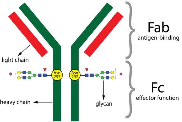
IgG antibodies are a very important component of the immune system as they protect the organism against invading pathogens. They are produced by B lymphocytes. The IgG antibody consists of two fragments - Fab and Fc (Figure 1). The Fab fragment (antigen binding fragment) is the part of the IgG molecule which specifically recognizes and binds different antigens like bacteria or viruses. The Fc fragment (crystallisable fragment) provides the effector function, which means that this fragment determines how the immune system responds or reacts to the presence of a specific antigen. Through the interaction of the IgG Fc fragment with activating or
3 inhibitory Fc gamma receptors (FcγR), which are expressed on the majority of innate immune effector cells such as mast cells, monocytes, macrophages, natural killer cells, neutrophils, eosinophils and dendritic cells, pro-inflammatory or anti-inflammatory effector pathways can be activated13,14. Furthermore, the Fc fragment of IgG can also interact with component complement C1q13 and activate the complement pathway with pro-inflammatory effects14. The Fc fragment of IgG is also involved in binding of IgG to the neonatal Fc receptor (FcRn) expressed on endothelial cells and monocytes15. The interaction of IgG with FcRn determines antibody half-life but is also potentially involved in the anti-inflammatory activity of IgG14,15. It is also known that the ability of IgG to interact with different Fc receptors or to activate complement varies depending on the IgG subclass14,16,17. In humans there are four different IgG subclasses, IgG1, IgG2, IgG3 and IgG4, which differ in the constant regions of their heavy chains and are named according to their relative abundance in plasma. In mice there are five different IgG subclasses: IgG1, IgG2a, IgG2b, IgG2c and IgG3, however, although similar in name, they are not direct homologues of the human proteins18,19.
Figure 1. Schematic representation of immunoglobulin G (IgG). IgG protein is composed of two heavy and two light chains. IgG protein can also be devided into two functional fragments: antigen-binding fragment (Fab) which binds antigen and crystallizable fragment (Fc) which is important for effector functions such as ADCC or complement activation. Each heavy chain of Fc fragment contains a covalently attached N-glycan to highly conserved N-glycosylation site located at position aparagine (Asn) 297. In addition, some IgG molecules can contain N-glycans in the Fab fragment (~20% of IgG molecules).
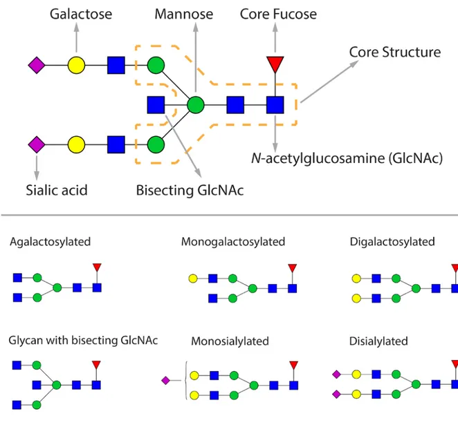
4 IgG contains a conserved N-glycosylation site at position asparagine (Asn) 297 on each heavy chain of the Fc fragment. In addition, around 20% of IgGs also have additional glycosylation sites in the Fab fragment20–24. All IgG glycans have a pentasaccharide core structure (consisting of two
N-acetylglucosamines (GlcNAc) and three mannose residues) which can be additionally modified with a core fucose, a bisecting GlcNAc, one or two galactoses and one or two sialic acids (Figure 2).
Figure 2. The composition of IgG glycans.
More than 30 different glycan structures can be found on human IgG25. Overall, human IgG
glycans are predominately biantennary structures and most of them are core-fucosylated (>90%). A bisecting GlcNAc is present in ~18% of human IgG glycans and sialic acid is found in ~25% of IgG glycans. Approximately 30% of human IgG glycans contain no galactose25. The largest and most complex IgG glycan structure is a core-fucosylated bianntenary glycan with two galactoses,
5 two sialic acids and with bisecting GlcNAc25. Furthermore, it is known that the Fab and Fc fragment are differentially glycosylated. In comparison to Fc glycans, IgG Fab glycans are generally more highly galactosylated, sialylated and have higher level of bisecting GlcNAc. On the other hand, Fc IgG glycans are more highly core-fucosylated than IgG Fab glycans and are mostly nonsialylated (neutral) structures20,26–28.
Contrary to the protein part of the IgG molecule, the synthesis of the glycan part is not regulated by a template. Rather, glycans are synthesised by the concerted action of many different proteins, including glycosyltransferases, glycosidases, nucleotide sugar transporters, transcription factors, and many other proteins29. Additionally, the biosynthesis of glycans is influenced by various environmental factors and physiological conditions29–32. Despite the absence of a direct genetic template, a relatively high heritability of IgG glycosylation has been reported, ranging from 30% to 80%, depending on the IgG glycan25,33.
While human IgG glycosylation has been well studied, data on IgG glycosylation in mice are rather scarce and obscure. The data available in the literature show that though most glycan structures found on mouse IgG are shared between human and mouse, IgG glycosylation in mice exhibits some different characteristics from those observed in humans. Mouse and human IgG glycosylation differs with respect to the type of sialic acid present at the terminus of sugar chains attached to IgG proteins; mouse IgGs contain sugars which terminate with N-glycolylneuraminic (Neu5Gc) acid while human IgGs have terminal N-acetylneuraminic acid (Neu5Ac)34–36. Further, glycan structures with terminal alpha 1–3 bound galactose are found on mouse IgG but not on human IgG34. Also, several inconsistencies are found within the literature with regards to some specific features of mouse IgG glycosylation. Some studies reported that the level of fucosylation was higher for mouse than for human IgG and that almost all mouse IgG glycans contained core fucose35–37, while others reported that fucosylation of mouse IgG was lower than in humans38. Further, the prevailing view in the field is that mouse IgG does not have bisecting GlcNAc34–37. However, more recent evidence suggests that mouse IgG glycans may contain bisecting GlcNAc38,39.
1.3 The impact of glycosylation on the structure and biological function of IgG It is well documented that IgG Fc glycosylation stabilizes the structure of the Fc fragment, which is required for binding to the Fc receptors and complement factors. Deglycosylation (i.e. the removal of glycans) of the Fc fragment results in decreased binding or complete loss of binding to
6 FcγR or to proteins involved in the complement pathways and thus leads to an inability of IgG to elicit effector functions including complement activation, antibody-dependent cellular phagocytosis (ADCP) and antibody-dependent cellular cytotoxicity (ADCC)40–43. It is also well known that differences in the Fc glycosylation pattern alter the conformation of the Fc fragment which in turn modulates the binding affinities of IgG for FcγR and complement factors enabling fine-tuning of the IgG Fc-mediated immune response42,44.
Core fucose
For example, the absence of core fucose on IgG glycans results in a stronger binding affinity of IgG to Fc gamma receptor IIIa (up to 50-fold increased binding) and leads to enhanced ADCC activity while the presence of core fucose reduces the ADCC activity of IgG45,46. Moreover, it
was found that interactions between IgG Fc glycans and glycans of the FcγRIIIa receptor have an
effect on binding affinity between IgG and FcγRIIIa where the presence of core fucose on glycans attached to Fc fragment sterically inhibited such interactions which resulted in decreased binding affinity for the receptor47.
Bisecting GlcNAc
The addition of bisecting GlcNAc to IgG glycans was reported to cause an increase in ADCC
through higher affinity for FcγRIIIa48. However, Shinkawa et al49 showed that the presence of
very high levels of IgG glycans with bisecting GlcNAc resulted in only a slight increase of ADCC activity and that high content of glycans lacking core fucose was more potent in enhancing ADCC when compared to bisected glycans. It is also known that the presence of bisecting GlcNAc inhibits the addition of core fucose50.
Sialic acid
The effector function of IgG can also be modulated by terminal sialic acid residues. Many studies have demonstrated that sialylation acts as a switch between pro-inflammatory and anti-inflammatory activity of IgG51,52 (reviewed in53–55). IgG antibodies which are not sialylated stimulate pro-inflammatory immune responses through interaction with Fcγ receptors42. On the other hand, IgGs which contain sialylated glycans exhibit anti-inflammatory properties. Moreover, several lines of evidence suggest that sialic acid residues of N-glycans attached to the IgG Fc fragment are responsible for the anti-inflammatory properties of intravenous immunoglobulin (IVIG) preparation but the exact mechanism by which sialylated IgGs exert anti-inflammatory activity is still unclear14,42,44,51–56. One of the proposed mechanisms suggests that sialylated IgGs interact with the C-type lectin receptor DC-SIGN which increases expression of
7 inhibitory receptor FcγRIIb and thus suppresses inflammation57–59. However, conflicting results
exist regarding the involvement of DC-SIGN in the anti-inflammatory activity of sialylated IgG and some other receptors which could recognize sialic acid and trigger anti-inflammatory pathways have been proposed53. Moreover, there is also some evidence that sialic acid can act in a receptor-independent manner to activate the anti-inflammatory response and that IVIG may recruit sialic acid-independent anti-inflammatory pathways44,53,54.
Galactose
Karsten et al60 have recently showed that galactosylated IgG also exerts anti-inflammatory activity by FcγIIB receptor- and Dectin-1-mediated inhibition of the complement pathway. On the contrary, IgG glycans which lack terminal galactose residues (agalactosylated glycans) posses the ability to interact with mannose binding lectin (MBL) and activate the lectin complement pathway61, and it has been shown that the pro-inflammatory activity of agalactosylated IgG is dependent on the presence of activating Fc receptors62. In addition, several lines of studies have
demonstrated that removal of terminal galactose residues from IgG glycans significantly reduces complement-dependent cytotoxicity (CDC)63,64. IgGs containing high-mannose glycans and
glycans with terminal GlcNAc residues (agalactosylated glycans) have been shown to bind to the mannose receptor (MR) which plays a potential role in antibody clearance65–68.
Role of glycans in activation of complement pathways
Banda et al69 have reported that the alternative complement pathway can also be initiated by IgG antibodies present in the form of immune complexes (and not just by the spontaneous hydrolysis of the complement component C3) and that this process is dependent on IgG N-glycans. The same study has also shown that agalactosylated IgG activates both the classical and alternative complement pathways more efficiently than the lectin pathway.
Role of IgG glycans in the IgG-FcRn interaction
The neonatal receptor (FcRn) transports IgG from mother to fetus and also, throughout life, regulates the serum half-life of IgG15. Although it was thought that the interaction between IgG and the neonatal Fc receptor (FcRn) is not influenced by Fc glycosylation, recent data suggest that glycans on IgG do affect the IgG-FcRn interaction70,71.
Fab glycans
There is emerging evidence that Fab glycosylation has a significant impact on the functional properties of IgG. Fab glycosylation can presumably affect antigen-binding affinity, antibody half-life, antibody aggregation and immune complex formation72.
8 1.4 Changes in IgG glycosylation have been observed in many diseases
A great number of studies reported significant changes in the composition of the IgG glycome in different diseases. Over 30 years ago, Parekh et al8 reported that the IgG glycosylation profile is changed in patients with rheumatoid arthritis and since then lots of work has been done on IgG glycosylation in rheumatoid arthritis28,73–76. These subsequent studies confirmed and extended initial observations and also demonstrated that the glycosylation pattern of IgG which is associated with rheumatoid arthritis is detectable before the onset of the disease77. Moreover, following the discovery of the correlation between decreased galactosylation and occurrence of rheumatoid arthritis, changes in IgG glycosylation have been observed in numerous other inflammatory and autoimmune diseases including psoriatic arthritis78, systemic lupus erythematosus79,80, inflammatory bowel disease81,82, Hashimoto's thyroiditis83, Sjögren's syndrome73,84, Lambert-Eaton myasthenic syndrome and myasthenia gravis85. In addition, aberrant IgG glycosylation was also demonstrated in neurodegenerative diseases86–88, infectious
diseases89–94, cancers95–106, periodontal disease107, small vessel vasculitis108 and in many other
diseases. Furthermore, it was also reported that IgG glycosylation changes in pregnancy27,109,
upon vaccination and depending on the type of vaccine used (different vaccines are needed for different pathogens)110–113, after anti-TNF therapy114, after treatment with different B cell stimulants (such as all-trans retinoic acid, CpG oligodeoxynucleotide, interleukin-21, etc.)115 and depending on hormonal status98,116.
1.5 Aging
Aging is most commonly described as a complex and continuous process characterized by the increasing accumulation of damage and changes with time that leads to progressive functional decline, increased susceptibility to disease and ultimately, death117,118. A large body of scientific literature exists on aging and many research efforts have been made in an attempt to unravel the underlying mechanism of aging. Multiple lines of evidence have been provided suggesting the existence of multiple mechanisms of aging. Recently, nine hallmarks of aging were proposed that characterize the process of aging and define the mechanisms that contribute to the aging process118. These hallmarks are: 1) Genomic instability (accumulation of DNA damage and disruption of nuclear architecture); 2) Telomere shortening; 3) Epigenetic alterations (alterations of histone modifications, DNA methylation, chromatin remodeling and transcriptional alterations); 4) Loss of proteostasis (decline in chaperons synthesis and activity of proteolytic
9 systems); 5) Deregulated nutrient sensing (deregulation of the nutrient sensing pathway: insulin and insulin growth factor 1 (IGF-1)-signaling, mammalian target of rapamycin (mTOR), adenosine monophosphate-activated protein kinase (AMPK) and sirtuins); 6) Mitochondrial disfunction (increase in reactive oxygen species (ROS) production, reduction in mitochondrial biogenesis and disruption of mitochondrial integrity); 7) Cellular senescence; 8) Stem cell exhaustions; 9) Altered intercellular communication (chronic low-grade inflammation (inflammaging)). Each of the proposed hallmarks fully or partly meets the following three criteria for designation as a hallmark of aging: 1) it should manifest during normal aging; 2) experimental aggravation should accelerate aging; 3) experimental amelioration should slow down normal aging and extend lifespan118. The hallmarks are interconnected, they co-exist and interact with each other during aging and together determine the aging phenotype118.
The genetic contribution to variation in human lifespan has been estimated to range from 15 to 30%, based on twin and population studies119. Environment and lifestyle also influence the aging
process. It has been shown that various environmental factors and lifestyle choices, such as diet, physical activity, stress, smoking, radiation, air pollution and many others, can accelerate or delay the progression of aging120–125.
1.6 Chronological and biological age
It is known that all organ systems decline in functionality with age and that this results in age-related changes in physical appearance, as well as in changes in physical and cognitive abilities124,126. Moreover, the age-associated physiological deterioration is a major risk factor for many common diseases, such as Alzheimer’s disease, diabetes, heart disease, and cancer 127–129. The rates of occurrence of these and many other age-related diseases increase dramatically with age128. However, although all people age (i.e. experience age-related changes) they do not age at the same rate130–133. People of the same chronological age can vary greatly in physical appearance, disability, health status and can possess different risks for age-associated diseases, or in other words, people of the same chronological age can vary considerably in their biological age131,133–135. Chronological age is a measure of time that has passed since birth. On the other hand, biological age, which is a measure of the health status and overall physiological state of an individual, is determined by physiology rather than chronology131,134–136. The existence of large interindividual differences in the rate and extent of physiological decline associated with aging, as well as differences in susceptibility to different age-related diseases has led to a need for
10 biomarkers of aging that can be used to predict, monitor, and provide insight into age-related physiological decline and disease130–132,134,135,137,138. Such biomarkers could be also used to guide lifestyle changes (for individuals at high risk of age-associated morbidity and mortality) or early treatment of age-related diseases134. Furthermore, biomarkers of aging could serve to monitor and evaluate interventions that may potentially slow the progression of age-related physiological decline, prevent or delay the onset of age-related diseases and extend healthy lifespan132,134. Moreover, given the increase in the elderly population throughout the world and the accompanying rise of age-related diseases and their economic burden (i.e. increase in healthcare costs)139,140, the urgent need for effective biomarkers of aging has become even more evident131.
1.7 Biomarkers of aging
The American Federation for Aging Research has proposed the following criteria for a biomarker of aging: 1) it must predict rate of aging (i.e. it should tell us exactly where a person is in their total lifespan) and be a better predictor of lifespan than chronological age alone; 2) it must monitor a basic process that underlies the aging process, not the effects of disease; 3) it must be able to be tested repeatedly without harming the person (e.g. blood test or an imaging technique); 4) it must be something that works in humans and in laboratory animals, such as mice (so that it can be tested in laboratory animals before being validated in humans)141,142. To date, a very large number of potential molecular and physiological biomarkers of aging have been proposed in the literature130–132,135,143. Among all candidate biomarkers of aging, telomere length and DNA methylation are probably the best studied.
1.7.1 Telomere length
Telomeres are DNA-protein complexes (tandem repeats of the TTAGGG sequence and associated protective proteins) located at the ends of chromosomes that protect the genomic DNA from degradation, unnecessary recombination and interchromosomal fusion during cell division. With each round of cell division, telomeres become shorter because telomerase, the enzyme responsible for maintaining telomere length, has very low or undetectable activity in many human cell types144,145. Both cross-sectional and longitudinal studies have shown that leukocyte telomere length decreases with age, but it appears that the correlation between telomere length and chronological age is rather weak with a correlation coefficient of only around -0.3146,147. There is high interindividual variability in telomere length at birth148 and among individuals of the same age149. Part of this variability is heritable149 and part is determined by environmental factors150,151.
11 Many, but not all, studies have demonstrated that the individuals with shorter leukocyte telomere lengths have a higher mortality rate145,147,152,153. Moreover, shorter telomeres have been associated with a variety of age-related conditions and diseases, including cardiovascular disease, Alzheimer’s disease, diabetes, various cancers, poor immune function and osteoporosis, although not all studies have confirmed these associations130,138,145,147,153,154. Also, telomere length has been inconsistently associated with measures of physical function, cognitive function and general health status152,153. Interindividual telomere length variability may partly explain such conflicting results152,155,156. Further, there is also evidence suggesting that telomere length and the rate of telomere shortening can reflect lifestyle habits/factors. It has been proposed that unhealthy lifestyle habits/factors such as smoking, lack of physical activity, obesity, stress, exposure to pollution, etc. can potentially increase the rate of telomere shortening, disease risk, and pace of aging. On the other hand, dietary restriction, appropriate diet and regular exercise can potentially reduce the rate of telomere shortening, disease risk, and pace of aging150,151,154,157. Mice have longer telomeres than humans and also higher telomerase activity in somatic cells, but the lifespan of mice is shorter than that of humans152,153,158–160. In addition, it seems that mouse
strains with longer telomeres do not live longer than mouse strains with shorter telomeres160.
However, telomere shortening with age has been observed in mice118,152,160. Furthermore, it has
been shown that knocking-out telomerase in mice leads to telomere shortening, but several generations of such mice appear to be phenotypically normal. Later generations of telomerase-deficient mice show reduced lifespan and some signs of premature aging, including reduced stress response, increased genetic instability and reduced tissue regeneration152,153,160–162. Besides this, it has been shown that reactivation or overexpression of telomerase in mice can reverse or delay aging118,152,153,160.
1.7.2 DNA methylation
DNA methylation refers to the presence or absence of a methyl group (5-methylcytosine) at CpG dinucleotides. In the genome, CpG dinucleotides tend to come in clusters called CpG islands, which are mostly found in or near gene promoters (~5% of all CpGs in mammalian genomes). CpG islands are mainly unmethylated. However, the majority of CpG dinucleotides are dispersed throughout the genome and are predominately methylated163–165. It has been shown that DNA methylation levels of certain CpG sites in the human genome are highly correlated with chronological age131,163,164,166,167. These CpG sites either get hypermethylated or hypomethylated with age and when combined these sites can accurately predict chronological age, with a
12 coefficient of correlation of up to 0.97166,167. Such a biomarker of age is often called the “epigenetic clock/age” or “DNA methylation age”131,163,167,168. Moreover, it has been demonstrated that DNA methylation-based biomarkers can accurately predict age across different tissues167. Besides being able to accurately predict chronological age, DNA methylation has also been found to be a good predictor of biological age. Studies have linked DNA methylation age acceleration (residuals of the DNA methylation age estimate regressed on chronological age)
and/or the difference between DNA methylation age and chronological age (Δage) to mortality,
physical and cognitive (in)ability and age-related diseases such as cancers, Alzheimer’s disease and Parkinson's disease131,168–171.Very recently, a DNA methylation age predictor has been identified in mouse172. However, the molecular mechanism underlying age-related changes in DNA methylation and the role of these changes in aging remain to be elucidated131,163,164. It has also been reported that telomere length and DNA methylation age estimates do not correlate to each other and that they are independent predictors of chronological age. It is therefore likely that telomeres and DNA methylation employ different aging mechanisms and describe different parts of the aging process173,174. Given the existence of multiple mechanisms of
aging and also the fact that all potential biomarkers of aging reported to date have some advantages and disadvantages, it has been proposed that a combination of different biomarkers of aging might measure biological age better than any individual marker134.
1.8 Aging in mice
Mice are the most commonly used animal model for studying different aspects of human biology and disease (about 61% of total animals used175) due to the their high level of genetic and physiological similarity to humans but also due to a number of other factors like low cost, relatively short lifespan, availability, genetic manipulability and the ability to control for environmental effects. The field of aging is not exception - the mouse has also been extensively used as an experimental animal in aging research. However, some aspects of aging are different between mice and humans. The average lifespan of laboratory mice is 1-3 years (depending on strain), whereas the average lifespan of humans is about 80 years (but this varies depending on the country)176. Several hypotheses have been proposed to explain the difference in lifespan between mice and humans, including differences in the mass-specific metabolic rate, production rate of reactive oxygen species (ROS), body size and DNA repair capacity177–180. As already mentioned earlier, mice have longer telomeres than humans and also have active telomerase enzyme in almost all their tissues159,181 but their lifespan is much shorter than that of humans,
13 implying that telomeres are not relevant to aging in mice. Although, there is also some evidence to suggest that telomeres may be relevant to aging in mice182 and also that long telomeres in mice potentially protect against age-dependent diseases such as cardiovascular disease183. Moreover, mice and humans differ in respect to age-related pathologies that they develop. For example, although quite common in elderly people, cardiovascular disease and Alzheimer’s disease are rare or nonexistent in old mice125. Additionally, in mice, mesenchymal and hematopoietic cancers prevail, whereas humans predominantly suffer from epithelial cancers177. Many mouse models of aging have been developed, including premature aging mouse models, mouse models of delayed aging and mouse models of human premature aging syndromes (i.e. Werner syndrome (WS) and trichothiodystrophy (TTD))125,159. Although mouse models have proven useful for studying aging, these models also have some limitations. First, mouse models of accelerated aging and premature aging syndromes display only a few characteristics of premature aging125. Second, most aging
studies in mouse models use inbred strains that might not be representative of a genetically diverse aging human population159,184. Finally, the genetic background of the strain used could
affect the results of aging studies. For example, many inbred strains exhibit some strain-specific pathologies which may interfere with the studied aging phenotype184.
1.9 Changes in IgG glycosylation with age: Studies in human populations
Exactly three decades ago, in 1988, Parekh et al.185 reported that IgG glycosylation changes with
age. By examining IgG glycosylation in a population of 151 healthy individuals of both sexes ranging in age from 1 to 70 years, they observed a decrease in agalactosylated (lacking galactose) glycans until the age of approximately 25 years, and afterwards an increase in the same group of glycans with age, while age-related changes of digalactosylated glycans were inverse to those observed for agalactosylated glycans. They also noted that the level of monogalactosylated glycans remained constant with age. However, they found no significant differences between the sexes in the galactosylation of IgG. Studies following this initial observation further confirmed the reported association between a change in IgG galactosylation and age, but some new links between IgG glycans and age were also established. In a subsequent study on 112 healthy individuals of both sexes (20-70 years old) the age–related increase in agalactosylated IgG gylcans was confirmed for both sexes186. In addition, the study also reported that male individuals showed slightly higher levels of IgG agalactosylation than females of the same age. In a study published several years later, in 1997187, IgG glycosylation was analyzed in 176 female and 227 male individuals (403 individuals in total) varying in age from 0 to 85 years. It was found that not
14 only galactosylated glycans but also bisecting GlcNAc containing glycans display age-dependent changes; both agalactosylated glycans and bisecting GlcNAc-containing glycans were shown to increase with increasing age. It was also observed that the level of agalactosylated glycans correlated with age better in females (rs=0.666) than in males (rs=0.327). Moreover, a difference
in the level of agalactosylated IgG glycans between males and females in their twenties was reported; the level of agalactosylated glycans was found to be lower in females than in males (similar to what had been reported previously by Tsuchiya et al.186). A year later, Shikata et al.188 published a study that investigated the glycosylation of IgG in a small cohort consisting of 43 female and 37 male healthy individuals ranging in age from 18 to 73 years. In a slight contrast to previous studies, a significant age-dependant increase in agalactosylated glycans and corresponding decrease in digalactosylated glycans were observed in females only. This inconsistency with previous studies could have arisen due to the small sample size of the study (only 37 male individuals) and consequent low statistical power. In female individuals a decrease in the level of monosialylated glycans with age was also reported. However, the level of IgG glycans with bisecting GlcNAc was found to increase with age in both sexes and this finding was consistent with that of a previous study by Yamada et al.187, while, on the other hand, the levels of
disialylated, monogalactosylated, or fucosylated IgG glycans did not change with age neither in males or females. In a more recent study, the age-dependent changes in IgG glycans were evaluated in a small sample of 62 individuals of both sexes aged between 22 and 79 years189. Consistent with earlier findings, results from this study demonstrated that the abundance of agalactosylated glycans increased with increasing age, while the abundance of digalactosylated glycans, as well as the abundance of monosialylated digalactosylated glycans, decreased with age. Additionally, in accordance with previous studies, the level of bisecting GlcNAc increased with age.
Furthermore, four studies with large simple sizes have explored the association between IgG glycans and age and they used different high-throughput methods to analyse IgG glycosylation. In the first of these studies published in 2010190, the glycosylation of IgG was analysed in 1287 offspring of long-lived siblings and in 680 partners of the offspring (age 30-79 years) from the Leiden Longevity Study. Six different IgG glycans were quantified by using MALDI-TOF (Matrix-Assisted Laser Desorption Ionization-Time of Flight) mass spectrometry analysis, which included two agalactosylated core fucosylated biantennary glycans (with and without bisecting GlcNAc), two monogalactosylated core fucosylated biantennary glycans (with and without bisecting GlcNAc) and two digalactosylated core fucosylated biantennary glycans (with and
15 without bisecting GlcNAc), and the relationship between each of these six glycans and age was explored. This study confirmed the previously reported age-related increase in agalactosylated glycan structures (with and without bisecting GlcNAc) and decrease in digalactosylated glycan structures (with and without bisecting GlcNAc). In addition, sex-related differences were observed at ages below 60 years. The level of agalactosylated glycans was lower in younger females (ages <60 years) than in males, but females also showed a more pronounced increase in agalactosylated glycans with increasing age, resulting in similar galactosylation for both sexes above the age of 60. It was also found that the ratio of presence to absence of bisecting GlcNAc in digalactosylated structures tended to increase with increasing age. Interestingly, in the same study, the authors also observed that at ages below 60, the offspring of nonagenarian siblings (who were assumed to have a higher susceptibility to become long-lived) had a lower level of agalactosylated core fucosylated biantennary glycan with a bisecting GlcNAc when compared to the partners of the offspring (who represented the general population and served as control subjects), indicating the potential of bisecting GlcNAc-containing agalactosylated glycan from human IgG as an early biomarker of human longevity. Moreover, given that the offspring were previously observed to be healthier than their partners (they showed lower prevalence of myocardial infarction, hypertension and diabetes mellitus)191, the authors speculated about the
possibility that the lower level of agalactosylated glycan with bisecting GlcNAc (compared to the average level of the same glycans observed in the general population) could be an indicator of better health status, thus raising the interesting possibility that this IgG glycans could be used as a biomarker of healthy aging. In the second of four large-scale studies published in 201125, the association of IgG glycosylation and age was investigated in a total of 2298 individuals of both sexes (age 18–100 years) from three isolated populations (from the Croatian island of Vis, the Croatian island of Korčula and the Northern Scottish Orkney Islands) using hydrophilic interaction liquid chromatography (HILIC). A strong association between the level of IgG galactosylation and age was observed (age explained 35% of the variance of agalactosylated glycans). In addition, an increase in bisecting GlcNAc content and a decrease in sialylation of IgG glycans were observed with increasing age, while core fucosylation of IgG did not change with age. However, this study did not examine age-related changes in IgG glycosylation in a sex-specific manner. In the third large-scale study published in 201298, the age-related changes in IgG glycosylaton were analysed in a cohort of 735 healthy Chinese individuals of both sexes (6-70 years old) using MALDI-FTICR (Matrix-Assisted Laser Desorption Ionization-Fourier-Transform Ion Cyclotron Resonance) mass spectrometry analysis. Fourteen individual IgG glycans and seven IgG glycosylation features (i.e. galactosylation, agalactosylation,
16 monogalactosylation, digalactosylation, bisecting GlcNAc, afucosylation and sialylation) were measured. Although many findings of this study were in accordance with previous studies (i.e. increase in agalactosylated and decrease in digalactosylated glycans with increasing age), there were also some new observations, especially those related to sex-specific differences in the pattern of age-related changes in IgG glycosylation, which had not been reported previously. It was found that in males the level of agalactosylated glycans began to increase in twenties, whereas in females the level of agalactosylated glycans began to increase after midlife. In addition, at ages around the twenties and midlife females exhibited a lower level of agalactosylated glycans and a higher level of galactosylated glycans than males, while around the age of 70 years the situation was reversed (i.e. levels of galactosylated glycans were lower and levels of agalactosylated glycans were higher in females than in males). Interestingly, females showed more dramatic changes in glycosylation during lifetime than males, which were especially visible between the ages of 18 to 24 and 43 to 48 years, the two age ranges which coincide with the end of puberty and the onset of menopause, respectively. In the fourth study published in 2013192, the glycosylation of IgG was investigated by MALDI-TOF mass
spectrometry in 1709 individuals of both sexes (age 18-98 years) from the Croatian island of Vis
and Korčula (these two population-based cohorts were also used in the large-scale study
published in 201125). Changes in levels of individual IgG glycans as a function of age were not explored; rather, this study focused on changes in four IgG glycosylation features, including sialylation, galactosylation, core fucosylation and the occurrence of bisecting GlcNAc, with respect to age. This study corroborated and extended the findings from the large-scale study published in 201298. The main findings of this study were that the age-related glycosylation changes in all examined glycosylation features were more pronounced in younger individuals (<57 years) than in older individuals (>57 years) and in females than in males. More specifically, in agreement with previous studies98, galactosylation and sialylation decreased with increasing age in both sexes and the most prominent decrease in the levels of both galactosylation and sialylation in females was observed around the age of 45 to 60 years, when females usually enter menopause. Furthermore, females showed higher levels of galactosylation and sialylation at younger age while males showed slightly higher levels at older age. The incidence of bisecting GlcNAc was found to increase with age but, interestingly, reached a plateau at older age. Additionally, the level of IgG core fucosylation decreased very slightly with increasing age but such association between core fucosylation and age was observed only in younger individuals. No sex differences were found for the bisecting GlcNAc and core fucosylation.
17 Very few studies have examined age-dependent changes in IgG glycosylation in children and adolescents, mainly because of difficulties in recruiting sufficient numbers of children and adolescents, and especially healthy children and adolescents. As already mentioned above, the study by Parekh et al.185 was the first to examine changes in IgG glycosylation with age in a cohort that consisted of both children and adults. The study found differences in the direction of age-related changes in IgG glycans between children and adults. In children agalactosylated glycans decreased with age while digalactosylated glycans increased with age and the opposite effect was observed in adults (i.e. agalactosylated glycans increased with age while digalactosylated glycans decreased with age). Successively, in a study on 164 healthy children and adolescents between 6 and 18 years of age (96 girls and 68 boys with a median age of 13 years), a decrease in the level of agalactosylated and core-fucosylated IgG glycans and an increase in the level of digalactosylated IgG glycans with age was reported193, but only in girls,
thus partially confirming the initial observation by Parekh et al.185. In boys, an increase in the incidence of bisecting GlcNAc in sialylated core-fucosylated structures with age was observed. In addition, this study revealed numerous differences in IgG glycans between girls and boys, especially at the onset of puberty. Another study on 609 children between 3 and 11 years of age (288 girls and 321 boys with a median age of 8 years) confirmed the previously reported age-dependent decrease in agalactosylated and increase in digalactosylated glycans, but also reported an age-dependent increase in monogalactosylated glycans and in glycans containing bisecting GlcNAc and a decrease in the level of core fucosylation and sialylation194. However, in disagreement with previous studies which demonstrated a tendency toward an age-dependent increase of galactosylation levels in children, one study of children and adolescents ranging in age from birth to 17 years (n=90, 44 girls and 46 boys) showed that galactosylation remained relatively constant throughout childhood and adolescence195. The same study also observed a decrease in fucosylation and sialylation and an increase in bisection between birth and 17 years of age, which were in accordance with the findings of previous studies.
A number of studies have investigated changes in the abundance of different glycan structures with age in total plasma or serum proteins instead of on individual proteins like IgG31,32,196–200. Two such studies from the same group found that individuals with premature aging syndromes (i.e. Werner syndrome and Cockayne syndrome) exhibited different a glycosylation pattern compared with that observed in age-matched controls, but which was rather comparable with the glycosylation pattern observed in very elderly (90 years and above)199,200. In addition, the same group of investigators reported that patients with dementia displayed the same glycosylation
18 features as individuals older than their respective controls, implying that glycans could be better than chronological age for estimating the biological age of an individual, and therefore could be used as an aging biomarker. Another study which analyzed glycans from total plasma proteins reported that increased body fat and blood pressure were associated with an increase in agalactosylated and a decrease in digalactosylated glycans and the same glycans were also shown to change with age; agalactosylated glycans increased and digalactosylated decreased with age31. Furthermore, in a more recent study which analyzed glycans derived from total plasma proteins, two glycan features were found to be associated with longevity, some indicators of overall health status (such as BMI, cholesterol and CRP) and also with incidence of myocardial infarction198. Therefore, studies on glycans from total plasma proteins have not only demonstrated that glycans undergo significant changes with age but also according to these studies it seems that based on glycans it is possible to distinguish between healthy and unhealthy aging. However, given that these observations were made on total plasma glycans which actually represent the sum of glycans which originate from many different plasma proteins, it is not clear whether these observations can also be applicable to IgG glycans.
Furthermore, changes in IgG glycans have been observed in many age-related diseases, including type 2 diabetes201, Alzheimer’s disease86 and several types of cancer including lung97, colorectal105 and prostate cancer100. In general, when compared to healthy individuals of the same age, patients with age-related disease showed increased level of agalactosylated glycans and decreased level of digalactosylated glycans. The same pattern of change in IgG glycans, as observed in patients suffering from age-related diseases, has been reported previously to also occur with increasing age.
1.10 Changes in IgG glycosylation with age: Studies in mice
Only a few studies exist in the literature that have investigated changes in IgG glycosylation with age in mice. In a study conducted by Bodman et al.202 seven different mouse strains were assessed at six distinct ages (2, 3, 4, 5, 6 and 8 months) for age-related changes in agalactosylated IgG glycans. An increase in the level of agalactosylated IgG glycans with age in six out of seven mouse strains was found. Another study examined age-related changes in glycosylation of total serum proteins in C57BL/6 mice aged between 3 and 25 months203. Levels of three glycans in total serum and in immunoglobulin (Ig)-depleted serum were found to change with increasing age: levels of agalactosylated and digalactosylated core-fucosylated bianatennary glycans
19 increased with age, while level of digalactosylated bianatenary glycans decreased. However, this same study did not examine age-related changes in glycosylation of the immunoglobulin (Ig) fraction.
1.11 Research problem and scope of the thesis
Based on the previous studies, indications exist that glycosylation of IgG changes with age and that IgG glycans could be used to evaluate overall age-related health status. However, previous studies that investigated glycosylation changes with age were characterized by several important limitations. First, most of the studies had relatively small sample sizes, meaning that the obtained results may not reflect a true effect204,205. It is only relatively recently that the high-throughput methods for analysis of IgG glycosylation have been developed that enable analysis of large number of samples in a short period of time. These methods include mass spectrometry (MS), capillary electrophoresis (CE) and liquid chromatography (LC)206. Second, many of the previous
studies were performed on total plasma glycans (i.e. glycans that originate from all plasma proteins and not only from IgG), thus, in addition to changes in glycosylation of only one, some or all of the plasma proteins, the observed differences reflected changes in the abundance of individual plasma proteins. Additionally, there were also studies that did not cover the entire adult lifespan and those that did not examine the factor of sex in the patterns of changes in IgG glycosylation with age. Furthermore, some previous studies investigated the age-related changes of only a few, most highly abundant, individual IgG glycans while other studies focused only on changes in specific glycosylation features shared by many individual IgG glycans such as galactosylation, sialylation, core fucosylation, etc. There were also inconsistencies between studies and also some isolated observations reported in only one of the studies but not in others.
In this thesis and the presented scientific papers we sought to perform a detailed analysis of changes in IgG glycosylation with age on a large number of individuals, over the entire adult life span and in several different populations. We also investigated changes in IgG glycoslation with age on an animal model. Finally, we aimed to explore the potential of glycans to be used as biomarkers of chronological and biological age.
20
2.
Glycans are a novel biomarker of
chronological and biological ages
Krištić Jasminka; Vučković Frano; Menni Cristina; Klarić Lucija; Keser Toma;
Bečeheli Ivona; Pučić-Baković Maja; Novokmet Mislav; Mangino Massimo; Thaqi
Kujtim; Rudan Pavao; Novokmet Natalija; Šarac Jelena; Missoni Saša; Kolčić Ivana; Polašek Ozren; Rudan Igor; Campbell Harry; Hayward Caroline; Aulchenko Yurii; Valdes Ana; Wilson James F.; Gornik Olga; Primorac Dragan; Zoldoš Vlatka; Spector Tim; Lauc Gordan.
The Journals of Gerontology. Series A, Biological sciences and medical sciences, 2014, 69(7):779-89.
32
3.
Profiling IgG N-glycans as potential
biomarker of chronological and
biological ages
A community-based study in a Han Chinese population
Yu Xinwei; Wang Youxin; Krištić Jasminka; Dong Jing; Chu Xi; Ge Siqi; Wang Hao; Fang Honghong; Gao Qing; Liu Di; Zhao Zhongyao; Peng Hongli; Pučić Bakovic Maja; Wu Lijuan; Song Manshu; Rudan Igor; Campbell Harry; Lauc Gordan; Wang Wei.
43
4.
Profiling and genetic control of the
murine immunoglobulin G glycome
Krištić Jasminka; Zaytseva Olga O.; Ram Ramesh; Nguyen Quang; Novokmet Mislav;
Vučković Frano; Vilaj Marija; Trbojević-Akmačić Irena; Pezer Marija; Davern
Kathleen M.; Morahan Grant; Lauc Gordan.
55
56 Glycosylation significantly affects structural and functional properties of immunoglobulin G (IgG), with multiple effects on the immune system207,208. In addition, numerous studies reported significant changes in the composition of the IgG glycome in different diseases7,208. These and other findings and observations in the field have pointed out that examination of glycosylation is of great interest in understanding the biology of IgG molecule, regulation of immune response and in understanding how changes in IgG glycosylation are linked to other processes in body in both physiological and pathological states. Characterization of IgG glycosylation in individuals from the general population is very useful as it forms a basis for all future studies on IgG glycosylation. Information about the variability of IgG glycosylation in the general population, and its dependency on age and sex is highly desirable and even indispensable.
We analyzed IgG glycoslyation in almost 6000 individuals from five different populations (four European populations and a Han Chinese population) with a very broad range of ages (from 16 to 100 years) which allowed us to gain a better understanding of the relationship between glycosylation of IgG and age. The levels of nearly all IgG glycans were found to significantly change with age. With increasing age the following changes were most pronounced: 1) an increase in levels of agalactosylated core-fucosylated biantennary glycans (with and without bisecting GlcNAc), 2) a decrease in levels of digalactosylated core-fucosylated biantennary glycans (with and without bisecting GlcNAc), and 3) a decrease in the level of monosialylated digalactosylated core-fucosylated biantennary glycan without bisecting GlcNAc. These observations can be summarized as an increase in levels of agalactosylation and a decrease in levels of digalactosylation and sialylation with increasing age and they are consistent with previous findings185,187. Interestingly, although slight differences in age-related IgG glycosylation changes between different populations were observed, overall patterns of changes in IgG glycans with age were very similar among all examined populations. This may imply the existence of a universal principle of changes in IgG glycosylation with age, regardless of population. In addition, our findings demonstrate that age and sex interact, so that glycosylation changes with age are more prominent in females than in males. In particular, we found that, in women, the most dramatic change in IgG glycosylation occurred between the ages of 40 and 60 years, which coincides with the menopause transition, a period characterized by hormonal changes. Interestingly, not only do hormones modulate IgG glycosylation209, but sex differences in hormonal status have been proposed as a possible explanation for sex differences in lifespan210. This indicates that the relationship between age, sex, hormonal status and changes in IgG glycosylation is quite complex as these factors are obviously not independent of each other.
57 The exact molecular mechanisms that underlie age-related changes in IgG glycosylation are not currently known. However, the consistency of changes between different populations, points to a tight regulation of age-related changes in IgG glycosylation. Several mechanisms have been proposed that may lead to age-related changes in IgG glycosylation. It has been suggested that a decrease in IgG galactosylation (and an increase in IgG agalactosylation) with age may be explained by a decrease in the expression and/or activity of beta-1,4-galactosyltransferase (B4GALT) enzyme that adds galactose to glycans185,200. However, the expression level and/or activity of galactosyltransferase in B lymphocytes have not been examined in the general population across a wide age range and correlated with glycan levels. Recently, one study measured the activity of galactosyltransferase in the plasma of 125 individuals ranging in age from 5 to 105 years211 and reported an increase in plasmatic galactosyltransferase activity with age but the level of enzyme activity did not correlate positively with the level of IgG galactosylation. The expression (mRNA and protein level) and enzyme activity of galactosyltransferase have been examined in B lymphocytes of rheumatoid arthritis patients and healthy controls212–216, cell culture systems217 and in mouse models214. These studies yielded
conflicting results, with some studies finding a negative relationship between the expression and/or activity of galactosyltransferase and levels of agalactosylated glycans216,217, while others
found no relationships212–215. Furthermore, it has been suggested that the beta-galactosidase enzyme, that removes galactose from glycans, may also be responsible for age-related changes in IgG glycosylation196,200. It has also been proposed that age-related changes in IgG glycosylation might result from expansion of specific clones of B lymphocytes185,218.
It is still unknown what purpose these age-related changes in IgG glycosylation serve. Agalactosylated glycans enable IgG to activate pro-inflammatory effector pathways through interaction with the mannose binding lecting (MBL)61 and Fcγ receptors62. On the other hand, galactosylated glycans enable IgG to exert anti-inflammatory activity through promotion of
association between FcγRIIB and Dectin-160. This suggests that IgG glycosylation changes that
occur with aging (i.e. an increase in agalactosylated glycans and corresponding decrease in digalactosylated glycans) would promote inflammation. A low-grade systemic chronic inflammatory status, called “inflammagingˮ, has been recognized as a hallmark of aging and is characterized by increased levels of pro-inflammatory molecules118,219. This age-related increase in pro-inflammatory status is suggested to be driven by lifelong antigenic load (e.g. microbial infections, damaged macromolecules and cells, changes in gut microbiota), age-related deregulation of the immune system (immunosenescence) and by numerous other factors such as
58 production of reactive oxygen species (ROS) and accumulation of senescent cells121,220–222. Chronic inflammation can contribute to the aging phenotype through promotion of tissue damage220,221 and is considered to be implicated in many age-related diseases121,221. Thus, age-related changes in IgG glycosylation may represent an important contributor to the initiation and/or maintenance of chronic inflammation associated with aging, as suggested by Dall’Olio et al.223. However, although the evidence suggests the existence of a relationship between glycosylation changes, chronic inflammation and aging222,223, the direction of a cause-and-effect relationship remains unclear. It is very likely that there is no simple cause-and-effect relationship, rather, that there is a complex interplay between all these factors.
The ability to accurately predict chronological age is very useful, especially in fields such as forensics. Our results demonstrate that IgG glycans are able to predict chronological age with correlation coefficients between the age predicted by IgG glycans and the chronological age ranging from 0.56 and 0.76 and an error of 9.7 years or higher depending on the population and number of glycans included in the prediction model. Although the precision of chronological age predictions based on glycans is rather moderate, and surely not precise enough for forensic purposes, it appears that glycans are more precise in the estimation of chronological age than telomere length which demonstrates correlation coefficient of around -0.3147. On the other hand, DNA methylation is probably the most accurate known predictor of chronological age with correlations between chronological age and predicted age higher than 0.9 and reported errors lower than 5 years166,167,224.
Biological age often differs from chronological age. It is known that individuals of the same chronological age vary in their health and physiological status. We have shown that the difference between the age predicted by IgG glycans and the true chronological age (i.e. according to their IgG glycans some individuals were predicted to be younger or older than their chronological age and also, some individuals showed larger deviation from chronological age than others) can, at least partly, be attributed to differences in biochemical and physiological parameters (e.g. lung function parameters, blood pressure, BMI, triglycerides and lipoproteins). Therefore, it appears that the age that is estimated based on IgG glycans to some extent reflects an individual's general health status. Future studies, in particular longitudinal studies, are needed to confirm the potential of glycans to be used as a biomarker of biological age. In particular, studies should determine whether changes in IgG glycans are modifiable (i.e. can they be slowed or reversed by interventions such as dietary changes or physical exercise), and also whether the rate of change in IgG glycans could be used to monitor health status and to predict age-related diseases.
59 Mice are the most commonly used animal model for studying different aspects of human biology, including protein glycosylation. In this thesis we have also examined IgG glycosylation in 589 mice from the Collaborative Cross (CC) cohort. The CC mouse population is derived from eight founder strains (five classical inbred strains and three wild-derived strains) and closely resembles genetic and phenotypic variability observed in human populations225,226. By analyzing IgG glycosylation in 95 different CC strains ranging in age from 20 to 80 weeks (most of the studied mice were in this age range) we found that the level of only one mouse IgG glycan changed with age. Interestingly, this glycan contains alpha-1,3-galactose that is not present in human IgG. To the best of our knowledge, only one prior study202 has investigated changes in IgG glycosylation with age in mice. The study analyzed levels of agalactosylated IgG glycans in seven strains of mice ranging in age from 2 to 8 months and it reported that levels of agalactosylated glycans (in six out of seven strains of mice) increased with age, thus confirming observations from human studies. On the contrary, we did not find a strong general trend toward a change in the level of agalactosylated glycans with age in the CC strains. This inconsistency may be due to: 1) differences in the age range that was covered (from 2 to 8 months in the study by Bodman et al. and from 20 to 80 weeks (which corresponds to approximately 5 to 20 months) in our study, and/or 2) possible differences in the antigenic and physical environmental conditions (for example, it is not clear whether in the study by Bodman et al.202 mice were kept in a specific pathogen-free environment, as were mice in our study). If the latter is true, then this would strongly support the “inflammaging hypothesisˮ which attributes to lifelong antigenic load the continuous increase of inflammation with age and consequential tissue damage that leads to health deterioration220,223. Future studies could explore the effects of antigenic challenge and/or different environmental factors on changes in IgG glycosylation during the lifetime of the mice.
In the end, it is worth discussing whether IgG glycans fulfill the aging biomarker criteria suggested by the American Federation for Aging Research141,142. The first criterion has been defined as follows: It must predict rate of aging and be a better predictor of lifespan than chronological age alone. Given our observation that levels of IgG glycans exhibit strong correlations with physiological parameters used in the assessment of the individual’s health status and that IgG glycan levels are associated with mortality (our unpublished data), it is highly probable that IgG glycans meet the first criterion for biomarkers of aging. The second criterion has been defined as follows: It must monitor a basic process that underlies the aging process. Given our observation that individuals from different populations follow the same pattern of change in IgG glycosylation through lifetime and the possible role of IgG glycans in the chronic
60 inflammation associated with aging-“inflammagingˮ, it is highly probable that IgG glycans also meet the second criterion for biomarkers of aging. The third criterion has been defined as follows: It must be able to be tested repeatedly without harming the person. IgG glycans are commonly analyzed in plasma or serum samples, thus, as glycan testing is minimally invasive, IgG glycans meet the third criterion for biomarkers of aging. The fourth criterion has been defined as follows: It must be something that works in humans and in laboratory animals. Since, as described in the previous paragraph, the only two studies (the study by Bodman et al.202 and our study) that have examined changes in IgG glycosylation with age to date have reported confilicting results regarding whether or not the levels of agalactosylated glycans change with age, future studies are necessary for addressing the fourth criterion. Taken together, it seems that IgG glycans have great potential as biomarkers of aging, either individually or in combination with other biomarkers of aging.
61
62 This thesis provides detailed information on the changes in IgG glycosylation through lifetime.
By examining IgG glycosylation in 5818 individuals from five different populations (four European populations and a Han Chinese population) with a very broad range of ages (from 16 to 100 years) we found that:
• The levels of all IgG glycans showed a significant association with age. The strongest association with age was observed in the level of galactosylation. IgG glycans that lack galactose (agalactosylated glycans) increased with age, whereas IgG glycans that contain two galactoses (digalactosylated glycans) decreased with age. Among other IgG glycans, the level of monosialylated digalactosylated core-fucosylated biantennary glycan also showed a strong association with age and its level decreased with age. These findings suggest that age-related changes in IgG glycosylation would increase the pro-inflammatory effector function of immunoglobulin G.
• All examined populations exhibited similar patterns of changes in IgG glycosylation with age.
• Men and women showed differences in the patterns of age-related changes in IgG glycosylation. Women exhibited more pronounced changes in IgG glycosylation with age than males, and especially between the ages of 40 and 60 years, an age range that coincides with menopause transition.
• The combination of several IgG glycans was able to explain from 30 to 58% of the variation in chronological age, depending on the population and number of IgG glycans included in the model. After accounting for chronological age, the remaining variation in IgG glycans was shown to be associated with differences in physiological parameters associated with biological age. Therefore, IgG glycans have the potential to be used as a biomarker of chronological and biological ages.
By examining changes in IgG glycosylation with age in nearly 600 mice belonging to 95 different Collaborative Cross strains and ranging in age from 20 to 80 weeks we found that only the level of IgG glycan containing alpha-1,3-galactose showed a significant association with age. Agalactosylated glycans did not show a general tendency to increase with age in mice.
63