Primerjava metod za detekcijo štirih pogostih povzročiteljev bolnišničnih okužb na bolnišničnih tekstilijah
Bebas
9
0
0
Teks penuh
(2) 18. Zdrav Var 2014; 53. DNK s PCR-metodo, saj smo po inkubaciji zaznali tudi vzorec z začetno koncentracijo 102 cfu/mL bakterijske suspenzije, inokulirane na tekstilno površino pred sušenjem. Končna koncentracija bakterijske suspenzije, ki smo jo zaznali na tekstilnem vzorcu, je bila do 10 cfu/mL. Ključne besede: bolnišnično pridobljene okužbe, Staphylococcus aureus, Klebsiella pneumoniae, Pseudomonas aeruginosa, Clostridium difficile, Morapex. 1 INTRODUCTION In 1920, Charles Winslow defined Public health as the science and art of preventing disease, prolonging life and promoting health through the organized efforts and informed choices of society, public and private organizations, communities and individuals (1) and the basic concept of this definition still stands today. Thus, one of the areas of public health includes preventing diseases transmitted from inanimate surfaces. Such surfaces, which are common in hospital areas, are: metal, glass, plastic, ceramics and textiles. It has been determined in published literature (2, 3) that the main sources of nosocomial or health-care associated infections are the individual patient, medical equipment or devices, the hospital environment, healthcare personnel, contaminated drugs, contaminated food and contaminated patient care equipment. Although the person-to-person transmission route is the most common, the role of the environment should not be ignored and hospital linen may contribute to the spread of nosocomial infections (4, 5). Microorganisms are able to survive on environmental surfaces for periods up to several weeks (6), providing a significant biotransfer / crosscontamination / crossinfection potential (7) that should not be overlooked. One of the possible sources of nosocomial pathogens can be inappropriately disinfected textiles (8). Published research shows that hospital textiles have been the possible source for infections of patients or hospital workers (9) with Staphylococcus aureus (10), methicilin resistant Staphylococcus aureus (MRSA) (11), Streptococcus pyogenes (12), vancomycin resistant enterococci (13), Bacillus cereus (14), Salmonella Typhimurium(15), Salmonellahadar (16), antibiotic resistant coliform bacteria (17), hepatitis A virus (18), Trichophyton interdigitale (19), Sarcoptes scabiei (20), Microsporum canis (21) and others. Research has also shown the survival of various microorganisms on textiles after laundering in hospital laundries where the following microorganisms have been detected: aerobic bacteria, total coliforms, Enterococcus faecium, Staphylococcus aureus, Pseudomonas aeruginosa, Klebsiella pneumoniae, Enterobacter aerogenes, Clostridium difficile spores (22-30).. It is therefore obvious that hospital textiles need to be ‘hygienically clean’, that is free of pathogenic microorganisms in concentrations sufficient to cause human illness (31). The concentrations or the infectious dose for pathogen bacteria can be from 1 to 100 cells, where the immune status of the individual plays an important role (32), therefore the low detection limit of bacteria is very important. Until now, the most commons methods used for sampling hospital textiles were: RODAC surface sampling, swabbing and destructive elution method (33). There is a published research (33) that includes a novel non-destructive method for implementing the elution method using aMorapex device. However, all these methods are based on classical incubation methods followed by phenotypic detection of microorganisms, which can take between 2 to 4 days to be completed. A faster and more reliable possibility is to use polymerase chain reaction (PCR) detection of microorganisms (from eluted samples used by the Morapex device). In our study, we compared this method with classical RODAC surface sampling and elution method with the Morapex device followed by cultivation on differential media. We used these three methods for detecting artificially inoculated textiles with various concentrations of potentially pathogenic bacteria: S. aureus, K. pneumoniae, P. aeruginosa and. C. difficile.. 2 METHODS 2.1 Textile swatches 100% cotton tabby weave textile (thread spacing warp/ weft 27 threads/cm; weight 190 g/m2) cut into square swatches (7x7cm) was used. The swatches were sterilized in an autoclave at 121°C for 15 min and then dried in an oven at 100°C for 2 hours. The swatches were transferred with sterile forceps to labelled petri dishes. Work was conducted in a laminar flow cabinet. 2.2 Microorganisms 48 hour cultures of S. aureus, K. pneumoniae and P. aeruginosa grown in nutrient broth were used. Before each experiment, viable counts of the cultures were. Brought to you by | University of Maribor Authenticated Download Date | 2/20/17 10:31 AM.
(3) Fijan S., Šostar Turk S., Rozman U. Comparison of methodes for detection of four common nosocomial pathogers on hospital .... made to enable the calculation of the number of cells inoculated onto the surfaces. Serial tenfold dilutions and viable plate counting using the appropriate selective mediums noted below for each microorganism were used. All work was carried out in a laminar flow cabinet. For C. difficile spore preparation, a five day old culture grown in anaerobic conditions on a blood agar plate was swabbed and resuspended in 1 mL sterile distilled water. This suspension was washed three times with fresh sterile water. The spore suspension was stored at 4°C until use in experiments. 2.3 Selective medium Selective agars were used as a medium for incubating the microorganisms after retrieving from swatches. For S. aureus, the Baird-Parker agar base (SigmaAldrich), with added egg-yolk tellurite emulsion (Fluka) (incubation 48 hours at 37°C), was used. For K. pneumonia, the HiCrome Klebsiella Selective Agar base (Fluka) (incubation 48 hours at 37°C) was used. Cetrimide agar base (Sigma-Aldrich) with added glycerol (Sigma) (incubation 48 hours at 37°C) was used for the detection of P. aeruginosa. Clostridium Difficile Agar Base (Fluka) with added Clostridium difficile Supplement (Fluka) (incubation under anaerobic conditions for 72 hours at 37°C) was used to determine the presence of C. difficile spores that were on the textile swatches after overnight drying. 2.4 Application of microorganisms on textile swatches On each swatch, 2 mL of a prepared suspension of microorganisms was applied. All work was conducted in a biosafety cabinet. Petri dishes with inoculated swatches were left in the laminar flow cabinet for 24 hours to allow the applied suspension to dry. The method has been described previously (8). 2.5 RODAC plate method The RODAC plates were prepared with selective mediums for each of the chosen bacteria. The RODAC plate was pressed onto the inoculated swatch and held for 3 s, followed by closing and placing into the incubator. After incubation, the colonies were counted and the cfu was calculated. 2.6 Non-destructive elution method using the Morapex device The swatch was placed between two plates of the Morapex device (33). 20 mL of test liquid (0.9% NaCl +0.2% Tween 80) was pressed through the textile swatch. 19. in three cycles of 10 s. The extract was collected in a tube and serial tenfold dilutions were prepared followed by viable plate counting using differential media for each microorganism. After incubation, colonies were counted and the cfu (colony forming units) was calculated. 2.7 DNA detection DNA extraction: Bacterial genomic DNA was extracted from the suspension of bacteria retrieved from swatches with the elution method using the Morapex device. PrepMan Ultra Sample Preparation Reagent (Applied Biosystems) was used in accordance with the manufacturer’s instructions. Extracted DNA was stored at -200C prior to PCR amplification. A negative control of DNA extraction was also conducted for each experiment. Bacterial genomic DNA extracted from an overnight culture in liquid broth was used as a positive control. Selection of primers: The target genes for the four chosen bacteria are shown in Table1. The following oligonucleotide primer pairs were used: egcAU (34) for S. aureus; ITS(35) for K. pneumoniae, gyrB (36) for P. aeruginosa and CD (37) for C. difficile. DNA amplification: PCR was performed using HotStarTaq DNA polymerase (Qiagen) following the manufacturers’ instructions where 9 µL of reaction mix was added to 1 µL of extracted DNA. The reaction mix was separately prepared for each chosen bacteria and consisted of 1 µL 10x PCR buffer with 15 mM MgCl2 (Qiagen), 0.2 mM of each dNTP (Sigma-Aldrich), 0.5 µM of each oligonucleotide primer (Omega) and 2.5 units of HotStarTag polymerase per reaction. Sensoquest S labcycler was used under the following amplification conditions: initial denaturation at 95°C for 15 min, followed by: 35 cycles for S. aureus (denaturation at 94°C for 45 s; annealing at 56°C for 45 s and extension at 72°c for 90 s); 40 cycles for P. aeruginosa (denaturation at 94°C for 1 min; annealing at 55°C for 1 min and extension at 72°C for 1 min); 40 cycles for K. pneumoniae (denaturation at 94°C for 1 min; annealing at 56°C for 1 min and extension at 72°C for 1 min); and 30 cycles for C. difficile (denaturation at 94°C for 45 s; annealing at 52°C for 1 min and extension at 72°C for 80 s). All PCR reactions were concluded with final extension for 10 min at 72°C. Each PCR reaction included positive controls directly from nutrient broths and negative controls containing sterile water. Detection of PCR amplicons: Agarose gel electrophoresis was performed to visualize amplified products with 1.2 % agarose gel (Sigma) in 0.5 TBE buffer (89 mM Tris base (Sigma), 89 mM Boric acid (Sigma-Aldrich) and 2 mM EDTA (Sigma-Aldrich) stained with SYBR Green I nucleic acid gel stain. Brought to you by | University of Maribor Authenticated Download Date | 2/20/17 10:31 AM.
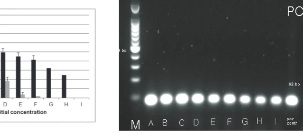
(4) 20. Zdrav Var 2014; 53. (Sigma-Aldrich) with a 100 bp ladder (Promega). Gels were visualized under UV illuminator Transiluminator Super-Bright (VilberLourmat) at 312 nm using a gel. images system Doc Print VX2 (VilberLourmat) to confirm the presence of the amplified DNA. Images processed by the Photo-Capt software.. Table 1. Primers used for PCR amplification. Tabela 1. Začetni oligonukleotidi za pomnoževanje DNK z metodo PCR. Target bacteria / Primer/začetni Primer 5’-------3’ tarčna bakterija oligonukleotid S. aureus K. pneumoniae P. aeruginosa C. difficile. egcAU (34). Product size / velikost baznega produkta (bp). f (5’- CTTCATATGTGTTAAGTCTTGCAGCTT-3’) r (5’-TTCACTCGCTTTATTCAATTGTTCTG-3’). 82. ITS (35). f (5’-ATT TGA AGA GGT TGC AAA CGA T-3’) r (5’-TTC ACT CTG AAG TTT TCT TGT GTT C-3’). 130. gyrB (36). f (5’-CCT GAC CAT CCG TCG CCA CAA C-3’) r (5’-CGC AGC AGG ATG CCG ACG CC-3’). 222. CD (37). f (5’-TTG AGC GAT TTA CTT CGG TAA AGA-3’) r (5’- CCA TCC TGT ACT GGC TCA CCT-3’). 3 RESULTS 3.1 Detection of S. aureus on textile swatches after 24 hour drying Figure 1 shows that the detection of S. aureus inoculated onto textile swatches at eight different concentrations. 157. levels (A to H) in descending order from 1010 cfu/mL to 102 cfu/mL yielded positive results after overnight drying for samples A to F (RODAC sampling method with incubation on selective agars); A to H (Morapex sampling method with incubation on selective agars); andA to I (Morapex sampling method with PCR).. Figure 1. Detection of different concentrations of S. aureus on textile swatches after 24 hour drying with three methods: RODAC with incubation (grey columns) and parallel Morapex with incubation (black columns) on upper figure and Morapex with PCR on lower figure. Slika 1. Detekcija različnih koncentracij S. aureus na tekstilnih krpicah po 24 urnem sušenju s tremi metodami: RODAC s kultivacijo (sivi stoplci), vzporedno Morapex s kultivacijo (črni stoplci) na zgornji sliki in Morapex s PCR na spodnji sliki. Initial bacterial concentration before overnight drying /začetna koncentracija suspenzije bakterij pred sušenjem: A (2.13 x 10¹⁰ cfu/mL); B (1.64 x 10⁹ cfu/mL); C (1.77 x 10⁸ cfu/mL); D (5.20 x 10⁶ cfu/mL); E (7.73 x 10⁵ cfu/mL); F (1.33 x 10⁵ cfu/mL); G (8.60 x 10⁴ cfu/mL); H (1.05 x 10³ cfu/mL); I (2.0 x 10² cfu/mL); M: (size marker / masni označevalec); positive control/pozitivna kontrola. 82 bp.. Brought to you by | University of Maribor Authenticated Download Date | 2/20/17 10:31 AM.
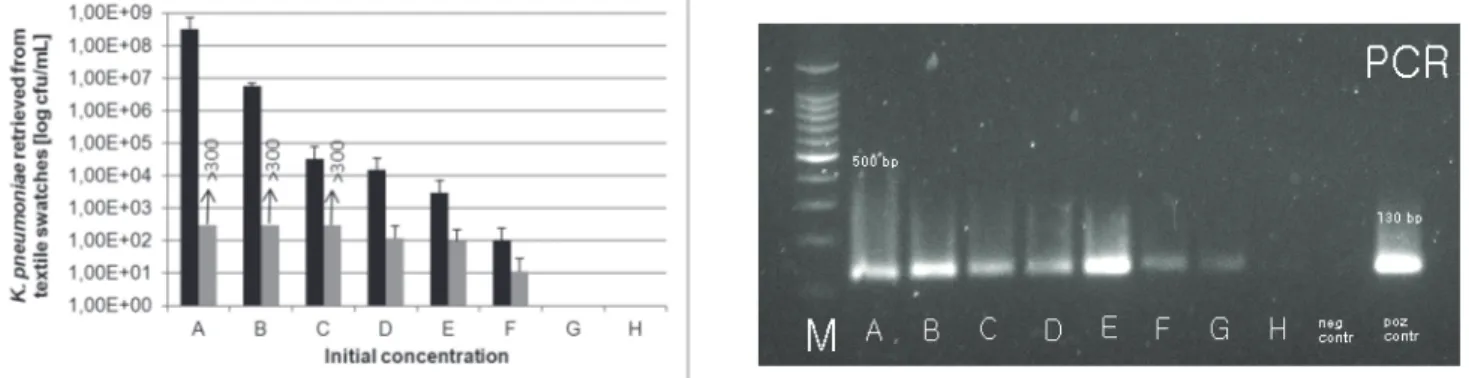
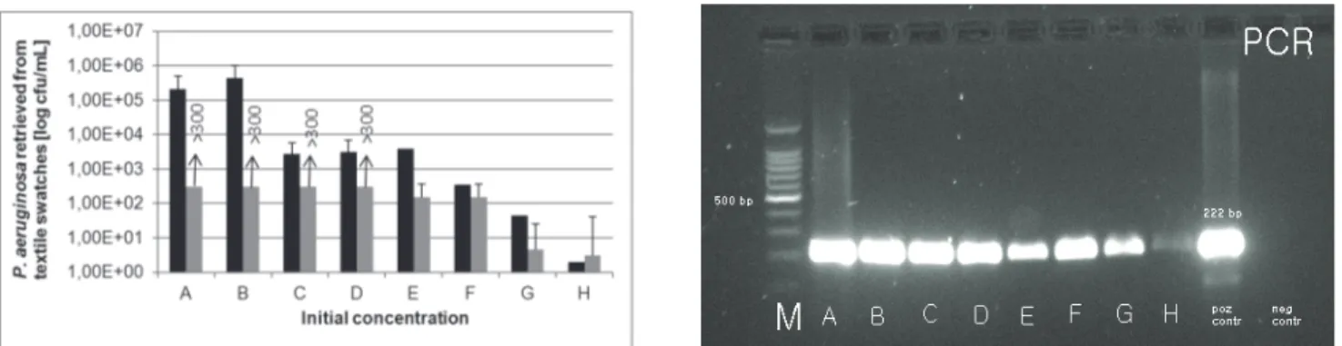
(5) Fijan S., Šostar Turk S., Rozman U. Comparison of methodes for detection of four common nosocomial pathogers on hospital .... 3.2 Detection of K. pneumoniae on textile swatches after 24 hour drying Figure 2 shows that the detection of K. pneumoniaei noculated onto textile swatches at eight different concentrations levels (A to H) in descending order from. 21. 1010 cfu/mL to 102 cfu/mL yielded positive results after overnight drying for samples A to F (RODAC sampling method with incubation on selective agars and Morapex sampling method with incubation on selective agars) andA to G (Morapex sampling method with PCR).. Figure 2. Detection of different concentrations of K. pneumoniae on textile swatches after 24 hour drying with three methods: RODAC with incubation (grey columns) and parallel Morapex with incubation (black columns) on upper figure and Morapex with PCR on lower figure. Slika 2. Detekcija različnih koncentracij K. pneumoniae na tekstilnih krpicah po 24 urnem sušenju s tremi metodami: RODAC s kultivacijo (sivi stoplci), vzporedno Morapex s kultivacijo (črni stoplci) na zgornji sliki in Morapex s PCR na spodnji sliki. Initial bacterial concentration before overnight drying / začetna koncentracija suspenzije bakterij pred sušenjem: A (2.58 x 10¹⁰ cfu/mL); B (1.04 x 10⁹ cfu/mL); C (2.90 x 10⁷ cfu/mL); D (1.30 x 10⁶ cfu/mL); E (4.00 x 10⁵ cfu/mL); F (4.05 x 10⁴ cfu/mL); G (9,88 x 10³ cfu/mL); H (2.00 x 10² cfu/mL); M: (size marker / masni označevalec); positive control/pozitivna kontrola: 130 bp. 3.3 Detection of P. aeruginosa on textile swatches after 24 hour drying Figure 3 shows that the detection of P. aeruginosa inoculated onto textile swatches at eight different. concentrations levels (A to H) in descending order from 1010 cfu/mL to 105 cfu/mL yielded positive results after overnight drying for samples A to H for all three methods.. Brought to you by | University of Maribor Authenticated Download Date | 2/20/17 10:31 AM.
(6) 22. Zdrav Var 2014; 53. Figure 3. Detection of different concentrations of P. aeruginosa on textile swatches after 24 hour drying with three methods: RODAC with incubation (grey columns) and parallel Morapex with incubation (black columns) on upper figure and Morapex with PCR on lower figure. Slika 3. Detekcija različnih koncentracij P. aeruginosa na tekstilnih krpicah po 24 urnem sušenju s tremi metodami: RODAC s kultivacijo (sivi stoplci), vzporedno Morapex s kultivacijo (črni stoplci) na zgornji sliki in Morapex s PCR na spodnji sliki. Initial bacterial concentration before overnight drying / začetna koncentracija suspenzije bakterij pred sušenjem: A (4.35 x 10¹⁰ cfu/mL); B (1.06 x 10⁹ cfu/mL); C (1.51 x 10⁸ cfu/mL); D (4.55 x 10⁷ cfu/mL); E (3.50 x 10⁷ cfu/mL); F (1.28 x 10⁶ cfu/mL); G (2.25 x 10⁵ cfu/mL); H (1.92 x 10⁴ cfu/mL); M: (size marker / masni označevalec); positive control/pozitivna kontrola: 222 bp. 3.4 Detection of C. difficile on textile swatches after 24 hour drying The detection of C. difficile spores inoculated onto textile swatches noted in figure 4 shows that at five different concentrations levels (A to E) in descending order from. 108 cfu/mL to 103 cfu/mL positive results after overnight drying were noted for samples A to D (RODAC sampling method with incubation on selective agars and Morapex sampling method with incubation on selective agars) andA to E (Morapex sampling method with PCR).. Figure 4. Detection of different concentrations of C. difficile on textile swatches after 24 hour drying with three methods: RODAC with incubation (grey columns) and parallel Morapex with incubation (black columns) on upper figure and Morapex with PCR on lower figure. Slika 4. Detekcija različnih koncentracij C. difficile na tekstilnih vzorcih po 24 urnem sušenju s tremi metodami: RODAC s kultivacijo (sivi stoplci), vzporedno Morapex s kultivacijo (črni stoplci) na zgornji sliki in Morapex s PCR na spodnji sliki. Initial sporal concentration before overnight drying / začetna koncentracija suspenzije bakterijskih spor pred sušenjem: A (2.29 x 10⁸ cfu/mL); B (2.23 x 10⁶ cfu/mL); C (1.57 x 10⁶ cfu/mL); D (1.16 x 10⁴ cfu/mL); E (3.80 x 10³ cfu/mL); M: (size marker / masni označevalec); positive control/pozitivna kontrola: 157 bp.. Brought to you by | University of Maribor Authenticated Download Date | 2/20/17 10:31 AM.
(7) Fijan S., Šostar Turk S., Rozman U. Comparison of methodes for detection of four common nosocomial pathogers on hospital .... 4 DISCUSSION Quick detection of microorganisms on textiles is important especially in hospital and other healthcare settings where many different kinds of textiles are used such as: bed sheets, blankets, towels, patient apparel, uniforms, gowns, drapes for surgical procedures (38). Contaminated textiles often contain high numbers of microorganisms from body substances, including blood, skin, stool, urine, vomitus and other body tissues and fluids. Although contaminated textiles in healthcare facilities can be a source of substantial numbers of pathogenic microorganisms, reports of healthcare associated diseases linked to contaminated textiles are few; therefore, the overall risk of disease transmission is very low. However, experience encourages infection control teams to take hospital textiles as a possible vehicle for transmission of infection very seriously in outbreaks that seem to have no obvious cause (39). Classical methods for the detection of microorganisms on textiles are RODAC sampling, swabbing or destructive elution methods (33) based on capturing microorganisms followed by incubation on nutrient or selective media to enable phenotypic determination of colony forming units of present microorganisms. In this research (33), it was found that wash-out or elution methods are much more efficient in detecting microorganisms on textiles than surface sampling such as RODAC sampling or swabbing, as these methods do not capture microorganisms that have penetrated into the three dimensional structure of textiles, which can cause false negative results. It takes at least 2 days to achieve results for these methods and in the case of infections in hospital settings, this time is much too long. PCR detection on the other hand yields results in less than four hours, thus being a much more efficient alternative. In our research, we compared the detection limit of chosen microorganisms (S. aureus, K. pneumoniae, P. aeruginosa and C. difficile) using three methods: RODAC sampling, elution method using the Morapex device followed by classical incubation on selective agars or by PCR detection. The Morapex device proved to be efficient for eluting microorganisms from textiles (33) without destroying the fabric and is therefore an efficient method for determining the hygiene of laundered hospital textiles. The basic principle is that the textile material is placed between two metal plates; the test liquid is pressed through the material and then collected in a tube. This test liquid was then used for two completely different methods. The first consisted of classical detection consisting of serial tenfold. 23. dilutions following the plating on selective medium and incubation, and the second method consisted of DNA extraction followed by PCR amplification and agarose gel electrophoresis. The first method yielded results in two days and the second within four hours, thus significantly shortening detection time. In all our experiments, the PCR method also proved to be the most sensitive method as detection of PCR fragments were positive at the lowest concentrations, while the results for the elution method followed by classical incubation and surface sampling with RODAC plates yielded detectable results at the second or third lowest concentrations, with RODAC plating being the least reliable method. The drying process lowered the concentration of all microorganisms by2 to 4 log steps. The lowest detection limits of our experiments with PCR method were reached at the initial bacterial concentration before overnight drying between 100 and 1000 cfu/mL. After overnight drying, this concentration was at least one log step lower, so the lowest possible concentration of detection with PCR methods was between 10 and 100 cfu/mL for all chosen bacteria. This coincides with results from a similar research (40) where the detection limit for different strains of Salmonella species was between 2 and 103 cfu/mL depending on the primer pairs used. Our results also prove that appropriate primer pairs for all four microorganisms were chosen. Another important observation is that no inhibition occurred during PCR amplification of high concentrated samples as has occurred in other research (41). Smith and co-authors (42) found that P. aeruginosa and S.aureus inoculated and then dried on a woven cotton fibre surface and a blood protein coagulum surface can survive over six months at room temperature. Although the viability was consistently higher on dried blood surfaces, the viability was next highest on cotton strings. For both of these environments, staphylococci appeared to lose viability between three and six months, while P. aeruginosa survived longer. The authors importantly conclude that such extended survival on blood and fibre surfaces, as observed in part, explains the difficulty in controlling colonization of patients and spread of these nosocomial pathogens. This conclusion can also be hypothesized for most pathogenic microorganisms. In our research, we found that P. aeruginosa was detectable with all three methods at the lowest initial bacterial concentration (104 cfu/mL), thus confirming survival on inanimate surfaces. S. aureus was also detected at the lowest initial bacterial concentration before overnight drying in sample I (102 cfu/mL) with the PCR method. Classical elution with incubation on. Brought to you by | University of Maribor Authenticated Download Date | 2/20/17 10:31 AM.
(8) 24. Zdrav Var 2014; 53. selective agar yielded a positive result for sample H, and RODAC plating proved least reliable as the lowest positive result was found in sample F. Klebsiella spp. are coliform bacteria ubiquitous in nature and natural habitants of environmental waters; they can also survive laundering as well as on inanimate surfaces such as hospital textiles (43-45). In our research, we found that an inoculation of 1000 cfu/mL (sample G) before overnight drying yielded positive results for PCR, while the classical elution method followed by incubation on selective medium yielded positive results after drying of 100 cfu/mL for sample F (initial bacterial concentration 104 cfu/mL). It has been reported that C. difficile [46] spores can survive temperatures and chemical treatment of typical hospital laundering cycles and that cross-contamination of C. difficile spores can occur on bed linen during a wash cycle. Therefore, the persistent nature of this organism must be considered by infection control personnel when implementing programs for laundering soiled and contaminated hospital linen (47). In our research, PCR yielded the lowest detectable survival of C. difficile spores on textiles after overnight drying in sample E with the lowest inoculated concentration before drying of (103 cfu/mL). Elution followed by incubation as well as RODAC plating yielded positive results (<50 cfu/mL) for sample (D) with initial concentration before drying of 104 cfu/mL.. classical incubation followed by phenotypic identification and an important method for the quick detection of microorganisms on textiles and enabling a direction of support in evaluating the cause of hospital acquired infections due tomicroorganisms on inanimate surfaces. However, a limitation of this method is that classical molecular methods are unable to discriminate between live and dead microorganisms, therefore special variants of real-time PCR reaction could be used such as ethidium bromide monoazide (EMA) PCR (50) or propidium monoazide (PMA) PCR (51) that distinguish viable from non-viable cells.. 5 CONCLUSION. 4.. The most common microorganisms found on healthcare associated textiles are Gram negative bacteria, coagulase negative staphylococci, Bacillus sp. and typical skin flora (48).In the study by Catano et al. (49), it was found that a large proportion of textiles in hospital settings (white coats, curtains and ties) as well as computer keyboards and cell phones were contaminated with bacterial pathogens that may act as reservoirs for bacterial pathogens that may be associated with healthcare-associated infections. They concluded that further research is needed to evaluate strategies to minimize the risk of patient-to-patient transmission of pathogens from other contaminated items. One of these strategies is to perform regular sanitary controls of all inanimate surfaces as well as implementing quick methods for the determination of cleanliness and hygiene. Elution of microorganisms using the Morapex device followed by PCR detection on the other hand yield results in less than four hours, thus being a much more efficient alternative than. 5.. Acknowledgements We are very grateful to SedoTreepoint GmbH, Germany, for the rental of the Morapex A device. This work was supported by the ARRS 1000-10-310152. References 1. 2.. 3.. 6. 7. 8.. 9. 10. 11. 12. 13.. Winslow CEA. The untitled fields of public health. Science 1920; 51: 23–33. Collins AS. Preventing healthcare—associated infections. In: Hughes RG. Patient safety and quality: an evidence-based handbook for nurses. Rockville: Agency for Healthcare Research and Quality, 2008: 547–75. Gastmeier P, Stamm-Balderjahn S, Hansen S, NitzschkeTiemann F, Zuschneid I, Groneberg K et al. How outbreaks can contribute to prevention of nosocomial infection: analysis of 1,022 outbreaks. Infect Control Hosp Epidem 2005; 26: 357–61. Bureau-Chalot F, Piednoir E, Camus J, Bajolet O. Microbiologic quality of linen and linen rooms in short-term care units. J Hosp Infect 2004; 56: 329–31. Dancer SJ. How do we assess hospital cleaning?: a proposal for microbiological standards for surface hygiene in hospitals. J Hosp Infect 2004: 56: 10–5. Wilks SA. The survival of Escherichia coli O157 on a range of metal surfaces. Int J Food Microbiol 2005; 105: 445–54. Verran J. Biofouling in food processing: biofilm or biotransfer potential? Inst Chem Eng Trans C: Food Bioprod Process 2002; 80: 292–8 Fijan S, Šostar-Turk S, Cencič A. Implementing hygiene monitoring systems in hospital laundries in order to reduce microbial contamination of hospital textiles. J hosp infect 2005; 61: 30-8. Fijan S, Šostar-Turk S. Hospital textiles, are they a possible vehicle for healthcare-associated infections? Int J Environ Res Public Health 2012; 9: 3330-43. Ndawula EM, Brown L. Mattresses as reservoirs of epidemic methicillin-resistant Staphylococcus aureus. Lancet 1991; 337: 488. Creamer E, Humphreys H. The contribution of beds to healthcare-associated infection: the importance of adequate decontamination. J Hosp Infect 2008; 69: 8–23. Brunton WA. Infection and hospital laundry. Lancet 1995; 345, 1574–5. Bonten MJM, Hayden MK, Nathan C, van Voorhis J, Matushek M, Slaughter S et al. Epidemiology of colonisation of patients. Brought to you by | University of Maribor Authenticated Download Date | 2/20/17 10:31 AM.
(9) Fijan S., Šostar Turk S., Rozman U. Comparison of methodes for detection of four common nosocomial pathogers on hospital .... 14.. 15. 16. 17. 18. 19. 20. 21. 22. 23. 24.. 25. 26. 27. 28. 29. 30. 31. 32. 33.. and environment with vancomycin-resistant Enterococci. Lancet 1996; 348: 1615–9. Sasahara T, Hayashi S, Morisawa Y, Sakihama T, Yoshimura A, Hirai Y. Bacillus cereus bacteremia outbreak due to contaminated hospital linens. Eur J Clin Microbiol Infect Dis 2011; 30: 219–26. Datta N, Pridie RB. An outbreak of infection with Salmonella Typhimurium in a general hospital. J Hyg (London) 1960; 58: 229–40. Standaert SM, Hutcheson RH, Schaffner WA. Nosocomial transmission of Salmonella gastroenteritis to laundry workers in a nursing home. Infect Control Hospl Epidem 1994; 15: 22–6. Kirby WMM, Corporon DO, Tanner DC. Urinary tract infections caused by antibiotic-resistant coliform bacteria. JAMA 1956; 162: 1–4. Keeffe EB. Occupational risk for hepatitis A: a literature-based analysis. J Clin Gastroenterol 2004; 38: 440–8. English MP, Wethered RR, Duncan EH. Studies in the epidemiology of Tinea pedis VIII: fungal infection in a long-stay hospital. Brit Med J 1967; 3: 136–9. Thomas MD, Giedinghagen DH, Hoff GL. An outbreak of scabies among employees in a hospital-associated commercial laundry. Infect Control 1987; 8: 427–9. Shah PC, Krajden S, Kane J, Summerbell RC. Tinea corporis caused my Microsporum canis: report of a nosocomial outbreak. Eur J Epidemiol 1988; 4: 33–8. Wilcox MH, Jones BL. Enterococci and hospital laundry. Lancet 1995; 345: 594. Orr KE, Holliday MG, Jones AL, Robson I, Perry JD. Survival of enterococci during hospital laundry processing. J Hosp Infect 2002; 50: 133–9. Fijan S, Koren S, Cencič A, Šostar-Turk S. Antimicrobial disinfection effect of a laundering procedure for hospital textiles against various indicator bacteria and fungi using different substrates for simulating human excrements. Diagn Microbiol Infect Dis 2007; 57: 251–7. Fijan S, Šostar-Turk S. Antimicrobial activity of selected disinfectants used in a low temperature laundering procedure for textiles. Fibres Textiles East Eur 2010; 18: 89–92. Altenbaher B, Šostar-Turk S, Fijan S. Ecological parameters and disinfection effect of low-temperature laundering in hospitals in Slovenia. J Clean Prod 2011; 19: 253–8. Walter WG, Schillinger JE. Bacterial survival in laundered fabrics. Appl Microbiol 1975; 29: 368–73. Smith JA, Neil KR, Davidson CG, Davidson RW. Effect of water temperature on bacterial killing in laundry. Infect Control 1987; 8: 204–9. Christian RR, Manchester JT, Mellor MT. Bacteriological quality of fabrics washed at lower-than-standard temperatures in a hospital laundry facility. Appl Environ Microbiol 1983; 45: 591–7. Hellickson LA, Owens KL. Cross-contamination of Clostridium difficile spores on bed linen during laundering. Amer J Infect Control 2007; 35: E32-3. Wetzler TF, Qquan TF, Schatzle K. Critical analysis of the microflora of toweling. Am J Public Health 1971; 61: 376–93. Greig JD. Infective doses and pathogen carriage. Food Safety Education Conference, Atlanta, Georgia, 2010. Rabuza U, Šostar-Turk S, Fijan S. Efficiency of four sampling methods used to detect two common nosocomial pathogens on textiles. Textile Res J 2012; 82: 2099-105.. 25. 34. Fusco V, Quero GM, Morea M, Blaiotta G, Visconti A. Rapid and reliable identification of Staphylococcus aureus harbouring the enterotoxin gene cluster (egc) and quantitative detection in raw milk by real time PCR. Int J Food Microbiol 2011; 144: 528–37. 35. Liu Y, Liu C, Zheng W, Zhang X, Yu J, Gao Q et al. PCR detection of Klebsiella pneumoniae in infant formula based on 16S–23S internal transcribed spacer. Int J of Food Microbiol 2008; 125: 230–5. 36. Motoshima M, Yanagihara K, Fukushima K, Matsuda J, Sugahara K, Hirakata Y et al. Rapid and accurate detection of Pseudomonas aeruginosa by real-time polymerase chain reaction with melting curve analysis targeting gyrB gene. Diag Microbiol Infect Dis 2007; 58: 53–8. 37. Balamurugan R, Balaji V, Ramakrishna BS. Estimation of faecal carriage of Clostridium difficile in patients with ulcerative colitis using real time polymerase chain reaction. Indian J Med Res 2008; 127: 472-7. 38. Sehulster LM, Chinn RYW, Arduino MJ, Carpenter J, Donlan R, Ashford D et al. Guidelines for environmental infection control in health-care facilities: recommendations from CDC and the Healthcare Infection Control Practices Advisory Committee (HICPAC). Chicago: American Society for Healthcare Engineering/American Hospital Association, 2004. 39. Brunton WA. Infection and hospital laundry. Lancet 1995; 345: 1574–5. 40. Oliveira SD, Santos LR, Schuch MDT, Silva AB, Salle CTP, Canal CW. Detection and identification of salmonellas from poultry-related samples by PCR. Veter microbiol 2002; 87: 25-35. 41. Fijan S, Steyer A, Poljsak-Prijatelj M, Cencic A, Sostar-Turk S, Koren S. Rotaviral RNA found on various surfaces in a hospital laundry. J Virol Methods 2008; 148: 66-73. 42. Smith SM, Eng RH, Padberg FT. Survival of nosocomial pathogenic bacteria at ambient temperature. J Med 1996; 27: 293-302. 43. Bagley S. Habitat association of Klebsiella species“. Infect Control 198; 65: 52–8. 44. Smith J.A., Neil K.R., Davidson C.G., Davidson R.W. Effect of water temperature on bacterial killing in laundry. Infect Control 1987; 8: 204–9. 45. Učakar V, Grilc E, Jeraj I. An investigation of a waterborne outbreak caused by microbiological contamination of the drinking water supply system. Zdrav Var 2012; 51: 112-9. 46. Hellickson LA, Owens KL. Cross-contamination of clostridium difficile spores on bed linen. Amer J Infect Control 2007; 35: E32-3. 47. Grilc E. Epidemiological surveillance of gastrointestinal communicable diseases in Slovenia from 1999 to 2009. Zdr varstvo 2012; 51: 155-62. 48. Cataño JC, Echeverri LM, Szela C. Bacterial contamination of clothes and environmental items in a third-level hospital in Colombia. Interdiscip Perspect Infect Dis 2012; 2012: 1-5. 49. Blaser MJ, Smith PF, Cody H.J, Wang WLL, LaForce F.M. Killing of fabric-associated bacteria in hospital laundry by lowtemperature washing. J Infect Dis 1984; 149: 48–57. 50. Wang, L, Mustapha A. EMA-real-time PCR as a reliable method for detection of viable Salmonella in chicken and eggs. J Food Sci 2010; 75: 134-9. 51. Kaushik, R, Balasubramanian, R. Discrimination of viable from non-viable Gram-negative bacterial pathogens in airborne particles using propidium monoazide-assisted qPCR. Sci Total Environ 2013; 18: 237-43.. Brought to you by | University of Maribor Authenticated Download Date | 2/20/17 10:31 AM.
(10)
Gambar
Dokumen terkait
Berbagai informasi berkaitan dengan program dan pengembangan sekolah dapat diakses melalui website SMA Negeri 1 kota Pematang siantar sehingga media ini dapat digunakan
(“the Company”) hereby announce that the Extraordinary General Meeting of Shareholders (“EGM”), that was convened on Wednesday, September 28, 2011 at The Ritz
Ketentuan Peserta Ujian Ulang LPTK IAIN Sunan Ampel SurabayaI. Ujian Ulang Tulis Tahap 1 dilaksanakan Hari Sabtu, 23
Pada hari ini Kamis tanggal Tiga Belas bulan September Tahun Dua Ribu Dua Belas, kami yang bertanda tangan dibawah ini Panitia Pengadaan Barang dan Jasa Dinas Bina Marga dan
menyatakan dengan sesungguhnya bahwa skripsi yang berjudul: Analisis Pengaruh Market Value Added, Economic Value Added, Operating Cash Flow dan Earnings Per Share Terhadap
Setelah mengikuti pembelajaran mata kuliah Perencanaan Instalasi Listrik peserta didik diharapkan mempunyai kemampuan perencanaan (analisis, Gambar, Rencana anggaran
definisi, penyebab, jenis-jenis dan karakteristik, serta berbagai teknik layanan khusus bagi anak kesulitan belajar.. Kemudian diakhiri dengan
Hasil penelitian dan pembahasan tentang Pengembangan Kompetensi Sosial Guru di SMP Negeri 2 Mojosongo Boyolali , dapat disimpulkan sebagai berikut: 1) Pengembangan
