262
Corresponding author: Yuki Hashimoto [email protected]
Received 2016 August 19 Accepted 2016 October 6
Abbreviations: AML, acute myeloid leukemia; APL, Acute promy-elocytic leukemia; ATRA, all-trans retinoic acid; LAMP, loop-me-diated isothermal amplification; PML, promyelocytic leukemia; RARα, retinoic acid receptor α; RIN, RNA integrity number; RT-LAMP, reverse transcription loop-mediated isothermal amplifica-tion; RT-PCR, reverse transcription polymerase chain reaction ABSTRACT
Background Acute promyelocytic leukemia (APL) is
a disease characterized by expression of promyelocytic
leukemia–retinoic acid receptorα (PML-RARα) chi-meric mRNA. Although APL is curable, early death due to hemorrhage is a major problem. Here, we report the development of a simple and rapid diagnostic method for APL based on reverse transcription loop-mediated iso-thermal amplification (RT-LAMP).
Methods An RT-LAMP primer set was designed to
detect three types of PML-RARα mRNA in a single reaction. Serial dilutions of plasmid DNA containing bcr1, bcr2, or bcr3 PML-RARα sequences and RNA extracted from bone marrow aspirates of 6 patients with APL were used to compare the results of RT-LAMP and nested PCR assays.
Results Plasmid DNA was amplified by RT-LAMP,
for which the reaction time was > 4 h shorter and the lower detection limit was higher than for nested RT-PCR. Six of 7 samples tested positive by both methods.
Conclusion We developed an RT-LAMP assay for
simple and rapid PML-RARα mRNA detection that may be clinically useful for point-of-care testing and APL diagnosis.
Key words acute promyelocytic leukemia; polymerase
chain reaction; promyelocytic leukemia–retinoic acid
receptorα; reverse transcription loop-mediated isother-mal amplification; t(15;17) chromosome translocation
Loop-mediated isothermal amplification (LAMP) is an established nucleic acid amplification method that re-quires a set of 4 specially designed, highly specific inner and outer primers.1 Additional loop primers can further
accelerate the reaction.2 The LAMP product is detected
by turbidity arising from magnesium pyrophosphate formed during the reaction.3 This method is very rapid
and directly amplifies the target region from an RNA sample, and has been used to detect various pathogens, including bacteria and viruses.4 In addition, LAMP has
now been developed as commercial kits, some of which have been adopted as officially recommended methods for routine pathogen examination and surveillance in Japan.5 However, there have been few applications of
LAMP to the field of hematological malignancy, al-though one notable exception is the detection of Wilms’
tumor-1 gene mRNA.6
Acute promyelocytic leukemia (APL) is a distinct subtype of acute myeloid leukemia (AML) accounting for approximately 10% of cases. APL is cytogenetical-ly characterized by balanced reciprocal translocation between chromosomes 15 and 17, which results in the fusion of the promyelocytic leukemia (PML) and
retino-ic acid receptorα (RARα) genes.7 The PML-RARα
chi-meric gene is detected in nearly all APL patients and is used as a marker for clinical diagnosis. In addition, the mRNA is detected by reverse transcription polymerase chain reaction (RT-PCR) and/or real time RT-PCR for minimal residual disease monitoring of APL.8–10 Three
different types of PML-RARα mRNA exist owing to different breakpoints in the PML gene, although these do not result in different treatment outcomes: the long-, variable-, and short-form types are derived from break-points at bcr1 (intron 5), bcr2 (exon 6), and bcr3 (intron 3), respectively, and occur at relative frequencies of 55%, 5%, and 40%, respectively.11
APL is the most curable form of AML because of an established molecular targeted therapy combining all-trans retinoic acid (ATRA) and arsenic trioxide.12 Before
starting the treatment, it is necessary to verify that the patient has the PML-RARα fusion gene.13 However,
ear-ly death—mostear-ly due to severe hemorrhage—remains
Development of Reverse Transcription Loop-Mediated Isothermal Amplification
for Simple and Rapid Detection of
Promyelocytic Leukemia–Retinoic Acid
Recep-tor
α
mRNA
Yuki Hashimoto,* Yuki Hatayama,* Nao Kojima,* Shota Morishita,* Satoko Matsumoto,* Yuzuru Hosoda,†‡ Ayako Hara* and Toru Motokura*†‡
*Division of Clinical Laboratory, Tottori University Hospital, Yonago 683-8504, Japan, †Department of Hematology, Tottori University Hospital, Yonago 683-8504, Japan and ‡Division of Clinical Laboratory Medicine, Department of Pathophysiological and Therapeutic Science, School of Medicine, Tottori University Faculty of Medicine, Yonago 683-8503, Japan
the main cause of APL treatment failure according to recent population-based studies carried out in developed countries.14 Some reports recommend early and timely
initiation of ATRA and provision of aggressive sup-portive care at the first suspicion of the disease, without awaiting molecular confirmation, in order to reduce hemorrhagic deaths in APL.14 An improved PML-RARα
mRNA detection test is also needed.
We report here the development of a simple reverse transcription loop-mediated isothermal amplification (RT-LAMP) method for rapid PML-RARα mRNA de-tection. The advantages of this method with respect to RT-PCR are also discussed.
MATERIALS AND METHODS
Samples
Three different plasmids—containing bcr1, bcr2, and bcr3 PML-RARα mRNA sequences—were purchased from Eurofins Genomics (Louisville, KY) for use as positive controls. Vector plasmid was pTAKN-2 and chimeric sequences were made of sequences derived from the Genbank sequence database (Accession num-bers are M73778, NM_000964, and AB067754). IVS-0035 RNA (Invivoscribe Technologies, San Diego, CA) was used as a negative control. Clinical samples used in this study were archived specimens from APL patients diagnosed by detection of APL cells and PML-RARα mRNA by nested RT-PCR at our hospital. RNA was extracted from patient bone marrow aspirates using the RNeasy Plus Mini kit (Qiagen, Valencia, CA) accord-ing to the manufacturer’s instructions; these were used for diagnosis, and then frozen at −80 °C until the study. RNA concentrations were measured and 260/280 nm absorbance ratios were determined with a Nanodrop 2000 spectrophotometer (Thermo Fisher Scientific, Waltham, MA) prior to RT-LAMP and nested RT-PCR
assays. The study protocol was approved by the Tottori University Hospital ethics committee (number 2326). Primer design
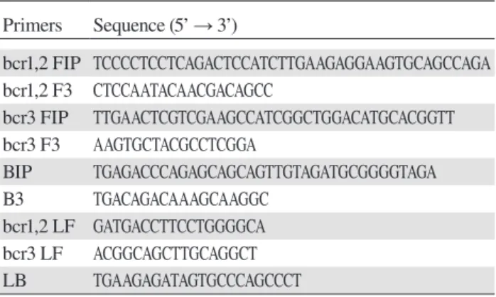
Primer sets for detection of PML-RARα mRNA by RT-LAMP (Table 1) were designed using PrimerExplorer ver. 4 software (Eiken Chemical, Tokyo, Japan). The primer sets were modified to detect the three types of
PML-RARα mRNA in a single tube—i.e., bcr1 and bcr2 were amplified with the same primer sets and an addi-tional set consisting of FIP, F3, and LF were included in one reaction to amplify bcr3 (Fig 1).
RT-LAMP
The RT-LAMP assay was carried out using the Loopa-mp RNA ALoopa-mplification kit (Eiken Chemical, Tokyo, Japan) under the following conditions: 1 μL sample (RNA or DNA) was mixed with 20 pmol each of FIP and BIP primers, 10 pmol each of LF and LB primers, 2.5 pmol each of F3 and B3 primers, 1 μL Enzyme Mix, and 12.5 μL of 2 × Reaction Mix, with distilled water added to obtain a final volume of 25 μL. For real-time turbidity monitoring, the reaction mixture was incubated at 68 °C for 60 min in an RT-160C Loopamp Realtime Turbidi-meter (Eiken Chemical). The specificity of LAMP was confirmed by restriction enzyme digestion using DdeI (Takara Bio, Otsu, Japan) under the following condi-tions: 5 μL LAMP product was mixed with 2 μL of 10 × K buffer (Takara Bio), 1 μL DdeI, with distilled water added to obtain a final volume of 20 μL, followed by incubation at 37 °C for 120 min. DdeI was inactivated at 70 °C for 15 min and the digestion products (10-μL aliquots) were mixed with loading buffer (Nippon Gene, Toyama, Japan) and separated by 1.5% agarose gel electrophoresis (100 V, 35 min) with 0.005% ethidium bromide using a Mupid mini gel electrophoresis system (Advance, Tokyo, Japan), with 5 μL Gene Ladder 100 (Nippon Gene) used as a molecular size marker.
Nested RT-PCR
The RT reaction was carried out using1 μL RNA sample and the ThermoScript RT-PCR system (Thermo Fisher Scientific) according to the manufacturers’ protocol. Three types of PML-RARα mRNA were detected using two forward primer sets designed according to published sequences.10 Specifically, bcr1 and bcr2 types were
amplified with the same primer set. The PCR reaction mixture was prepared using AmpliTaq Gold with 10 × PCR Gold Buffer and MgCl2 (Thermo Fisher Scientific)
and the GeneAmp dNTP mix (Thermo Fisher Scientific) under the following conditions: 1 μL plasmid DNA, 2 μL cDNA, or 1 μL first PCR product was mixed with Table 1. Primers for detection of PML-RARα mRNA
by RT-LAMP Primers Sequence (5’ → 3’) bcr1,2 FIP TCCCCTCCTCAGACTCCATCTTGAAGAGGAAGTGCAGCCAGA bcr1,2 F3 CTCCAATACAACGACAGCC bcr3 FIP TTGAACTCGTCGAAGCCATCGGCTGGACATGCACGGTT bcr3 F3 AAGTGCTACGCCTCGGA BIP TGAGACCCAGAGCAGCAGTTGTAGATGCGGGGTAGA B3 TGACAGACAAAGCAAGGC bcr1,2 LF GATGACCTTCCTGGGGCA bcr3 LF ACGGCAGCTTGCAGGCT LB TGAAGAGATAGTGCCCAGCCCT
PML-RARα, promyelocytic leukemia–retinoic acid receptorα; RT-LAMP, reverse transcription loop-mediated isothermal ampli-fication.
264
3
4
5
6
3
PML
RARα
bcr 1
3
4
5
6
3
bcr 2
3
3
bcr 3
DdeI
DdeI
DdeI
BIP
BIP
BIP
B3
B3
B3
bcr3 FIP
bcr3 F3
bcr1,2 FIP
bcr1,2 F3
bcr1,2 F3
bcr1,2 FIP
Fig. 1. Schematic representation of primer design for detection of PML-RARα mRNA by RT-LAMP and cutting sites of DdeI restriction enzyme assay. Exon numbers of the PML gene (dark) and the RARα gene (light) are shown. PML, promyelocytic leukemia; RARα, retino-ic acid receptorα; RT-LAMP, reverse transcription loop-mediated isothermal amplifi cation.
2.5 μL 10 × PCR Gold buffer, 2.25 nmol MgCl2
solu-tion, 500 pmol dNTP mix, 0.2 μL AmpliTaq Gold DNA polymerase, and 0.25 pmol each forward and reverse primers, with distilled water added to obtain a final volume of 25 μL. The forward primers were as follows: 5’-AGTCAGTGCCCGGGGCACAC-3’ for bcr1 and bcr2 and 5’-AGCTGCTGGAGGCTGTGGACG-3’ for bcr3 in the fi rst PCR; and 5’-AGTGTACGCCTTCTC-CATCAAAG-3’ for bcr1 and bcr2 and 5’-TGTGCTG-CAGCGACTCCGCA-3’ for bcr3 in the second PCR. The reverse primers were as follows: 5’-AGGGCTGG-GCACTATCTCTTC-3’ for bcr1, bcr2, and bcr3 in the first PCR; and 5’-CAGAACTGCTGCTCTGGGTCT-CAAT-3’ for bcr1, bcr2, and bcr3 in the second PCR. Reaction conditions on the MultiGene Mini Personal Thermal Cycler (Labnet International, Edison, NJ) were as follows: 94 °C for 5 min; 35 cycles at 94 °C for 30 s, 62 °C for 1 min, and 72 °C for 1 min; and 72 °C for 7 min. The nested PCR products (10-μL aliquots) were mixed with loading buffer and separated along with 5 μL of molecular size marker by 1.5% agarose gel elec-trophoresis (100 V, 35 min) with 0.005% ethidium bro-mide.
RNA integrity number (RIN) analysis
The quality of clinical RNA samples (expect for S-4,
for which the amount was insuffi cient) was evaluated by RIN as previously described15 on an Agilent 2100
Bio-analyzer (Agilent Technologies, Santa Clara, CA). RESULTS
Specifi city of amplifi cation by RT-LAMP
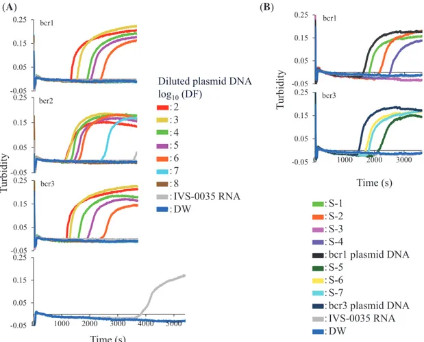
We performed the RT-LAMP assay with two concen-trations of plasmid DNA followed by DdeI restriction enzyme analysis. All plasmid DNAs were detected by RT-LAMP in 60 min (data not shown, Fig. 2A) and yielded the expected products after digestion with DdeI (Fig. 3), which differed for each breakpoint. Non-spe-cifi c amplifi cation products obtained after 60 min from
PML-RARα-negative RNA were not digestible by DdeI (Fig. 3).
Sensitivity of the RT-LAMP assay
The lower detection limit of the RT-LAMP assay was determined using10-fold serial dilutions of bcr1, bcr2, and bcr3 plasmid DNA; the result was compared to that obtained by nested RT-PCR. RT-LAMP was able to de-tect 880, 78, and 800 copies/tube of bcr1, bcr2, and bcr3 plasmid, respectively (Fig. 2A), as compared to 0.88, 7.8, and 80 copies/tube, respectively, by nested RT-PCR (Figs. 4A–C). The specifi city of all RT-LAMP products was confi rmed by DdeI restriction enzyme digestion.
-0.05 0.05 0.15 0.25 (A) (B) -0.05 0.05 0.15 0.25 -0.05 0.05 0.15 0.25 bcr1 bcr2 bcr3 Time (s) Turb idity ー:S-1 ー:S-2 ー:S-3 ー:S-4 ー:bcr1 plasmid DNA ー:S-5 ー:S-6 ー:S-7 ー:bcr3 plasmid DNA ー:IVS-0035 RNA ー:DW -0.05 0.05 0.15 0.25 bcr1 -0.05 0.05 0.15 0.25 0 1000 2000 3000 bcr3 Time (s) Turb idity -0.05 0.05 0.15 0.25 0 1000 2000 3000 4000 5000
Diluted plasmid DNA log10 (DF) ー:2 ー:3 ー:4 ー:5 ー:6 ー:7 ー:8 ー:IVS-0035 RNA ー:DW
Dde I
−
+
−
+
−
+
−
+
−
+
M
LAMP
bcr1 bcr2 bcr3 neg RNA DW M
Dde*I
100 200 300 400 500 100 200 300 400 500
Fig. 2. Detection of PML-RARα in plasmid DNA and clinical RNA samples by RT-LAMP. (A) Ten-fold serial dilutions of bcr1, bcr2, and bcr3 plasmid DNAs were used as template; the starting amounts were 8.8, 7.8, and 8.0 × 10⁸ copies/tube, respectively. DF of the indicated plasmid DNA is shown as a logarithmic value. (B) Clinical samples from APL patients (S-1 through -7) were analyzed by RT-LAMP. IVS-0035 RNA and distilled water were used as negative controls. A 100-fold dilution of corresponding plasmid DNAs served as a pos-itive control. The amplification reaction was carried out at 68 °C for indicated times. APL, acute promyelocytic leukemia; DF, dilution factor; PML-RARα, promyelocytic leukemia–retinoic acid receptorα; RT-LAMP, reverse transcription loop-mediated isothermal ampli-fication.
Fig. 3.DdeI restriction enzyme digestion of RT-LAMP products. Products of bcr1, bcr2, and bcr3 plasmid DNA amplification were incubated with (+) or without (−) DdeI at 37 °C for 120 min, followed by deactivation at 70 °C for 15 min and sepa-ration by 1.5% agarose gel electrophoresis (100 V, 35 min). The amounts of bcr1, bcr2 and bcr3 plasmid DNAs were 8.8, 7.8, and 8.0 × 10⁶ copies/tube, respectively. The expected sizes of digested RT-LAMP products are the followings: 468 bp, about 234 bp (stem and loop structure) and 83 bp for bcr1; 232 bp, about 116 bp (stem and loop structure) and 83 bp for bcr2; and 139 bp and 121 bp (merged in Figure) for bcr3. DW, distilled water (negative control); M, 100-bp ladder size marker; neg RNA, IVS-0035 RNA (negative control); RT-LAMP, reverse transcription loop-mediated iso-thermal amplification.
266
11)tube 10)tube 0.88)copies/tube LAMP bcr1 880)copies/tube 1000 ))M)))))))1))))))))2))))))))3)))))))4))))))))5)))))))6)))))))7))))))))8))))))))9))))))10))))))11)))))DW
(
A
)
M 0 1 2 3 4 5 6 7 8 9 10 DW(
B
)
M 0 1 2 3 4 5 6 7 8 9 10 11 DW(
C
)
M 0 1 2 3 4 5 6 7 8 9 10 11 12 13 14 DW log10 (DF)Fig. 4. Nested RT-PCR amplification using 10-fold serial dilutions of plas-mid DNA. Plasplas-mid DNAs contain-ing (A) bcr1, (B) bcr2, and (C) bcr3 sequences (starting amounts: 8.8 × 10⁸, 7.8 × 10⁸, and 8.0 × 10⁸ copies/ tube, respectively) were diluted by the indicated DF and amplified by nested RT-PCR. Amplification products were separated by 1.5% agarose gel electrophoresis (100 V, 35 min). DF, dilution factor; DW, distilled water; M, 100-bp ladder size marker; RT-PCR, reverse transcription polymerase chain reaction.
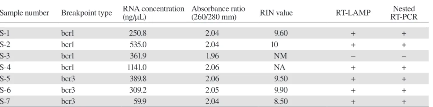
Table 2. Comparison between RT-LAMP and nested RT-PCR using clinical samples
Sample number Breakpoint type RNA concentration(ng/μL) Absorbance ratio(260/280 mm) RIN value RT-LAMP RT-PCRNested
S-1 bcr1 250.8 2.04 9.60 + + S-2 bcr1 535.0 2.04 10 + + S-3 bcr1 361.9 1.96 NM – – S-4 bcr1 1141.0 2.06 NA + + S-5 bcr3 389.8 2.06 9.50 + + S-6 bcr3 309.2 2.05 9.90 + + S-7 bcr3 59.9 2.04 8.50 + +
NA, not available; NM, not measured due to pattern deformation; RIN, RNA integrity number; RT-LAMP, reverse transcription loop-me-diated isothermal amplification; RT-PCR, reverse transcription polymerase chain reaction; Samples 5 and 6 were derived from the same patient: S-5, at the initial visit and S-6, at the time of relapse.
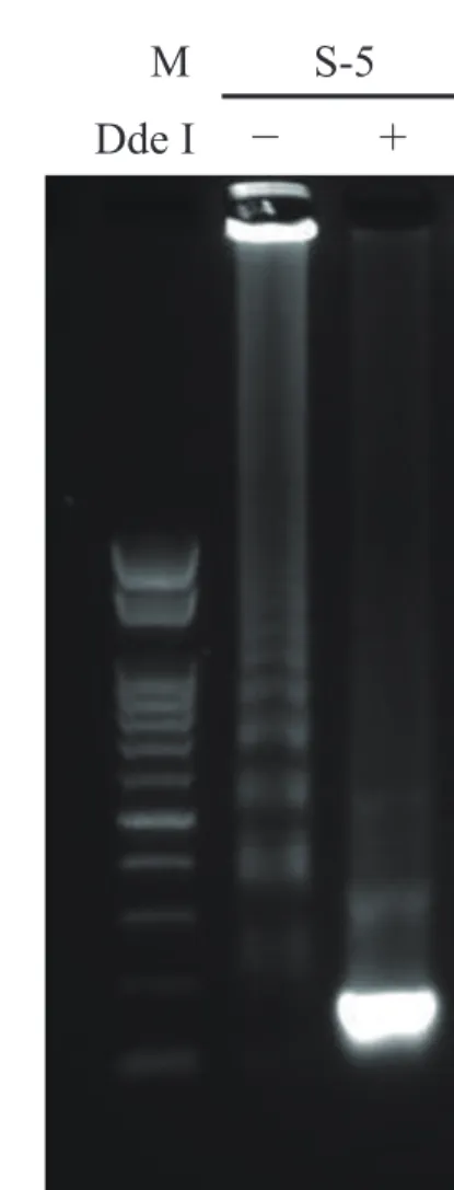
(A)
M S-1
Dde I
− +
(B)
M S-5
Dde I
− +
Fig. 5. Confirmation of bcr1 and bcr3 of clinical samples after RT-LAMP assay. The RT-LAMP products of clinical sample RNAs of S-1 (A) and S-5 (B) were incubated with (+) or without (−) DdeI at 37 °C for 120 min, followed by deactivation at 70 °C for 15 min and separation by 1.5% agarose gel electrophoresis (100 V, 35 min). M, 100-bp ladder size marker; RT-LAMP, reverse transcription loop-me-diated isothermal amplification.
RT-LAMP of clinical samples
Clinical samples were analyzed by RT-LAMP and nest-ed RT-PCR; these includnest-ed four bcr1- and three bcr3-type PML-RARα RNAs. The plasmid DNAs were used as positive controls, and IVS-0035 RNA and distilled water were used as negative controls. Three bcr1- and three bcr3-type RNAs were detected by RT-LAMP (Fig. 2B); the products were confirmed by DdeI restriction en-zyme digestion (Fig. 5), and the results were the same as those obtained by nested RT-PCR (Table 2). One sample yielded no product with both assays; this RNA had been stored for the longest period (about 9 years) and was likely degraded, as indicated by RIN analysis of RNA quality.15, 16
DISCUSSION
Each type of PML-RARα sequence was specifically amplified by RT-LAMP; moreover, each PML-RARα breakpoints as well as non-specific amplification could be distinguished by digesting the products with DdeI. The sensitivity of the RT-LAMP assay was inferior to that of nested RT-PCR by 10 fold for bcr2 and bcr3 and by 1000 fold for bcr1. Some RT-LAMP assays have higher sensitivity than nested RT-PCR,17–20 while others
have lower sensitivity.21, 22 This depends on the primers
used, which determine the amplification efficiency and has a significant effect on the sensitivity of gene ampli-fication. In this study, we selected 5 primer sets from more than 1000 candidates listed by PrimerExplorer ver. 4. It is virtually impossible to test all candidates, al-though a primer set with better amplification efficiency
268 may exist.
RT-LAMP and nested RT-PCR yielded similar results in the analysis of clinical samples. This sug-gests that RT-LAMP has clinical utility as a diagnostic test, especially at initial presentation when a sufficient amount of PML-RARα mRNA is available.
The nested RT-PCR assay requires about 6 h from RNA extraction to target mRNA detection. In contrast,
PML-RARα mRNA was amplified in 30 min by RT-LAMP, and is therefore more useful for early diagnosis and determination of an appropriate intervention. In ad-dition, the amount of sample that is lost with RT-LAMP is minimal because the extracted RNA is used directly in the amplification reaction even in one tube. This sim-plifies the procedure, thereby reducing the labor and cost of laboratory testing.
Spinelli et al. recently reported another assay using RT-LAMP to detect PML-RARα chimeric gene.23
Us-ing fluorescence, the assay appeared to be more rapid and sensitive than our assay. However, the detection of fluorescence requires a dedicated devise and their assay needs 2 reaction tubes to detect 3 PML-RARα mR-NAs. In addition, primer sequences were not described. Therefore, our assay has advantages of simple procedure and low cost, and could readily be applied in developing countries. Such simplicity can lead to the development of an automated system that can be used for point-of-care testing and diagnosis of APL.
In conclusion, we developed an RT-LAMP assay for detection of PML-RARα mRNA in clinical samples from APL patients. This assay is more simple and rapid than nested RT-PCR, and can contribute to early diagno-sis of APL.
Acknowledgments: The authors would like to thank Mr. Yasuharu Sasaki for technical advice and Editage (www.editage.jp) for En-glish editing.
This study was supported by a grant from Seeds Research Proj-ect at Tottori University Hospital.
The authors declare no conflict of interest. REFERENCES
1 Notomi T, Okayama H, Masubuchi H, Yonekawa T, Watanabe K, Amino N, et al. Loop-mediated isothermal amplification of DNA. Nucleic Acids Res. 2000;28:E63. PMID: 10871386. 2 Nagamine K, Hase T, Notomi T. Accelerated reaction by
loop-mediated isothermal amplification using loop primers. Mol Cell Probes. 2002;16:223-9. PMID: 12144774.
3 Mori Y, Nagamine K, Tomita N, Notomi T. Detection of loop-mediated isothermal amplification reaction by turbidity derived from magnesium pyrophosphate formation. Biochem Biophys Res Commun. 2001;289:150-4. PMID: 11708792. 4 Dhama K, Karthik K, Chakraborty S, Tiwari R, Kapoor S,
Kumar A, et al. Loop-mediated isothermal amplification of
DNA (LAMP): a new diagnostic tool lights the world of diag-nosis of animal and human pathogens: a review. Pak JBiol Sci. 2014;17:151-66. PMID: 24783797.
5 Mori Y, Notomi T. Loop-mediated isothermal amplification (LAMP): a rapid, accurate, and cost-effective diagnostic meth-od for infectious diseases. J Infect Chemother. 2009;15:62-9. PMID: 19396514.
6 Morishita S, Tani H, Kurata S, Nakamura K, Tsuneda S, Sekiguchi Y, et al. Real-time reverse transcription loop-medi-ated isothermal amplification for rapid and simple quantifica-tion of WT1 mRNA. Clin Biochem. 2009;42:515-20. PMID: 19297684.
7 Kakizuka A, Miller WH, Umesono K, Warrell RP, Frankel SR, Murty VV, et al. Chromosomal translocation t(15;17) in human acute promyelocytic leukemia fuses RAR alpha with a novel putative transcription factor, PML. Cell. 1991;66:663-74. PMID: 1652368.
8 Miller WH, Kakizuka A, Frankel SR, Warrell RP, DeBlasio A, Levine K, et al. Reverse transcription polymerase chain reaction for the rearranged retinoic acid receptor alpha clarifies diagnosis and detects minimal residual disease in acute promyelocytic leukemia. Proc Natl Acad Sci USA. 1992;89:2694-8. PMID: 1372989.
9 van Dongen JJ, Macintyre EA, Gabert JA, Delabesse E, Rossi V, Saglio G, et al. Standardized RT-PCR analysis of fusion gene transcripts from chromosome aberrations in acute leukemia for detection of minimal residual disease. Report of the BIOMED-1 Concerted Action: investigation of minimal residual disease in acute leukemia. Leukemia. 1999;13:1901-28. PMID: 10602411.
10 Tobal K, Liu Yin JA. RT-PCR method with increased sen-sitivity shows persistence of PML-RARA fusion transcripts in patients in long-term remission of APL. Leukemia. 1998;12:1349-54. PMID: 9737682.
11 Gallagher RE, Willman CL, Slack JL, Andersen JW, Li YP, Viswanatha D, et al. Association of PML-RAR alpha fusion mRNA type with pretreatment hematologic characteristics but not treatment outcome in acute promyelocytic leukemia: an intergroup molecular study. Blood. 1997;90:1656-63. PMID: 9269786.
12 Lo-Coco F, Avvisati G, Vignetti M, Thiede C, Orlando SM, Iacobelli S, et al. Retinoic acid and arsenic trioxide for acute promyelocytic leukemia. New Engl JMed. 2013;369:111-21. PMID: 23841729.
13 Wang ZY, Chen Z. Acute promyelocytic leukemia: from high-ly fatal to highhigh-ly curable. Blood. 2008;111:2505-15. PMID: 18299451.
14 Breccia M, Coco FL. Thrombo-hemorrhagic deaths in acute promyelocytic leukemia. Thromb Res. 2014;133:S112-S6. PMID: 24862130.
15 Schroeder A, Mueller O, Stocker S, Salowsky R, Leiber M, Gassmann M, et al. The RIN: an RNA integrity number for assigning integrity values to RNA measurements. BMC Mol Biol. 2006;7:3. PMID: 16448564.
16 Imbeaud S, Graudens E, Boulanger V, Barlet X, Zaborski P, Eveno E, et al. Towards standardization of RNA quality as-sessment using user-independent classifiers of microcapillary electrophoresis traces. NucleicAcids Res. 2005;33:e56. PMID: 15800207.
17 Lee S, Kim JH, Choi JY, Jang WC. Loop-mediated isother-mal amplification assay to rapidly detect wheat streak mosaic virus in quarantined plants. Plant Pathol J. 2015;31:438-40. PMID: 26674930.
18 Zhang Q, Liu S, Yang H, Zhu L, Wan X, Li X, et al. Reverse transcription loop-mediated isothermal amplification for rapid and quantitative assay of covert mortality nodavirus in shrimp. J Invertebr Pathol. 2015; Sep 9. doi: 10.1016/j.jip.2015.09.001. PMID: 26363123.
19 Liu DF, Liu CG, Tian J, Jiang YT, Zhang XZ, Chai HL, et al. Establishment of reverse transcription loop-mediated isother-mal amplification for rapid detection and differentiation of ca-nine distemper virus infected and vaccinated animals. Infect Genet Evol. 2015;32:102-6. PMID: 25769803.
20 A r u n r ut N, Suebsi ng R, Wit hyachu m na r n k u l B, Kiatpathomchai W. Demonstration of a very inexpensive, turbidimetric, real-time, RT-LAMP detection platform using shrimp Laem-Singh virus (LSNV) as a model. PLoS ONE.
2014;9:e108047. PMID: 25255231.
21 Hanaki K, Ike F, Hatakeyama R, Hirano N. Reverse transcrip-tion-loop-mediated isothermal amplification for the detection of rodent coronaviruses. J Virol Methods. 2013;187:222-7. PMID: 23123121
22 Kiatpathomchai W, Jareonram W, Jitrapakdee S, Flegel TW. Rapid and sensitive detection of Taura syndrome virus by re-verse transcription loop-mediated isothermal amplification. J Virol Methods. 2007;146:125-8. PMID: 17643501.
23 Spinelli O, Rambaldi A, Rigo F, Zanghì P, D’Agostini E, Ami-carelli G, et al. Simple, rapid and accurate molecular diagnosis of acute promyelocytic leukemia by loop mediated amplifica-tion technology. Oncoscience. 2015;2:50-8. PMID: 25815362.