R E S E A R C H A R T I C L E
High Expression of FcγII (CD32) Receptor on Monocytes in
Dengue Infected Patients
Umi Solekhah Intansari
1,, Harina Salim
2, Usi Sukorini
1, Mohammad Juffrie
31Department of Clinical Pathology and Laboratory Medicine, Faculty of Medicine, Universitas Gadjah Mada/Dr. Sardjito General Hospital, Jl. Farmako Sekip Utara, Yogyakarta, Indonesia
2Department of Clinical Laboratory, Ade Muhammad Djoen Hospital, Jl. Pattimura No. 1, Tanjung Puri, Sintang, Indonesia 3Division of Gastroenterology and Hepatology, Department of Child Health, Faculty of Medicine, Universitas Gadjah Mada/
Dr. Sardjito General Hospital, Jl. Farmako Sekip Utara, Yogyakarta, Indonesia
Corresponding author. E-mail: [email protected]
Received date: Jan 30, 2018; Revised date: Jul 12, 2018; Accepted date: Jul 31, 2018
B
ACKGROUND: Pathogenesis of severe dengue infection has not been elucidated. Immune complex of pre-existing antibodies and heterotypicdengue virus bind to FcγII (cluster of differentiation (CD32)) receptor (FcγIIR) on monocyte facilitates entry
and replication of dengue virus. Aim of this study was to
evaluate the expression of FcγIIR on monocytes in patients
infected with dengue and in healthy subjects.
METHODS: This study used a cross-sectional design that included patients infected with dengue who were hospitalized in Dr. Sardjito General Hospital, Panembahan Senopati Hospital, and Sleman Hospital, who met the inclusion criteria and selected consecutively. Examinations
were completed using a lyse, no-wash method of flow
cytometry. Computerized statistical analysis was conducted
and was considered to be significant if p<0.05.
RESULTS: Sixty-five study subjects were divided into healthy subjects (24 subjects) and patients with dengue infection (41 subjects). There were no significant differences in hemoglobin (Hb) and hematocrit (Hct) values between
the groups, but differences were found in the number of leukocytes, absolute number of monocytes and platelet
count (p<0.001, 0.002 and <0.001, respectively). The mean
expression of FcγIIR monocytes in patients with dengue infection (208.77±32.06 median fluorescent intensity (MFI)) and the healthy subjects (124.03±47.76 MFI) with
p<0.0001.
CONCLUSION: The mean expression of FcγIIR
monocytes in patients with dengue infection was higher than in healthy subjects.
KEywORDS: dengue infection, FcγII (CD32) receptor monocyte, flow cytometry
Indones Biomed J. 2018; 10(3): 256-62
Abstract
Introduction
Dengue is an arbovirus that causes infection with any one of four related dengue viral serotypes. It is currently the most important mosquito-borne viral pathogen affecting humans,
and it is emerging as a major threat to global health.(1) The
best estimates available indicate that some 3 billion people live in parts of the world where they are at risk for infection
and that approximately 40 million symptomatic episodes
and approximately 20,000 deaths occur each year as a
result of dengue. Currently, neither vaccines nor specific
therapies are available for this disease, although both areas are currently the focus of intense research efforts. With expert supportive care, mortality rates have been reduced to very low levels, down to less than 1% in many centres of
excellence for those with severe infections.(2) From 1968 until 2009, the World Health Organization (WHO) declared
that Indonesia was the country with the highest rate of
Dengue virus (DENV) has four serotypes: dengue (DEN)-1, DEN-2, DEN-3 and DEN-4, which target the liver, spleen, kidneys, lungs and bone marrow.(4) After being bitten by an infected mosquito, DENV enters the body and replicates within the mononuclear phagocytic cells (such as macrophages/monocytes). The incubation period of DENV infection is 7-10 days. The viraemic phase is followed by
fever at which time the patient becomes infectious. This stage can be followed by a convalescent phase or progress to the plasma leakage phase that may cause DHF/dengue
shock syndrome (DSS), that without proper treatment can be fatal.(1,5)
One of the central hypotheses proposed three decades
ago for the pathogenesis of DHF/DSS was antibody-dependent enhancement. Pre-existing antibodies to one serotype form immune complexes with heterotypic
serotypes. These immune complexes bind to the FcγII (cluster of differentiation (CD)32) receptor (FcγIIR) on
monocytes/macrophages that facilitates the entry, replication
and spread of DENV, increasing the disease severity.(4)
Dengue infection is divided into four grades of
severity according to the WHO 2011 criteria. The presence of thrombocytopenia (<100,000 cells/mm3) along with
haemoconcentration (increased hematocrit (Hct) ≥20%)
differentiates DHF from grades I and II of dengue fever
(platelets <150,000 cells/mm3 and increased Hct 5-10%),
while circulatory failure is considered DHF grade III and
severe shock indicates grade IV or DSS.(6)
Monocytes and macrophages have long been suspected
of being the primary target cells of DENV infection. A
study by Kou, et al., stated that the FcγIIR expressed by
monocytes plays an important role in the initial steps of
immune enhancement in patients infected with DENV. (7,8) Fcγ receptors are immunoglobulin (Ig) receptors for
IgG that provide the essential network for cellular effector mechanisms and humoral immunity, as well as playing
an important role in immune function. FcγIIR is an IgG receptor that binds the IgG1 subtype-4 with low affinity
and is involved in a number of immune responses including: cytotoxicity mediated by antibody-dependent cells;
clearance of immune complexes; release of inflammatory
mediators; and regulation of the formation of antibodies.
(9,10)
Single point mutations at the gene position 131
(arginine (R131) or histidine (H131)) encoded by FcγIIAR appear to bind IgG subclasses. The FcγIIA-R/ R131 genotype receptor binds IgG1/3 and the FcγIIA-H/ H131 genotype receptor binds IgG2 efficiently. Based on this finding, FcγIIAR polymorphisms may alter FcγIIAR
Methods
This was an observational analytical cross sectional study that was conducted in the Pediatric Department of Dr. Sardjito Hospital, Yogyakarta, Panembahan Senopati
Hospital, Bantul, Sleman Hospital, Sleman, Clinical
Laboratory Installation of Dr. Sardjito Hospital, Yogyakarta and the Faculty of Medicine at Universitas Gadjah Mada, Yogyakarta from September 2012 to January 2013. The inclusion criteria were patients with dengue infection
included fever (body temperature ≥38°C) for less than five days, and the IgM/IgG dengue/nonstructural protein 1 (NS1)/polymerase chain reaction (PCR) were positive,
whereas the exclusion criteria were if the sampling was not feasible; if the data were incomplete; and if there were co-infection with other infectious diseases obtained by anamnesis, communication with clinicians and medical records. The inclusion criteria for the control group was
a normal complete blood count (CBC) result, and the
exclusion criteria was a history of rheumatoid arthritis. This study used ethylene diamine tetra acetate
(EDTA) blood and FcγIIR expression was determined in 24 hours after sampling because sample stability only 48 hours. Expression of monocytes with FcγIIR in whole blood was determined using monoclonal antibody CD32 fluorescein isothiocyanate (FITC) (Cat No. #552883), CD14 PE (Cat No. #562691) and CD45 PerCP (Cat No. #347464) with a BD FACS Calibur flow cytometry (BD Biosciences, San Jose, CA, USA) and Cell-Quest software (BD Biosciences). The calibration test of the flowcytometer was carried out
prior to the examination of the sample material by using
CaliBRITE beads FACS Comp (BD Bioscience). Coefficient
of variance of intra-assay precision on the expression of
FcγIIR monocyte was 3.41%.
The collected data were checked for completeness and accuracy, then coded and tabulated and inserted into the computer. The subject characteristic data were presented descriptively in the form of mean and standard intersection for continuous data, while the categorical data functions and may be associated with the variability of the immune response associated with the pathogenesis of
dengue.(11) Earlier study showed that FcγIIR expression higher in secondary infection than primer.(12)
Nowadays, there is no data about the level of FcγIIR
expression in healthy individual. The objective of this study
were presented in the form of frequency and proportion. A mean test and proportion test between the characteristics of research subjects was performed.
Statistical analyses were performed using SPSS v.17.0 (SPSS Inc, Chicago, Illinois, USA). Continuous data were
summarized using means for normally distributed data
and median for abnormally distributed data. Ordinal and
categorical data were summarized with ratios or proportions.
Between-group differences were assessed using the
Mann-Whitney test or independent T-test depend on normality.
Statistical significance was considered if p<0.05.
This study was approved by Faculty of Medicine
Universitas Gadjah Mada Ethical Commission Board with certificate number KE/FK/256/EC.
Results
Sixty-five subjects were included in this study, and among these 41 patients had dengue infection and 24
were healthy subjects. In the healthy subjects group, 11
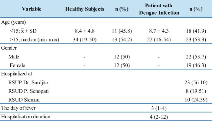
participants (45.8%) were ≤ 15 years old and 13 participants (54.2%) were > 15 years; 12 participants (50%) were male and 12 participants (50%) were female. Among the patients in the dengue infection group, 18 participants (41.9%) were ≤15 years old were 23 participants (53.3%) were > 15 years old, also 22 participants (53.7%) were male and 19 participants (46.3%) were female (Table 1).
Healthy Subjects n (%) Patient with
Dengue Infection n (%)
≤15; x ± SD 8.4 ± 4.8 11 (45.8) 8.7 ± 4.3 18 (41.9)
>15; median (min-max) 34 (19-50) 13 (54.2) 22 (16-54) 23 (53.3)
Male - 12 (50) - 22 (53.7)
Female - 12 (50) - 19 (46.3)
RSUP Dr. Sardjito 23 (56.10)
RSUD P. Senopati 8 (19.51)
RSUD Sleman 10 (24.39)
3 (1-4)
4 (2-12) Hospitalisation duration
Variable
Age (years)
Gender
Hospitalized at
The day of fever
Table 1. Study subjects’ demographic and clinical data.
x: mean; SD: standard deviation; min: minimum; max: maximum.
CBC test showed that there is no significant differences in hemoglobin (Hb) and Hct values, but found significant differences in the numbers of leukocytes,
the absolute number of monocytes, and platelet counts
(p<0.001, 0.002 and <0.001, respectively) (Table 2).
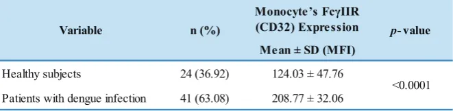
There was a significant difference in FcγIIR expression
on monocytes between healthy subjects and patients with dengue infection, p<0.0001 (Table 3).
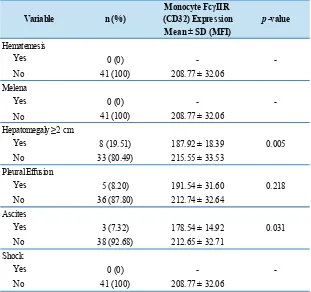
There were no cases of hematemesis/melena or shock in the patients in the dengue infection group. There
was a significant difference of FcγIIR between groups in
hepatomegaly vs. non-hepatomegaly and ascites vs. non-ascites, p=0.005 and 0.031, respectively (Table 4).
Of the forty-one patients with dengue infection, 26
people were diagnosed with dengue fever and 15 with DHF.
There was no significant difference between the groups in the mean FcγIIR expression of the monocytes, Hb, number
of leukocytes, the absolute number of monocytes and the
day of fever. However, there was a significant difference in the increased value of the Hct and platelet counts (p=0.004
and 0.004, respectively) (Table 5).
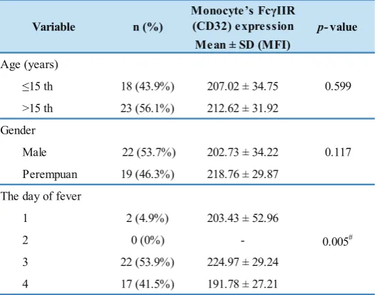
There was no significant difference in the expression of FcγIIR on monocytes by age group and gender, but a significant differences in the day of fever (p=0.005) between
Table 2. Haematological characteristics.
Independent sample T-test/*Mann-Whitney, significant if p< 0.05. abs: absolute
Table 3. Monocyte FcγII (CD32) receptor expression.
Variables Healthy Subjects
(n=24)
Patient with Dengue Infection
(n=41)
p-value
Hb (g/dL); x ± SD 13.20 ± 1.50 13.74 ± 2.03 0.235
Hct (%); x ± SD 39.81 ± 4.02 39.98 ± 5.58 0.886 ∑ leukocyte (103/µL); median (min-max) 7.91 (4.30-10.47) 3.57 (1.10-10.50) <0.001* ∑ abs. Monocyte (103/µL); median (min-max) 0.52 (0.15-1.87) 0.24 (0.07-0.86) 0.002* ∑ platelet (103/µL); x ± SD 263.58 ± 47.76 89.78 ± 48.41 <0.001
Monocyte’s FcγIIR (CD32) Expression
Mean ± SD (MFI)
Healthy subjects 24 (36.92) 124.03 ± 47.76
Patients with dengue infection 41 (63.08) 208.77 ± 32.06
Variable n (%) p-value
<0.0001
Independent T-test, significant if p<0.05.
Discussion
Ribonucleic acid (RNA) of DENV has been isolated from the bone marrow in patients infected with DENV,
indicating that the bone marrow and hematopoietic system
were also the target of DENV. In addition, DENV was
also recently isolated from polymorphonuclear neutrophil
(PMN), monocytes/macrophages and dendritic cells.(13)
Leukopenia and monocytopoenia in this study was similar with the study that that conducted by Kalayanarooj, et al.,
in Bangkok in children with a fever less than 72 hours, for whom neutropenia and monocytopoenia signified dengue
infection vs. other fever illness.(14) Similar results were
also obtained in a study from the Philippines in which 215 dengue patients had routine haematology checked on
the fourth or fifth day of fever, which found 136 patients (63.3%) had leucopoenia.(15)
Funahara, et al., proved that the DENV antigen can
affect platelets directly without going through the immune
responses.(16) A recent study conducted by Ghosh, et al., showed that DENV can directly interact and activate
platelets. RNA and DENV particles had also been detected in platelets in patients infected with DENV, indicating that DENV was able to replicate in platelets by an unknown mechanism.(13)
Hepatomegaly was more frequently found in patients with DHF compared to dengue fever. Hepatic dysfunction could occur due to the direct effects of the infection or
because of an immune response.(17) Interaction between
the immune response and the impact on the integrity and function of the endothelial cells caused increased vascular
permeability and plasma leakage.(18) Monocytes produced
cytokines that caused the activation of endothelial cells that expressed adhesion molecules such as vascular cell
adhesion molecule (VCAM)-1 and intercellular adhesion molecule (ICAM)-1.(19) Pleural effusion and ascites were
suspected to have occurred because of the increased vascular
permeability.(20)
The pilot study was conducted by Durbin, et al.,
during August 2005 - February 2006 in Nicaragua in children with DENV infection by using peripheral blood mononuclear cell (PBMC) obtained from most of the cells
containing the dengue antigen expressed phenotype typical of activated monocytes, even at primary dengue infection.
FcγIIR expression on monocytes in patients with DHF vs.
dengue fever were significantly different, p=0.01.(21) In
contrast with this study’s results that showed no significant differences in FcγIIR expression on monocytes between
dengue fever vs. DHF groups, the expression of FcγIIR
on monocytes in dengue patients was likely influenced by
Table 4. Comparison of the FcγII (CD32) receptor expression on monocytes of patients with dengue infection based on clinical symptoms of dengue infection.
Monocyte FcγIIR (CD32) Expression
Mean ± SD (MFI)
Yes 0 (0)
-No 41 (100) 208.77 ± 32.06
Yes 0 (0)
-No 41 (100) 208.77 ± 32.06
Yes 8 (19.51) 187.92 ± 18.39
No 33 (80.49) 215.55 ± 33.53
Yes 5 (8.20) 191.54 ± 31.60
No 36 (87.80) 212.74 ± 32.64
Yes 3 (7.32) 178.54 ± 14.92
No 38 (92.68) 212.65 ± 32.71
Yes 0 (0)
-No 41 (100) 208.77 ± 32.06 Shock
0.005 Variable
-Hematemesis
Melena
Hepatomegaly ≥2 cm
Pleural Effusion
Ascites
0.218
0.031
-n (%) p-value
-Independent T-test, significant if p<0.05.
DF (n=26) DHF (n=15)
FcγIIR(CD32) (MFI); x ± SD 217.05 ± 33.38 198.22 ± 29.33 0.069
Mean Hb (g/dL); x ± SD 13.41 ± 1.74 14.30 ± 2.42 0.228
Hct (%); x ± SD 39.27 ± 4.85 41.21 ± 6.66 0.335
Δ Hct (%); x ± SD 4.35 ± 1.81 6.87 ± 2.47 0.002
Increase Hct (%); x ± SD 12.22 ± 5.56 19.78 ± 8.06 0.004
Leukocytes count (103/µL); median (min-max) 3.28 (1.10-10.50) 4.10 (1.20-7.83) 0.317* ∑ abs. monocytes (103/µL); median (min-max) 0.33 (0.07-0.86) 0.22 (0.08-0.85) 0.192* Platelet count (103/µL); x ± SD 105.77 ± 45.44 62.07 ± 41.3 0.004
The day of fever; median (min-max) 3 (1-4) 4 (3-4) 0.053* Variable Patie nt with de ngue infe ction p-value
Independent T-test/*Mann-Whitney, significance if p<0.05.
Table 5. FcγII (CD32) receptor expression on monocytes, haematology variables and the day of fever based on the degree of severity.
response to dengue infection caused this study used subjects
with various day of fever. Other study confirm that FcγIIR
expression on monocytes was not related to clinical severity. Dengue infection is the result of a multifactorial interaction between agents, the host, and the environment.
On the one hand, the immune response can be beneficial,
but it may also be detrimental, which prompted the interest of the authors interested in understanding the involvement of genetic factors in these infections, namely genetic
certain genetic polymorphisms may provide protection to individuals who suffer from DHF preventing DSS. Loke, et al., found that the variant homozygous arginine
at position 131 of the FcγRIIA genes have low capacity
for opsonization IgG2 antibodies that provide protection
for the DHF.(9) Furthermore, the interaction between the complex and the virus-antibody FcγRIIA (DENV/IgG1/ IgG3) was associated with the formation of an efficient
phagolysosome to eliminate immune complexes and control
the spread of virus.(11) antibody-dependent enhancement (ADE) hypothesis states that non neutralizing antibodies in
secondary infection may enhances dengue infection. Higher
FcγIIR expression in dengue patients indicate secondary than primary infection.(12)
Table 6. Monocyte FcγII (CD32) receptor expression in patients with dengue infection group based on age, gender, and the day of fever.
Monocyte ’s FcγIIR
Perempuan 19 (46.3%) 218.76 ± 29.87
1 2 (4.9%) 203.43 ± 52.96
Variable n (%) p-value
0.599
0.117
Independent T-test /#one way ANOVA, significant if p<0.05.
Conclusion
The mean expression of FcγIIR (CD32) on monocytes in
patients with dengue infection was higher than in healthy
subjects. FcγIIR expression was not significantly different
between dengue fever and DHF.
References
1. Malavige, GN, Fernando S, Fernando, DJ, Seneviratne SL. Dengue
viral infections. Postgrad. Med. J. 2004; 80: 588-601
2. Martina BEE, Koraka P, Osterhaus ADME. Dengue virus pathogenesis: an integrated view. Clin Microbiol Rev. 2009; 22: 564-81.
3. Kementerian Kesehatan RI. Demam berdarah dengue. Buletin
Jendela Epidemiologi. 2010; 2: 1-15.
4. Blackley S, Kou Z, Chen H, Quinn M, Rose RC, Schlesinger JJ, et al.
Primary human splenic macrophages, but not T or B cells, are the principal target cells for dengue virus infection in vitro. J Virol. 2007; 81: 13325-34.
5. Clyde K, Kyle JL, Harris E. Recent advances in deciphering viral and host determinants of dengue virus replication and pathogenesis. J
Virol. 2006; 80: 11418-31.
6. World Health Organization. Comprehensive Guideline for Prevention
and Control for Dengue and Dengue Haemorrhagic Fever. Revised
and expanded edition. Geneva: WHO; 2011.
7. Wati S, Li P, Burrell CJ, Carr JM. Dengue virus (DV) replication in
monocyte-derived macrophages is not affected by tumor necrosis
factor alpha (TNF-alpha), and DV infection induces altered responsiveness to TNF-alpha stimulation. J Virol. 2007; 81: 10161-71.
8. Kou Z, Quinn M, Chen H, Rodrigo WW, Rose RC, Schlesinger JJ,
et al. Monocytes, but not T or B cells, are the principal target cells
for dengue virus (DV) infection among human peripheral blood mononuclear cells. J Med Virol. 2008; 80: 134-46.
9. Torsteinsdottir I, Arvidson NG, Hallgren R, Håkansson L. Monocyte activation in rheumatoid arthritis: increased integrin, Fcγ and
complement receptor expression and the effect of glucocorticoids.
Clin Exp Immunol. 1999; 115: 554-60.
10. Veri MC, Gorlatov S, Li H, Burke S, Johnson S, Stavenhagen
J, et al. Monoclonal antibodies capable of discriminating the
human inhibitory FcγRIIB (CD32B) from the activating FcγIIA (CD32A): biochemical, biological and functional characterization. Immunology. 2007; 121: 392-404.
11. García G, Sierra B, Pérez AB, Aguirre E, Rosado I, Gonzalez N,
et al. Asymptomatic dengue infection in a Cuban population
12. Intansari US, Sukorini U, Sari SI. FcγRII (CD32) Monocytes in
primary and secondary dengue infection. Indones J Clin Pathol Med
Lab. 2015; 22: 42-7.
13. Hottz E, Tolley ND, Zimmerman GA, Bozza FA. Platelets in dengue
infection. Drug Discov Today Dis Mech. 2011; 8: 33-8.
14. Kalayanarooj S, Vaughn DW, Nimmannitya S. Early clinical and laboratory indicators of acute dengue illness. J Infect Dis. 1997; 176: 313-21.
15. Rubio GD, Torno LL. Association of leukocyte and thrombocyte counts as a predictor of bleeding outcomes among dengue patients.
Phil J Microbiol Infect Di. 2007; 36: 33-8.
16. Soegijanto S. Patogenesa dan Perubahan Patofisiologi Infeksi Virus
Dengue. Surabaya: Airlangga University Press; 2008.
17. Seneviratnea SL, Malavige GN, de Silva HJ. Pathogenesis of liver
involvement during dengue viral infections. Trans R Soc Trop Med
Hyg. 2006; 100: 608-14.
18. Srikiatkhachorn A. Plasma leakage in dengue haemorrhagic fever.
Thromb Haemost. 2009; 102: 1042-9.
19. Anderson R, Wang S, Osiowy C, Issekutz IC, et al. Activation of endothelial cells via antibody enhanced dengue
virus infection of peripheral blood monocytes. J Virol. 1997; 71: 4226-32.
20. Suharti C. Dengue Hemorrhagic Fever in Indonesia: The Role of Cytokines in Plasma Leakage, Coagulation and Fibrinolysis [Dissertation]. Netherland: Nijmegen Universiteit; 2001.
21. Durbin AP, Vargas MJ, Wanionek K, Hammond SN, Gordon A,
Rocha C, et al. Phenotyping of peripheral blood mononuclear cells during acute dengue illness demonstrates infection and increased activation of monocytes in severe cases compared to classic dengue