of Anxiety: Gene–Environment Interactions
Vaishali P. Bakshi and Ned H. Kalin
The study of the neural substrates underlying stress and anxiety has in recent years been enriched by a burgeoning pool of genetic information gathered from rodent studies. Two general approaches have been used to characterize the interaction of genetic and environmental factors in stress regulation: the evaluation of stress-related behav-ioral and endocrine responses in animals with targeted deletion or overexpression of specific genes and the evaluation of changes in central nervous system gene expression in response to environmental perturbations. We review recent studies that have used molecular biology and genetic engineering techniques such as in situ hybrid-ization, transgenic animal, and antisense oligonucleotide gene-targeting methodologies to characterize the function of corticotropin-releasing hormone (CRH) system genes in stress. The effects of genetic manipulations of each ele-ment of the CRH system (CRH, its two receptors, and its binding protein) on stress-related responses are summa-rized. In addition, the effects of stress (acute, repeated, or developmental) on CRH system gene expression are de-scribed. The results from these studies indicate that experimentally engineered or stress-induced dysregula-tion of gene expression within the CRH system is associ-ated with aberrant responses to environmental contingen-cies. These results are discussed in the context of how CRH system dysfunction might contribute to stress-related psychopathology and are presented in conjunction with clinical findings of CRH system dysregulation in psychi-atric illness. Finally, future research strategies (i.e., high-throughput gene screening and novel gene-targeting meth-odologies) that may be used to gain a fuller understanding of how CRH system gene expression affects stress-related functioning are discussed. Biol Psychiatry 2000;48:
1175–1198 © 2000 Society of Biological Psychiatry
Key Words: CRH, CRF, mRNA, knockout, transgenic, antisense, stress
Introduction
T
he study of the neural substrates underlying stress-related disorders has been an active area of research over the past several decades. Significant efforts have focused on identifying genetic factors that might contrib-ute to the development of stress-related psychopathology. Several investigators have utilized rodent models to study interactions between specific genes and environmental contingencies that may interact in the regulation of stress-related functioning. The use of preclinical animal models has permitted controlled evaluation of genetic and envi-ronmental factors contributing to stress-related phenom-ena. The present article discusses some of this recent preclinical literature, but is not intended to be a compre-hensive review of the broad literature on animal models of stress and anxiety disorders, which has been summarized elsewhere (D’Aquila et al 1994; Gyertyan 1992; Martin 1998). Rather, the focus will be on recent approaches that have been used to specifically target certain gene products and evaluate their role in stress-related phenomena in rodents.Among the various peptide and neurotransmitter sys-tems that have been implicated in the regulation of stress is the corticotropin-releasing hormone (CRH) system. Corticotropin-releasing hormone is a 41–amino acid pep-tide that was originally discovered as a novel hypotha-lamic factor controlling the release of pituitary pro-opiomelanocortin peptides (Guillemin and Rosenberg 1955; Saffran et al 1955; Vale et al 1981). It has since been additionally found to play an important role in coordinat-ing the various components of the stress response (De Souza 1995; Dunn and Berridge 1990; Kalin 1997; Koob and Heinrichs 1999). A plethora of clinical and preclinical data indicate that the CRH system is involved in mediating behavioral, autonomic, neuroendocrine, and immune re-sponses to stress. Interestingly, several studies indicate that alterations in the CRH system might also be associ-ated with stress-relassoci-ated psychopathology such as depres-sion and anxiety disorders (for recent reviews, see Arbore-lius et al 1999; Mitchell 1998). It was initially found that depressed patients have elevated levels of cerebrospinal fluid (CSF) CRH relative to control subjects (Nemeroff et From the Department of Psychiatry, School of Medicine, University of Wisconsin,
Madison.
Address reprint requests to Vaishali P. Bakshi, Ph.D., University of Wisconsin, Department of Psychiatry, 6001 Research Park Boulevard, Madison WI 53719. Received July 28, 2000; revised October 11, 2000; accepted October 16, 2000.
© 2000 Society of Biological Psychiatry 0006-3223/00/$20.00
al 1984), an observation that has been replicated in a number of studies (Arato et al 1986; Banki et al 1987; France et al 1988; Widerlov et al 1988). Elevated CSF CRH has also been noted in other stress-related illnesses such as posttraumatic stress disorder, Tourette’s syn-drome, and obsessive– compulsive disorder (Baker et al 1999; Bremner et al 1997; Chappell et al 1996; Fossey et al 1996). Recent preclinical studies in nonhuman primates have indicated that increased CSF CRH levels are strongly correlated with extreme right frontal brain electrical activ-ity, a feature that has previously been found to correspond to extreme fearful behavior in rhesus monkeys (Kalin et al 1998a, 2000). Increases in CRH immunoreactivity as well as CRH messenger RNA (mRNA) have also been reported in the hypothalamic paraventricular nucleus of depressed patients (Raadsheer et al 1995), and decreased levels of CRH receptors have been reported in the frontal cortices of suicide victims. The latter finding is syntonic with receptor downregulation in response to chronically ele-vated transmitter levels (Nemeroff et al 1988). Eleele-vated brain CRH levels resulting from exposure to a stressor or due to exogenous administration of the peptide lead to increases in measures of stress and anxiety in rodents and in nonhuman primates; when administered in high doses to rhesus monkeys, CRH induces a depressionlike behavioral phenomenon (Kalin 1985). Conversely, the effects of acute stressors are blocked by administration of CRH antagonists into the brain, again suggesting an important role for the CRH system in the regulation of stress-related functioning (Kalin 1985; Koob and Heinrichs 1999). Thus, a large body of clinical and preclinical evidence indicates that the CRH system is critically important in the regula-tion of stress-related responses and that dysfuncregula-tion of this system may play a role in stress- and anxiety-related psychopathology.
Overview of the Corticotropin-Releasing Hormone System
Corticotropin-releasing hormone and the related endoge-nous peptide agonist urocortin (Vaughan et al 1995) bind to the two cloned CRH receptors, designated CRH1 and
CRH2(Chen et al 1993; Lovenberg et al 1995; Perrin et al
1995), and to the CRH-binding protein (CRH-BP; Potter et al 1991). The CRH-BP has been postulated to function as an endogenous buffer for the actions of the CRH family of ligands at their receptors (Potter et al 1992). Corticotropin-releasing hormone, its receptors, and its binding protein are expressed in key structures of the hypothalamic– pituitary–adrenal (HPA) axis, and thereby participate in mounting the neuroendocrine response to environmental perturbations. Corticotropin-releasing hormone that is syn-thesized in the paraventricular nucleus of the
hypothala-mus (PVN) is secreted into the median eminence, from where it travels into the pituitary and stimulates CRH1
receptors. This stimulation leads to the release of adreno-corticotropin hormone (ACTH) into the blood stream; ACTH in turn stimulates the secretion of glucocorticoids (i.e., corticosterone) from the adrenal cortex. Glucocorti-coids prepare the body for acute reactions to stress and also serve to shut off the stress response by inhibiting further activity of the HPA system (Kaplan 1992).
The various elements of the CRH system are widely and heterogeneously expressed in cortical, limbic, and brain stem structures in rodents, and in these regions are thought to regulate behavioral responses to stress. In addition to its dense concentration within the PVN, CRH is found in high abundance in the amygdala, neocortex, and brain stem. Corticotropin-releasing hormone 1 receptors are localized within the PVN and pituitary and are thus thought to be the primary mediators of CRH-induced adrenocorticotropin release. Moreover, high levels of CRH1receptors occur in
the neocortex. In rodents, CRH2 receptors are nearly
absent from those structures and are instead found in high abundance within subcortical structures such as the lateral septum and the ventromedial hypothalamus. This distribu-tion is complementary to that of the CRH1receptor and
has been previously suggested to underlie a putative functional dissociation between the CRH receptor sub-types. However, in primate species, CRH2 receptors are
more widely distributed and occur in higher densities in cortical regions (Sanchez et al 1999). Urocortin is another endogenous ligand that activates CRH receptors and has a five- to 20-fold higher affinity for CRH receptors than CRH. Terminals immunoreactive for urocortin are found in multiple brain regions that contain CRH2 receptors
(Bittencourt et al 1999; Kozicz et al 1998). Finally, CRH-BP is found within several CRH- and CRH recept-or– expressing regions such as the neocortex, hippocam-pus, pituitary, amygdala, and brain stem (Chalmers et al 1996). Thus, there are multiple sites at which environmen-tal stressors might impinge simultaneously on the various elements of the CRH system and thereby influence the expression and function of this critical stress hormone under a variety of conditions.
environmental contingencies and may offer some insight into the systems that become dysregulated in stress and anxiety disorders.
The efforts to identify and characterize genetic– envi-ronmental interactions in the regulation of stress have followed two general directions in rodent studies: the evaluation of stress-related behaviors in animals that have had a particular gene product over- or underexpressed using transgenic or antisense oligonucleotide approaches, and the characterization of gene expression changes in response to environmental perturbations. In this review we briefly summarize these areas with regard to the CRH system. Moreover, we include a discussion of the advan-tages and potential pitfalls of each of these approaches. The basic findings derived from these preclinical research strategies will also be discussed in the context of prelim-inary results about genetic contributions to anxiety that have been gleaned from postmortem and gene polymor-phism studies in humans. Finally, some speculation re-garding possible future directions of research in these areas will be provided, with an emphasis on novel high-throughput genetic screening methods and novel gene product targeting techniques that may be applied to the study of genetic– environmental interactions in stress-related functioning in rodents.
Effects of CRH System Gene Targeting on
Stress-Related Behaviors: Use of Transgenic
Mice and Antisense Oligonucleotides
A major approach for studying gene– environment inter-actions in the regulation of stress has focused on charac-terizing the change in an organism’s interaction with its environment following either overexpression or underex-pression of a particular gene product. The role of CRH system genes in stress-related functioning has accordingly been studied using transgenic (gene overexpressing) or knockout (gene deleted) mice and antisense oligonucleoti-de–treated rodents (gene underexpressing). To identify a stress-related behavioral phenotype in these animals, a variety of behavioral paradigms have been employed. Briefly, all of the paradigms presented in this section measure the animal’s ratio of approach versus avoidance behaviors by presenting a choice between an environment that feels safe (usually a dark, enclosed, small space) and an environment that seems novel but risky (usually bright, wide open, large spaces). The entries into and amount of time spent in the safe environment relative to the risky environment are used as an index of the animal’s stress level (an increase in exploratory behaviors toward and into the risky environment indicate a relatively low level of stress). A number of paradigms including the elevated plus
maze (comprised of safer closed, dark arms vs. riskier
open, bright arms), the open field (consisting of a darker wall-bordered peripheral portion vs. a brighter open-center section), a light– dark transition box (consisting of an exploratorium divided into halves, one that is dark and one that is bright), and a defensive withdrawal apparatus (comprised of a small dark chamber that is inside of a brightly lit open field) have been frequently used and validated as paradigms that are sensitive to detecting shifts in an animal’s approach/avoidance-based conflict (File 1990).
Genetically Altered Mice
Transgenic and knockout mice are now widely used in the ongoing effort to understand the contributions of specific genes to psychopathology. The detailed methodology for the generation of these animals and their use in neuro-science research has been reviewed recently (Picciotto and Wickman 1998). Briefly, genetic alterations are intro-duced in the embryonic stage such that the mouse devel-ops with the mutation, thereby putatively providing a model for congenital abnormalities that may contribute to anomalous functioning. With this strategy, all of the components of the CRH system have been successfully targeted and studied for their role in mediating stress-related behavioral effects (Contarino et al 1999b; Hein-richs 1999). It should be noted that, in the case of transgenic mice, expression of the gene of interest is not necessarily dependent on the endogenous promoter for that gene; hence the transgene can be spliced together with a different promoter and can thus be expressed in whatever tissues that promoter is active. In CRH-overexpressing mice, a metallothionene promoter, which drives wide-spread gene expression throughout the brain and the peripheral organs, was used; gene overexpression was thus achieved not only in brain regions where CRH is endog-enously found, but also in other CNS regions and in peripheral organs (testes, heart, and lung), where CRH is not endogenously found (Stenzel-Poore et al 1992). Inter-estingly, two different forms of CRH-BP– overexpressing mice have been engineered. In one, CRH-BP gene expres-sion was driven by a promoter that localized transcription to the the pituitary (Burrows et al 1998), and in the other, CRH-BP was expressed with the metallothionene pro-moter throughout the CNS and periphery (liver, kidney, and spleen), in regions where it is not normally found (ectopic expression) (Lovejoy et al 1998).
CRH OVEREXPRESSERS AND KNOCKOUTS. To
effects associated with acute CRH administration, but also to show marked long-term alterations in endocrine and immune function that are associated with disease states involving hypercortisolemia. Thus, CRH overexpressers have increased basal levels of plasma stress hormones (ACTH and corticosterone), and develop symptoms of Cushing’s syndrome such as muscle atrophy, fat accumu-lation, fur loss, and thin skin (Stenzel-Poore et al 1992). Overexpression of the CRH gene also results in a marked decrease in immune functioning, as evidenced by overall reductions in cell numbers and tissue weight in immune system organs such as the spleen and thymus gland and a failure to mount immunoglobulin antibody responses to immune challenge (Boehme et al 1997).
Behaviorally, CRH-overexpressing mice exhibit a profile that is consistent with increased levels of stress, such as reduced baseline and stress-induced exploration of a novel environment, and decreased activity and time spent in the open arms of an elevated plus maze (Stenzel-Poore et al 1994). These effects are potently blocked by administration of the CRH receptor antagonista-helical CRF. Corticotropin-releasing hormone transgenic mice also show a profound decrease in sexual behaviors and significant deficits in learning (Heinrichs et al 1996, 1997a, 1997b). This stresslike behavioral profile seems to be a reproducible and reliable phenomenon in CRH-overexpressing mice, given that the multiple reports generated over the last 6 years have all indicated that these mice exhibit the same behavioral deficits as do rodents who have received CRH administrations (Koob and Heinrichs 1999).
Interestingly, deletion of the CRH gene seems to alter certain endocrine measures, but stress-related behavioral function in the resulting knockout mouse is relatively unaf-fected. The major endocrine effect of this gene deletion is a decrease in plasma glucocorticoid levels; this deficiency is thought to contribute to developmental abnormalities in the lungs that can be fatal (Muglia et al 1999). However, with corticosterone replacement, normal patterns of growth, fer-tility, and longevity are seen in CRH knockout mice (Muglia et al 1995). Normal levels of ACTH are found in the pituitary and plasma, but the circadian pattern for this hormone is disrupted in CRH knockouts (Muglia et al 1997, 2000). Stress-induced activation of the HPA axis is preserved in mice without the CRH gene, albeit the level of activation is significantly decreased (Jacobson et al 2000). It is possible that this relative sparing of HPA axis function is related to increases in PVN arginine vasopressin (AVP) mRNA that are seen in CRH knockout mice (Muglia et al 2000). There is an increase in the inflammatory response to external challenge, and a decrease in basal and restraint stress–induced epineph-rine levels (Jeong et al 2000; Karalis et al 1999).
In terms of behavioral effects, CRH knockout mice appear to be nearly indistinguishable from their
geneti-cally unaltered wild-type control mice. Baseline locomo-tor, exploratory, stereotypic, startle, and operant learning behaviors are unaffected by CRH gene deletion (Dunn and Swiergiel 1999; Weninger et al 1999a). Moreover, stress-induced responses in feeding and freezing behavior and paradigms such as the elevated plus maze, open field, and multicompartment chamber (to measure stereotyped re-sponding) are no different in CRH knockout mice relative to wild-type control mice (Weninger et al 1999a, 1999b). Stress-induced behavioral effects in CRH knockout mice are blocked by CRH receptor antagonists, indicating that even in the absence of CRH, stress-induced behavioral effects are still being mediated through CRH receptors. It has thus been suggested that an alternative CRH-like ligand such as urocortin could be subserving these effects. In one recent study (Weninger et al 2000), it was found that basal levels of urocortin mRNA in the Edinger– Westphal nucleus were markedly higher in CRH knockout mice than in wild-type control mice. It is worth noting, however, that this increased level of urocortin gene ex-pression in CRH knockout mice is not always seen (Weninger et al 1999a). It is also possible that an entirely different CRH-like ligand (perhaps yet to be discovered) contributes to stress-related behavioral functioning in these animals (Dunn and Swiergiel 1999; Weninger et al 1999a). It is noteworthy that a large number of different paradigms were employed in the behavioral analysis of these animals, and that the results from each of these tests were remarkably consistent with each other. This consis-tency underscores the reliability of the behavioral profile reported in these studies of CRH knockout mice.
CRH RECEPTOR KNOCKOUTS. To examine the rela-tive contributions of the two known CRH receptors to stress-related functioning, investigators have developed knockout mice that are deficient for either the CRH1or the
CRH2 receptor genes. Mice with a null mutation for the
CRH1 receptor have normal baseline levels of plasma
ACTH, probably due to a maintenance of ACTH levels by AVP through vasopressin receptors (Turnbull et al 1999). In response to stress, however, HPA axis activation is significantly blunted in CRH1knockout mice, which also
have adrenal gland atrophy (Smith et al 1998; Timpl et al 1998). Plasma cytokine responses to local inflammation are also exaggerated in CRH1 knockout mice relative to
wild-type control mice (Turnbull et al 1999).
Perhaps most notable, however, are the behavioral sequelae of the CRH1receptor null mutation.
reduced level of anxiety in CRH1 knockout mice
(Con-tarino et al 1999a; Smith et al 1998; Timpl et al 1998). Moreover, CRH1knockout mice appear to be immune to
the anxiogenic effects of ethanol withdrawal (Timpl et al 1998). These “anxiolytic” effects occur in the absence of overall changes in activity levels (Contarino et al 1999a). Finally, no overall changes in body weight or food intake are observed in CRH1 knockout mice, but the circadian
pattern of normal feeding behavior is subtly altered in these animals (Muller et al 2000). When challenged with an intracerebroventricular (ICV) infusion of CRH, CRH1
knockout mice fail to show increased locomotor activity, but do exhibit the decrease in feeding that is typically observed with CRH administration; this finding has led to the suggestion that CRH1 receptors contribute to the
hyperactivity associated with CRH, but do not participate in CRH-mediated alterations in ingestive behavior (Con-tarino et al 2000). It should be noted that this “reduced anxiety” profile in CRH1knockout mice has been
consis-tently observed by separate labs and across a number of different behavioral paradigms, suggesting that it is a fairly robust and reliable phenomenon. Taken together, these results support the notion that CRH1receptors play
an important role in the expression of stresslike behavioral responses and that blockade of these receptors may lead to reduced baseline anxietylike states. Based on these hy-potheses, recent efforts have been directed toward the development of CRH1receptor antagonists for the
treat-ment of stress-related psychiatric disorders such as depres-sion. The first clinical trial of such a compound was recently completed, and revealed that administration of a CRH1receptor antagonist significantly reduced depression
and anxiety scores in depressed patients (Zobel et al 2000). Although these results are preliminary and need to be replicated in a larger study, they indicate that the CRH1
receptor may represent a promising new target for the future development of anxiolytics and antidepressants.
In comparison to the CRH1 knockout mice, CRH2
knockout mice seem to display a different and also less consistent phenotype. Three recent reports detail the en-docrine and behavioral effects of the CRH2null mutation
(Bale et al 2000; Coste et al 2000; Kishimoto et al 2000). Although certain aspects of the knockout phenotype are observed by all three labs, other facets seem to be more variable. For example, none of the groups found any alteration in baseline locomotor activity levels in CRH2
knockouts. An increase in stress-induced ACTH and corticosterone levels was observed in the mutant mice (Bale et al 2000; Coste et al 2000), although basal levels of these hormones did not appear to be affected by the gene deletion. The inflammatory response to thermal injury is elevated in knockouts (Kishimoto et al 2000). Despite an increase in basal blood pressure in the mutants,
the hypotension that is normally produced by systemic urocortin administration was absent in CRH2 knockouts
(Bale et al 2000; Coste et al 2000).
Behaviorally, although some CRH2 knockout mice
showed decreased open arm entries in the elevated plus maze (Bale et al 2000; Kishimoto et al 2000), other CRH2
null mutants failed to exhibit any change in this measure of stress (Coste et al 2000). Moreover, in one study, entries into the center of an open field were decreased in CRH2
knockouts (Bale et al 2000), but in another, entries into the center were increased (Kishimoto et al 2000). In these latter animals, however, the amount of time spent in the brightly lit compartment of a light– dark transition box was decreased. Thus, part of the behavioral profile of CRH2
knockouts is suggestive of increased stresslike responding, but other aspects of the behavioral profile indicate either no alteration of stress-related responding (Bale et al 2000; Coste et al 2000) or a decrease in anxietylike behaviors (Kishimoto et al 2000). In terms of feeding behavior, which has been postulated to involve CRH2 receptors,
CRH2 knockout mice are relatively insensitive to the
anorectic effects of urocortin relative to wild types, sug-gesting that the CRH2 receptor is one of the major
mediators of urocortin-induced effects on ingestive behav-ior (Coste et al 2000). Corticotropin-releasing hormone 2 knockout mice also show a deficit in deprivation-induced feeding, again suggesting that this receptor may be impor-tant in mediating feeding responses (Bale et al 2000). In summary, though it certainly seems that CRH2 receptors
play a role in ingestive behavior, the precise nature of that regulation remains to be determined. It should be men-tioned that, to date, there are no studies reporting on urocortin-overexpressing or urocortin knockout mice. As mentioned above, the behavioral profile of CRH2
knock-out mice is not as clear-cut and consistent across labs as are the behavioral profiles of some of the other CRH system transgenic animals.
Moreover, developing with a missing gene may lead to an induction of compensatory systems, making the inter-pretation of results with transgenic and knockout mice difficult. For example, in CRH2 knockout mice, basal
levels of urocortin and CRH gene expression are elevated, perhaps indicating a possible long-term compensatory response of the system to CRH2deletion (Bale et al 2000;
Coste et al 2000), and suggesting a putative mechanism for the observed increases in anxietylike behaviors in these genetically altered mice. It should be noted, however, that the behavioral profile of these animals is the opposite of that of animals that have received acute administration of CRH antagonists to block CRH2receptors (Radulovic et al
1999). Our group has recently found that antagonizing CRH2receptors in the lateral septum, a region that is enriched
stress-induced defensive behavior in rats (Bakshi et al 1999). Thus, the timing of the gene deletion may drastically influ-ence the nature of the behavioral phenotype that ensues. Future studies utilizing novel inducible-knockout technolo-gies may help in clarifying the developmental versus acute role of CRH2receptors in stress-related functioning (Stark et
al 1998).
CRH-BP OVEREXPRESSERS AND KNOCKOUTS.
Be-sides the two cloned CRH receptors, the other major target to which CRH and related ligands can bind is CRH-BP. This entity is thought to reversibly bind CRH-like ligands but, unlike the CRH receptors, not to activate intracellular postsynaptic events as a result of the association. Associ-ation of CRH ligands with the binding protein thereby represents a putative mechanism through which the level of ligand for the CRH receptors to bind to is effectively reduced. It has thus been suggested that the CRH-BP is an endogenous “buffer” for the actions of CRH and related peptides at CRH receptors (Potter et al 1992). Transgenic and knockout mice have been generated that respectively overexpress or fail to express the CRH-BP. In CRH-BP– overexpressing mice, basal and stress-induced ACTH and corticosterone levels are the same as those in wild types, regardless of whether the gene overexpression is restricted to the pituitary gland (Burrows et al 1998) or occurs in areas where CRH-BP is not normally expressed (Lovejoy et al 1998). Alterations in HPA axis activity (either basal or stress induced) are also absent in CRH-BP knockout mice (Karolyi et al 1999).
Although manipulations of the CRH-BP gene fail to change HPA axis function, these manipulations do pro-duce effects on behaviors known to be modulated by CRH. Thus, in CRH-BP overexpressers, there is enhanced body weight gain relative to wild-type control mice; in contrast, CRH-BP knockouts have reduced body weight gain over several weeks (Karolyi et al 1999; Lovejoy et al 1998). In addition to effects on weight gain, CRH-BP knockout mice exhibited decreases in open arm entries and open arm time in an elevated plus maze and showed a decrease in the number of exits from a safe box in a defensive withdrawal/open field paradigm (Karolyi et al 1999), consistent with findings that exogenous CRH administra-tion alters these behaviors in a similar fashion (Koob and Heinrichs 1999). Thus, CRH-BP knockout mice display increased stresslike behavioral responses. Interestingly, CRH-BP overexpressers failed to show any change in anxietylike behaviors, but did show an increase in overall activity levels, which is generally associated with height-ened levels of CRH (Burrows et al 1998). Given that mRNA levels of CRH and vasopressin were found to be significantly elevated in CRH-BP– overexpressing mice,
it is possible that these putative compensatory changes in CRH system gene expression contribute to the increase in activity levels seen in these mice (Burrows et al 1998). It is surprising, however, that if a compensatory increase in CRH expression does indeed underlie the hyperactivity ob-served in CRH-BP– overexpressing mice, stresslike be-haviors are not also elevated in these animals, since doses of CRH that produce hyperactivity also lead to increases in stresslike behaviors (Koob and Heinrichs 1999).
Knockdown of Specific Gene Products with Antisense Oligonucleotides
An alternative method of gene targeting that allows for temporal specificity of the gene manipulation is that of antisense oligonucleotide administration (Neumann 1997; Wahlestedt et al 1993). In this approach, synthetic strands (usually 15–21 bases) of DNA or RNA that are comple-mentary to the mRNA from the gene of interest are microinfused directly into the brain, usually in adult animals. Thus, the specific contributions of that particular gene product to behavioral functioning can be evaluated in a specific brain region in the absence of developmental confounds. Although conceptually compelling, this tech-nique has been plagued with numerous methodological problems (see below). This method has been used by several groups to further characterize the role of various components of the CRH system in stress-related functioning.
CRH KNOCKDOWN. A handful of studies have
exam-ined the behavioral and neuroendocrine sequelae of reduc-ing CRH peptide levels via antisense targetreduc-ing of CRH gene transcripts (Heinrichs 1999). In general, the effects of decreasing CRH tone through this method are consistent with the behavioral effects that have been associated previously with the CRH system (Koob and Heinrichs 1999)—namely, reducing CRH levels through antisense application results in behavioral effects that are in the opposite direction from those that are normally seen with administration of CRH itself. Thus, ICV administration of CRH antisense oligonucleotides has been reported to decrease stress-induced HPA axis activation, reduce stress-induced CRH gene expression in the PVN, prevent stress-induced avoidance behavior in an elevated plus maze, and increase short-term feeding behavior (Hulsey et al 1995; Skutella et al 1994a, 1994b). Microinfusion of CRH antisense directly into the PVN has similarly been found to decrease HPA axis function and CRH immuno-reactivity in the median eminence (Neumann 1997; Wu et al 1997), and intrahippocampal administration of CRH antisense has been found to decrease grooming behavior (Wu et al 1997). Reduction of CRH levels through antisense administration has also been reported to affect learning and memory processes as assayed in paradigms of passive and active avoidance (Skutella et al 1994a; Wu et al 1997). Therefore, it appears that decreasing the trans-lation of CRH gene transcripts results in a behavioral profile that is syntonic with reduced levels of stress and anxiety in rats.
CRH RECEPTOR KNOCKDOWN. From the differen-tial anatomic distributions of CRH1and CRH2receptors, it
has been hypothesized that these receptors mediate
differ-ent behavioral effects of CRH system ligands. Although there has been some progress in the development of antagonists that are selective for the CRH1 receptor
(McCarthy et al 1999), the CRH2 receptor remains an
elusive target for drug design. Thus, the antisense gene knockdown strategy has been used by several investigators to assess the relative contributions of CRH1 and CRH2
receptors to stress-related functioning. Results from these investigations have yielded some preliminary evidence for a possible functional dissociation between CRH1 and
CRH2receptors (Steckler and Holsboer 1999). It has been
shown that ICV or intra-amygdala infusions of CRH1
antisense oligonucleotides significantly decrease CRH- or stress-induced avoidance behaviors in tests such as open field, elevated plus maze, or defensive withdrawal, sug-gesting that reducing CRH1receptors in regions such as
the amygdala can decrease stress-related behavioral re-sponses (Heinrichs et al 1997a; Liebsch et al 1995, 1999; Skutella et al 1998). This profile of results is consistent with the reduced-anxiety phenotype that has been ob-served for CRH1knockout mice.
The results obtained with CRH2 receptor antisense
treatments are less clear-cut. One group has shown that ICV administration of CRH2 antisense oligonucleotides
results in a 32% decrease in hypothalamic CRH2receptors
and a marked reduction in CRH or urocortin-induced anorexia (Smagin et al 1998). In the same study, it was found that pretreatment with the CRH1-selective
antago-nist NBI27914 failed to alter feeding responses to the CRH receptor agonists. Therefore, it has been suggested that CRH2but not CRH1receptors regulate alterations in
ingestive behavior. As summarized above, an important role for CRH2 receptors in ingestive behavior has also
been indicated by studies of CRH2knockout mice. Other
studies have reported either no effect on indices of stress with CRH2antisense treatment or increased immobility in
a forced swim test with CRH2targeting; it has thus been
suggested that CRH2receptors do not mediate anxiety per
se but rather play a role in “coping” responses (Heinrichs et al 1997a; Liebsch et al 1999). It should be noted, however, that in studies that report a lack of an effect on “anxietylike” behaviors with CRH2 antisense treatment
(Heinrichs et al 1997a; Liebsch et al 1999), the level of receptor knockdown was either not measured or was very small (approximately a 10% reduction in receptor levels in the lateral septum of CRH2antisense–treated rats). Thus, it
is difficult to definitively conclude without further exper-imentation that CRH2 receptors do not exert effects on
indices of stress or anxiety in rodents. Recent studies from our lab and others indicate that when CRH receptors in CRH2 receptor– containing brain regions are acutely
Radulovic et al 1999). With the advent of truly selective CRH2 receptor ligands and the development of novel
gene-targeting strategies, the precise role of CRH1 and
CRH2receptors in stress-related functioning will be more
clearly elucidated. To the best of our knowledge, no studies to date have utilized the antisense oligonucleotide approach to examine the role of CRH-BP in behavioral responses to stress.
CAVEATS OF ANTISENSE OLIGONUCLEOTIDE
AP-PROACHES. In addition to the concerns raised above regarding antisense study design, a number of other problems have been reported to occur with antisense oligonucleotide administration. Neurotoxic events including cell infiltration, morphological abnormalities, and widespread reductions in nontargeted proteins have been described following oligonu-cleotide administration. These effects are especially common with the phosphorothioate-backbone oligonucleotides, which have been chemically modified to enhance temporal stability (Broberger et al 2000; Neumann 1997; Skutella et al 1994c). Thus, it appears that the “window of opportunity” between efficacy and toxicity is very narrow when using the oligonu-cleotide infusion approach. Furthermore, similar behavioral effects have been obtained with either an increase, a decrease, or no change in the protein levels of the gene target, indicating that antisense-induced alterations in behavior are not necessarily related to changes in the targeted gene product (Liebsch et al 1995; Skutella et al 1998). This phenomenon mandates the careful evaluation of gene-prod-uct protein levels for every experiment in which behavioral data are collected, and also brings into question whether observed behavioral effects can truly be attributed to changes in the expression of the gene of interest. Given that protein levels were not examined in several of the aforementioned studies (Liebsch et al 1999; Neumann 1997; Skutella et al 1994c; Wu et al 1997), it is difficult to conclude that the reported behavioral effects can be attributed specifically to the targeted gene product. Thus, some caution is perhaps warranted when using antisense oligonucleotides to study gene function. It is worth noting that new chemistries are currently under development to minimize toxicity and max-imize efficacy; it is possible that some of these modified probes will be free of the aforementioned problems and might indeed prove to be better research tools (Broberger et al 2000; Ho et al 1998).
Alterations in CRH System Gene Expression
Induced by Environmental Manipulations:
Effects of Acute, Repeated, or
Developmental Stressors
One of the primary approaches for evaluating how envi-ronmental contingencies interact with genetic factors is to
study the effects of environmental stressors on gene expression patterns in the central nervous system. This approach has been widely used in the past decade to gain a fuller understanding of how specific environmental events can activate or inhibit the transcription of various genes that have been hypothesized to play a role in the regulation of stress-related functioning. The typical design of such studies is to expose an animal to a discrete stressor and, after a prescribed delay, sacrifice the animal and process the brain tissue for analysis of gene expression. The other side of the coin is to investigate how naturally occurring genotypic differences contribute to stress re-sponsivity. To examine this possibility, investigators have examined differences in gene expression between strains of rodents that differ in their response to stress. To date, the most commonly employed methods for measuring gene expression are Northern blotting, RNase protection assays, and in situ hybridization, all of which gauge the mRNA levels of a particular gene product, albeit with different levels of quantitative and anatomic precision.
Using this approach, several investigators have charac-terized the effects of strain differences and environmental manipulations on the various components of the CRH system. The following sections briefly summarize the most commonly cited effects on CRH system gene expres-sion that have been reported to occur in response to different types of environmental contingencies.
Strain Differences in CRH System Gene Expression
and stress-induced levels of PVN CRH mRNA relative to the Wistar and Sprague–Dawley strains (Imaki et al 1998; Krukoff et al 1999; Mansi et al 1998). Spontaneously hypertensive rats also have elevated levels of CRH1gene
expression in the PVN relative to Wistars (Imaki et al 1998). It has thus been hypothesized that this increased sensitivity of CRH system gene expression in the HPA axis contributes to the development of the hypertensive phenotype in these animals. Lewis rats, which are suscep-tible to athritis as a result of low circulating levels of corticosterone, show a blunted HPA axis response to stress and have lower levels of basal and stress-induced CRH gene expression in the PVN than several other rat strains (Rivest and Rivier 1994; Sternberg et al 1992).
Finally, in comparing lean (Fa/?) and obese (fa/fa) Zucker rats, it has been found that the obese strain has lower baseline CRH2mRNA levels in the ventromedial
hypothalamus, and that obese rats have higher levels of stress-induced CRH gene expression in the PVN and other sites (Richard et al 1996; Timofeeva and Richard 1997). To the best of our knowledge, urocortin gene expression in these animals has not been reported. Strain differences in CRH receptor gene expression have been observed in mice as well; C57 mice have higher baseline and stress-induced increases in CRH1
receptor mRNA levels than DBA mice (Giardino et al 1996). Taken together, these findings indicate that different rodent strains, as a consequence of their distinct genetic makeups, display different baseline levels of expression of CRH system genes. Moreover, these various strains display differential responsiveness to stress with regard to CRH system gene regulation. The study of various rat strains may thus help to identify the neurogenetic differences that contribute to individual differences in stress susceptibility, and thereby further characterize the interaction between genes and environmental conditions in the etiology of anxiety. Although such information is useful, it remains to be determined whether or not the specific genetic differences identified above actually underlie the dif-ferent behavioral phenotypes. It is probable that a number of genes in addition to those described above are differentially expressed across different rodent strains. Which other genes differ across strains, and of these, which ones contribute to the behavioral pheno-type? It is also unclear whether the differential gene expression patterns are the cause or the result of the different phenotypes observed in the separate strains. Future studies in which behavioral phenotypes are assessed after the application of novel gene targeting techniques to selectively disrupt or restore gene func-tion in these rodent strains will aid in clarifying these issues.
Effects of Acute Stress
The most widely reported phenomenon in the study of CRH system gene expression is the increase in CRH gene transcription within the PVN in response to stress. The most commonly cited stress-induced gene expression changes involving the CRH system are summarized in Table 1. This list is not an exhaustive compendium of all studies that have examined CRH system gene expression, but rather is intended to outline some of the major environmental perturbations that lead to alterations in the regulation of CRH system genes.
A wide variety of psychologic, physical, physiologic, and immune stressors have been found to modulate the expression of the CRH gene in the PVN. Briefly, increases in CRH gene expression in the PVN can be elicited by restraint or immobilization (a more severe form of re-straint stress in which the animal’s limbs are immobilized) stress, foot shock, hypovolemia, hypoglycemia, and cyto-kines. Corticotropin-releasing hormone gene expression in the amygdala has also been studied and has been reported to increase primarily in response to stressors that contain a significant psychologic component (restraint, or exposure of unmanipulated rats to rats that have just received a foot shock). Recent studies from our laboratory indicate a novel anatomic substrate for stress-induced CRH gene regulation. A large distribution of CRH mRNA has been found within parts of the thalamus; exposure of rats to acute restraint stress significantly elevates this signal, suggesting that the thalamus may be another important site involved in the processing of stress-related information (Hsu and Kalin 2000). The gene expression of other components of the CRH system are less well studied, but increases in CRH1 receptor mRNA levels have been
observed in the PVN in response to restraint, immobiliza-tion, immune, or osmotic stress. Finally, restraint stress increases transcription of the CRH-BP gene in the pitu-itary and also in limbic brain regions such as the amyg-dala; food deprivation increases CRH-BP gene expression in the amygdala and regions of the hypothalamus.
A large body of literature also indicates that CRH system gene expression is regulated in a complex manner by glucocorticoids; this information is beyond the scope of this review but has been thoroughly reviewed elsewhere (Schulkin et al 1998). Briefly, in the PVN, acutely administered corticosterone decreases CRH mRNA levels, whereas, in the amygdala, corticosterone administration increases CRH mRNA (Makino et al 1994; Shepard et al 2000). Increases in CNS CRH peptide levels (via ICV infusion or through acute stress) also have been found to upregulate the expression of the CRH and the CRH1
genes, but not the CRH2gene (Imaki et al 1996; Jezova et
Pharmacologically induced alterations in the expression of CRH and related genes in the PVN have also been reported with the acute administration of a variety of
drugs. Cocaine or N-methyl-D-aspartate antagonists have
been found to increase CRH mRNA levels in the PVN, and ethanol or cocaine induces CRH1 receptor gene
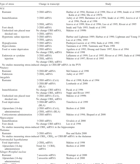
Table 1. Induced Alterations in CRH System Gene Expression
Type of stress Change in transcript Study
PVN
Restraint 1CRH mRNA Harbuz et al 1994; Herman et al 1998; Hsu et al 1998; Imaki et al 1995b, 1996; Kalin et al 1994; Ma et al 1997b
Immobilization 1CRH mRNA Aubry et al 1999; Bartanusz et al 1994; Imaki et al 1992; Jezova et al 1999; Kiss et al 1996; Pacak et al 1996
1CRH1mRNA Jezova et al 1999; Kiss et al 1996; Luo et al 1995; Rivest et al 1995 Foot shock 1CRH mRNA Imaki and Vale 1993; Imaki et al 1991
Unshocked rats placed near shocked rats
No change CRH mRNA,
1CRH1mRNA
Makino et al 1999b
Hypertonic saline injection 1CRH mRNA
1CRH1mRNA
Harbuz and Lightman 1989; Harbuz et al 1990; Lightman and Young 1988, 1989 Luo et al 1994, 1995
Hypoglycemia 1CRH mRNA Paulmyer-Lacroix et al 1994; Suda et al 1988, 1992 Hypovolemia 1CRH mRNA Tanimura et al 1998; Tanimura and Watts 1998 Food or water deprivation 2CRH mRNA
No change CRH2mRNA
Aguilera et al 1993; Hwang and Guntz 1997; Kiss et al 1994 Makino et al 1998
Endotoxin or cytokine injection
1CRH mRNA
1CRH1mRNA No change CRH2mRNA
Laflamme et al 1999; Lee et al 1995; Rivest et al 1995; Suda et al 1990 Makino et al 1997; Rivest et al 1995
No studies measuring stress-induced changes in CRH-BP mRNA in the PVN
Pituitary
Restraint 1CRH-BP mRNA McClennen et al 1998 Endotoxin 1CRH1mRNA Aubry et al 1997
Amygdala
Restraint 1CRH mRNA (CeA)
1CRH-BP mRNA (BLA)
Hsu et al 1998; Kalin et al 1994 Lombardo et al 2000
Immobilization No change CRH mRNA No change CRH1mRNA
Pacak et al 1996 Nappi and Rivest 1995 Unshocked rats placed near
shocked rats
1CRH mRNA (CeA), No change CRH1mRNA
Makino et al 1999b
Food deprivation 1CRH-BP mRNA (BLA)
Timofeeva et al 1999
Alprazolam (14-day administration)
2CRH mRNA (CeA),
2CRH1mRNA (BLA)
Skelton et al 2000
Corticosterone administration 1CRH mRNA Makino et al 1994; Shepard et al 2000
Hippocampus
Immobilization 1CRH mRNA Givalois et al 2000 Foot shock No change CRH mRNA Lee et al 1996 No studies measuring stress-induced CRH1mRNA in the hippocampus
Thalamus
Restraint 1CRH mRNA Hsu and Kalin 2000 No studies measuring stress-induced CRH1, CRH2, or CRH-BP mRNA in the thalamus
Ventromedial hypothalamus
Food deprivation 2CRH2mRNA Makino et al 1998 Alprazolam (14-day
administration)
Trend for1CRH2 mRNA
Skelton et al 2000
Edinger-Westphal nucleus
Restraint 1urocortin mRNA Weninger et al 2000 Alprazolam (14-day
administration)
1urocortin mRNA Skelton et al 2000
CRH or CRH2gene deletion
1urocortin mRNA Bale et al 2000; Coste et al 2000; Weninger et al 2000
expression in this structure (Givalois et al 2000; Lee and Rivier 1997; Zhou et al 1996). In contrast, clinically effective antidepressants have been found to decrease CRH gene expression in the PVN (Brady et al 1992). Stress-induced increases in PVN CRH gene expression can be blocked by benzodiazepines, but not by serotonin depletion (Harbuz et al 1993; Imaki and Vale 1993; Imaki et al 1995a). Acute or subchronic (2 weeks) administration of benzodiazepines decreases CRH1 gene expression in
the amygdala and cortex, but ethanol increases transcrip-tion of this gene in the PVN (Lee and Rivier 1997; Skelton et al 2000). The expression of the CRH2receptor gene is
decreased by food deprivation but increased by benzodi-azepine administration. Urocortin gene expression in the Edinger–Westphal nucleus (where the majority of cell bodies that synthesize urocortin are found) is similarly increased by benzodiazepine administration.
Taken together, these findings provide many different pieces of information regarding how the CRH system changes in response to acute environmental perturbations. The increase in PVN CRH mRNA after exposure to acute stress appears to be a reliable phenomenon because it has been reported with a wide variety of stressors and by several different labs. Stress-induced elevations in CRH mRNA in the amygdala are less well characterized and seem to occur under a more limited set of circumstances, but have been reported by at least three different labora-tories. Information about other CRH system transcripts within other brain regions is much more limited. It remains to be seen which of the other preliminary results regarding stress-induced alterations in CRH system gene expression are reproduced in future studies.
One problem with the state of knowledge about CRH system gene expression is that in the majority of studies cited in this review, only one component of the CRH system was examined (usually just the CRH gene within the PVN). Thus, much remains unknown regarding other brain regions and CRH system gene transcripts. Also, as discussed below, subtle methodological variability can significantly influence the outcome of the studies. We are thus left with a large number of separate findings regard-ing stress-induced gene expression that have been ob-tained under different experimental conditions and focus on disparate themes. A challenge that remains is to assemble these separate pieces of information about stress-induced CRH system gene expression into a comprehen-sive story that will clarify how the system as a whole responds to different types of environmental perturbations. Future studies in which the multiple components of the CRH system are all studied at once and in various brain regions will aid in understanding how the individual elements of the CRH system coordinate their activity in integrating the stress response.
Effects of Repeated or Chronic Stress
It is clear that several types of environmental perturbations are able to acutely modulate the expression of CRH system genes (vide supra). In a clinical context, however, it is likely that conditions of prolonged or repeated stress are a major contributing factor to the development of stress-related psychopathology. Thus, attention has been focused on the effects of repeated or chronic stressors on expression of CRH system genes. In general, the repeated stress regimen involves presentation of an acute stressor to adult rats once a day for a period of 6 –14 days total. Upon termination of this repeated regimen, rats are sacrificed either 24 hours after the final stress presentation (to gauge possible changes in basal CRH system gene expression) or after presentation of an acute stress challenge (to deter-mine changes in responsivity to acute stress in animals that have had prior exposure to repeated stress). It should be noted that the term chronic stress is distinct from repeated
stress and refers to a condition in which animals are
continuously in the presence of the stressful stimulus (as opposed to discrete bouts presented intermittently) for a prolonged period of time (several weeks). Through studies of repeated or chronic stress, it is possible to understand some of the neural genetic mechanisms underlying regu-latory responses to ongoing stress and the eventual mech-anisms underlying stress-related disorders.
CRH GENE EXPRESSION. As outlined in Table 1, the most common perturbation that has been used to study stress-related CRH gene function is that of restraint or immobilization stress. To examine how CRH system gene expression is regulated by repeated exposure to environ-mental perturbations, the effects of a repeated restraint or immobilization stress regimen on levels of CRH mRNA have been examined.
stressor presentation in rats that have had a prior history of exposure to stress (Imaki et al 1991). Basal CRH gene expression levels in the PVN are also increased after repeated exposure of rats to other types of stressors such as foot shock or hypertonic saline injections or to a repeated stress regimen that consists of several different types of stressors presented in an alternating and random order (Herman et al 1995; Kiss and Aguilera 1993; Sawchenko et al 1993). It is of interest to note that, although acute restraint stress increases CRH gene expression within the amygdala, repeated exposure to the restraint fails to affect basal CRH mRNA levels within this region (Hsu et al 1998; Kalin et al 1994; Mamalaki et al 1992). Neverthe-less, rats that have experienced chronic social stress by becoming subordinate animals in a visible burrow system dominance hierarchy can show blunted HPA axis activity; basal CRH mRNA levels in these rats are decreased in the PVN but elevated in the amygdala (Albeck et al 1997). Hence, the latter study demonstrates that basal CRH mRNA levels in the amygdala can indeed be affected by certain forms of long-term stress.
Changes in Responsivity to Acute Stressors after a History of Repeated Stress Exposure of rats to a regimen of repeated restraint stress prevents the acute stress– induced elevation in CRH mRNA that is normally seen in the amygdala of rats that have not experienced prior stress (Mamalaki et al 1992). We have found in recent studies that this profile of gene expression is identical for the CRH-BP gene in the amygdala; a single episode of restraint stress in naive rats increases CRH-BP mRNA, but this acute stress–induced effect is not observed in rats that have previously been exposed to restraint stress for 12 consecutive days (Lombardo et al 2000). This dampening of the CRH mRNA response to acute stress in animals with a history of previous stress has also been seen in the PVN, particularly if the acute challenge utilizes the same stress that was employed in the repeated regimen (homo-typic) (Ma et al 1997a, 1999). It should be noted, however, that at least one study (Mamalaki et al 1992) failed to demonstrate this effect. It is possible that the failure to see CRH gene expression changes in response to the acute homotypic stressor indicates that the system has under-gone habituation to that particular type of stressor. When animals are challenged with a heterotypic (different type) stressor after a repeated stress regimen, however, a high level of CRH gene transcription is seen in the PVN (Ma et al 1999). It has thus been suggested that after repeated stress, the CRH system may habituate, but other peptide systems are recruited to respond to the acute (homotypic) stressor challenge. It has been found in a number of studies that PVN gene expression levels of AVP, the other major releasing factor for ACTH, are dramatically increased
after a repeated stress regimen. Interestingly, preliminary evidence indicates that formerly CRH-expressing neurons begin to show increased levels of AVP transcription. Taken together, these findings have led to the suggestion that repeated stress may cause a shift in the peptidergic mechanisms that regulate HPA axis responsivity to sub-sequent stress (Bartanusz et al 1993; Ma et al 1997a; Ma and Lightman 1998; Makino et al 1995b). Thus, it may be that CRH gene transcription is strongly activated by exposure to novel stressors but, as the stressor becomes familiar, the CRH system habituates and, instead, the AVP system begins to respond to the stressor. Subsequently, when a new type of stress is presented, the CRH system becomes activated again. This theoretical mechanism would be consistent with the finding that heterotypic but not homotypic stressors can increase CRH gene expres-sion in rats that have had prior exposure to stress.
CRH RECEPTOR GENE EXPRESSION. The effects of
repeated stress on CRH receptor gene expression are not as well characterized as are those regarding the expression of the CRH gene. As seen in Table 1, an increase in CRH1
receptor mRNA in the PVN appears to be consistently observed after exposure to an acute session of immobili-zation or restraint stress (Aguilera et al 1997; Bonaz and Rivest 1998; Makino et al 1995a). A similar increase in basal CRH1PVN gene expression has been reported with
repeated stress (Makino et al 1995b) but was not corrob-orated by other groups (Aguilera et al 1997; Bonaz and Rivest 1998). Two weeks of restraint stress has been found to decrease CRH1mRNA in the pituitary (Makino et al
1995a). However, this effect was not replicated by a different group (Aguilera et al 1997). In certain strains of mice, cortical levels of CRH1mRNA increase in response
to a regimen of repeated restraint stress (Giardino et al 1996). In rats, however, CRH1 mRNA levels in several
extrahypothalamic regions including the cortex, amygdala, and hippocampus are not affected by repeated restraint stress (Iredale et al 1996; Makino et al 1995a). In contrast, when the repeated stress consists of a variable, unpredict-able, multimodal regimen, a significant reduction in CRH1
gene expression is observed in the cortex and a significant increase in this transcript is seen in the hippocampus of rats (Iredale et al 1996). Finally, to the best of our knowledge, only one study to date has reported the effects of repeated immobilization stress on CRH2 gene
expres-sion levels. A small (roughly 12%) decrease in CRH2
of change (if any) is produced in CRH system gene expression.
It can be gathered from these studies that the state of knowledge about CRH receptor gene regulation in re-sponse to stress is less consistent and less well character-ized than that about the CRH gene. It seems that the findings with repeated stress and CRH1receptor mRNA
levels are not easily reproduced across laboratories. In general, much less is known about the different experi-mental conditions that influence the expression of the CRH receptor genes. Some of the aforementioned discrep-ancies in the literature may derive from subtle method-ological differences between labs (i.e., rat strain, nature of the stressor, hybridization conditions, probe selection). Nonetheless, these results provide good starting points to refine our knowledge about CRH receptor gene transcrip-tion; as additional labs attempt to reproduce these findings, it will become more apparent which results are the most robust and reliable.
Effects of Developmental Stress
Perhaps the most significant environmental factor during the early development of mammals is the interaction between the infant and its mother. A large body of literature indicates that, in animals and humans, separation of an infant from its mother during this early developmen-tal phase is a significant stressor that markedly and negatively affects the subsequent emotional development of the infant (Bowlby 1973; Carlson and Earls 1997). In nonhuman primates, long-term maternal separation can result in profound alterations in stress-related behavioral responses in the separated offspring. The seminal work of Harlow and colleagues (Harlow et al 1964) indicates that nonhuman primates that have undergone long-term mater-nal separation as infants display enhanced fear-related behavioral responses and appear socially withdrawn. Neu-roendocrine studies in rhesus monkeys indicate that an infant’s stress hormone levels are negatively correlated with the number of offspring the mother had, suggesting that, when mothers are less experienced, cortisol levels in their (early born) infants are high; elevated cortisol levels also correspond to increased fearful behavioral responses in the infants (Kalin et al 1998b). Similarly, in rats, disturbing the prenatal environment by stressing the mother can lead to increases in CRH gene expression in the fetal PVN, increases in CRH content in the amygdala of adult offspring, and potentiation of stresslike behavioral responses in those rats whose mothers had undergone stress during pregnancy (Cratty et al 1995; Fujioka et al 1999; Takahashi et al 1992). These findings support the notion that mother–infant interactions may be a critical factor in determining the future fearful disposition of the
offspring. Given the importance of the CRH system in modulating fear and stress-related responses, and the critically important nature of the interaction between mother and offspring, several investigators have studied the effects (short and long term) of maternal separation on CRH system gene expression in rodents.
STRESS HYPORESPONSIVE PERIOD AND MATERNAL
DEPRIVATION. The perinatal developmental stage in rats includes an early “stress hyporesponsive period” (SHRP; from postnatal day 3 to day 14) that is character-ized by a diminished HPA axis response to stress (for a review, see Rosenfeld et al 1992). To determine if hypo-responsiveness to stress during this period reflects a deficit in CRH gene transcription, the effects of maternal sepa-ration during the SHRP on CRH system gene expression have been studied. It has been found that presentation of a mild stressor such as an intraperitoneal injection of iso-tonic saline is not sufficient to “overcome” the SHRP and recruit an activation of the HPA axis in rat pups (Dent et al 2000). The more intense stress of maternal separation, however, produces a significant increase in HPA axis activity if the deprivation takes place for 24 hours. Despite the HPA axis stimulation, this stressor seems to cause a decrease in the level of CRH gene expression in the PVN (Dent et al 2000; Smith et al 1997; van Oers et al 1998a), which is opposite to the profile that is seen in acutely stressed adult rats. When maternally separated rats are additionally challenged with a second stressor, however, these animals exhibit elevations in PVN CRH gene tran-scription (Dent et al 2000; Yi and Baram 1994). Similarly, increases in CRH gene expression are observed in the amygdala of rat pups that have received repeated expo-sures to a maternal separation/cold stress protocol (Hatal-ski et al 1998). Nevertheless, the extent to which HPA axis activation in maternally deprived pups is related to CRH gene expression remains unclear. Although CRH anti-serum administration can prevent elevations in corticoste-rone that are seen in pups after a combined maternal deprivation/cold stress exposure (Yi and Baram 1994), a dissociation between deprivation-induced alterations in CRH mRNA levels and HPA axis activity has also been reported in maternally deprived rat pups (Smith et al 1997). Interestingly, the aforementioned adverse effects of maternal deprivation can be prevented by providing the deprived pups with certain aspects of the maternal behav-ioral repertoire (i.e., stroking) (van Oers et al 1998b). This phenomenon has also been noted for maternal depriva-tion–induced decreases in ventromedial hypothalamic CRH2 receptor gene expression (Eghbal-Ahmadi et al
pups that are maternally deprived at postnatal day 3 have elevated stress-induced HPA axis responses relative to nondeprived control pups when they are tested after the SHRP (van Oers et al 1998a; Workel et al 1997). When rats that are maternally deprived during the SHRP are challenged with a stressor 2 weeks later, they exhibit an increase in CRH gene expression in the PVN relative to nondeprived rats (van Oers et al 1998a), suggesting that early postnatal environmental perturbations can have long-lasting effects on CRH system gene expression.
LONG-LASTING EFFECTS OF MATERNAL
DEPRIVA-TION STRESS INTO ADULTHOOD. Maternal separation
has also been found to produce long-term changes in CRH system gene expression into adulthood. Interestingly, the nature of the separation determines the direction of the long-term changes, as has been reviewed in detail recently (Anisman et al 1998; Francis et al 1999b; Heim and Nemeroff 1999). Thus, brief periods of separation from the mother (3–15 min per bout, once a day, for roughly 2 weeks) result in a profile indicative of diminished anxiety, whereas more protracted separations (3 hours or more) have the opposite effect, resulting in increased stresslike responses. In an elegant series of studies by Meaney, Plotsky, and colleagues, the long-term effects of these different types of maternal separation have been described, and the behavioral and neuroendocrine mechanisms un-derlying these long-term effects have been characterized. It was initially found that rat pups that underwent very short periods of separation from their mothers (termed
handling) had decreased basal levels of hypothalamic
CRH mRNA and median eminence CRH immunoreactiv-ity as adults relative to undisturbed control rats (Plotsky and Meaney 1993). As adults, these handled rats also displayed significantly lower elevations of stress-induced corticosterone levels and blunted CRH release from the median eminence relative to control rats. It has since been found that the mechanism underlying this reduction in stress-related functioning in handled rat pups involves the type of maternal behavior that is displayed after the pups are returned to the mother (Liu et al 1997), confirming earlier hypotheses that maternal behavior is the critical component in the developmental milieu of the infant (Levine 1957). A brief removal of rat pups from the dam results in a significant increase in the amount of licking, grooming, and arched-back nursing (LG-ABN) that the mother lavishes upon the pups when they are returned; the total amount of time spent nursing and being with the offspring is not affected, but rather the quality of the interaction between mother and pup is altered. In nonsepa-rated pups, individual differences in LG-ABN predict HPA axis responsivity in adulthood such that mothers that engage in high levels of LG-ABN have offspring that, as
adults, show reduced HPA axis activation in response to stress and have decreased levels of CRH mRNA in the PVN (Liu et al 1997). Pups that are born to mothers that naturally exhibit high levels of LG-ABN grow up into adults that display low anxietylike behaviors (increased exploration of novel environments) and, relative to low– LG-ABN offspring, have decreased levels of CRH recep-tors in brain regions such as the locus coeruleus that are thought to mediate stress responses (Caldji et al 1998). Taken together, these findings suggest that increased nurturance by the mother can lead to a toned-down stress-responsive system in the offspring.
In contrast, longer periods of maternal separation seem to have the opposite effect on stress-related functioning in adulthood. Rat pups that are separated from the mother for 3 or more hours (investigators have often used a 24-hour separation) show increased CRH system gene expression, exaggerated HPA axis responses to stress, and increased stresslike behaviors in paradigms such as the elevated plus maze (Plotsky and Meaney 1993; Rots et al 1996; Wigger and Neumann 1999). Other intense stressors such as an endotoxin insult during the perinatal stage are also able to produce marked elevations in basal CRH gene expression and lead to an exaggerated stress-induced HPA axis response in adulthood (Shanks et al 1995). Long-lasting dysregulation of the CRH system has also been reported in nonhuman primates exposed to adverse rearing conditions during infancy. Coplan and colleagues (Coplan et al 1996, 2000) found that CSF levels of CRH are basally and chronically elevated in adult bonnet macaques whose mothers were exposed for 3 months to a variable foraging demand (VFD), in comparison to mothers confronted with either a high but predictable or low but predictable foraging demand. Infants reared by VFD-exposed mothers have been found to subsequently display abnormal affili-ative social behaviors in adulthood (Andrews and Rosen-blum 1994). Moreover, it has recently been found that maternal styles can be passed down through a “non-genomic” mode of transmission such that offspring adopt the maternal style of the dam that fostered them, regardless of whether or not that dam is the biological mother (Francis et al 1999a). It is likely that similar nongenomic transmission of stress responsivity occurs in primates. For example, in rhesus monkeys, there is a correlation between birth order and cortisol such that earlier off-spring have higher basal cortisol concentrations (Kalin et al 1998b).
distur-bances may be more influential than either acute or repeated stressors in producing long-term alterations in CRH system gene expression. Indeed, of all the CRH system gene expression studies presented in this review, the studies that report on the effects of developmental stressors perhaps tell the most complete and compelling story about how stress may lead to traitlike disrupted functioning in adulthood. Moreover, the effects of perina-tal stress have been characterized at multiple levels of analysis within a given study (several brain regions, several gene transcripts, and concurrent behavioral analy-ses) and have been studied across different species. Al-though further work is needed to clarify the mechanisms through which early developmental stressors produce their long-lasting effects, these studies can serve as a valuable heuristic model for designing future experiments in the area of stress-induced CRH system gene expression changes. As stated earlier, one of the major weaknesses of many of the studies in the previous sections is that they examined only one element of the system in a single brain region. Moreover, few studies have integrated molecular and cellular analyses with behavioral observations, as has been done in the aforementioned studies of perinatal stress, particularly with maternal separation. Thus, by taking the multilevel, integrative approach of these mater-nal separation studies, one might come to better under-stand the effects of other types of stressors on CRH system gene expression as well.
Caveats of Stress-Induced Gene Expression Studies
It is important to note that although the aforementioned sections have outlined some of the major environmental contingencies that influence the gene expression of the CRH system, further studies are necessary to evaluate the functional implications of these reported alterations in mRNA levels. The careful measurement of protein levels is a critical step in understanding how environmental effects on gene expression are translated into actual changes in the organism’s behavior. Several studies indi-cate that some of the aforementioned stress-induced changes in CRH system gene expression are in fact accompanied by alterations in the levels of the correspond-ing proteins. For example, as summarized above, pro-longed separation of rat pups from their mothers has been found to cause increases in CRH mRNA that are apparent well into adulthood (Plotsky and Meaney 1993). Cortico-tropin-releasing hormone immunoreactivity in hypotha-lamic and extrahypothahypotha-lamic structures is also increased in maternally separated rats (Ladd et al 1996; Plotsky and Meaney 1993). On an endocrine and behavioral level, maternally separated rats exhibit a hypersensitive HPA axis response to stress and display an increase in
anxiety-like behavior in approach–avoidance conflict tasks when tested as adults (Rots et al 1996; Wigger and Neumann 1999). It remains to be determined, however, if the alterations in stress-related functioning are a direct result of the reported alterations in gene and protein expression within the CRH system. Future studies in which the CRH system is targeted (pharmacologically or through novel gene transfer approaches) during development or in adult-hood will help in ascertaining the functional importance of the mRNA and protein expression changes that have been reported. For example, would administration of a CRH antagonist during maternal separation prevent the devel-opment of the stress-sensitive phenotype in adulthood? Alternatively, once such a phenotype develops, would CRH system antagonism block the expression of stresslike behavioral responses? It should also be noted that alter-ations in the level of expression of the CRH peptide have been seen in the absence of CRH mRNA changes (Hauger et al 1994), indicating that mRNA and protein levels are not necessarily regulated in the same manner by a partic-ular manipulation or event. It is possible that posttranscrip-tional events that are not yet fully understood are affected by environmental perturbations and may thus alter the functional impact of observed increases or decreases in mRNA levels.
In addition, as stated previously, it is important when evaluating the aforementioned studies to keep in mind that a number of methodological issues influence conclusions regarding the strength and, perhaps, anatomic pattern of the signal being measured. The type and duration of the stressor, the poststress delay before animal sacrifice, the strain and age of the subjects, and the time of day for testing and sacrifice are just among a few of the many factors that can profoundly affect the nature of stress-induced gene expression changes. For example, recent studies from our lab indicate that there is a different poststress time course for the activation of CRH gene transcription in the PVN versus the thalamus such that PVN mRNA levels are elevated immediately but thalamic levels are increased only after several hours (Hsu and Kalin 2000). One can imagine that if only a single immediate poststress time point were examined, the stress-induced increase in thalamic CRH signal would be entirely missed. Thus, there are a number of meth-odological sources for potential variability in reported findings; it is quite possible that separate research groups might therefore reach different conclusions about the stress-induced activation of a particular gene transcript if the methodology differs somewhat between the two labs.
![Toxicological Data on Ingredients: Sodium benzoate: ORAL (LD50): Acute: 4070 mgkg [Rat]. 1600 mgkg [Mouse]. 2000](data:image/gif;base64,R0lGODlhAQABAIAAAP///wAAACH5BAEAAAAALAAAAAABAAEAAAICRAEAOw==)