www.elsevier.com / locate / bres
Research report
Chronic lithium and sodium valproate both decrease the concentration
of myo-inositol and increase the concentration of inositol
monophosphates in rat brain
a a b c a
T. O’Donnell , S. Rotzinger , T.T. Nakashima , C.C. Hanstock , M. Ulrich ,
a ,
*
P.H. Silverstone
a
Department of Psychiatry, University of Alberta, Edmonton, Alberta, Canada T6G 2B7 b
Department of Chemistry, University of Alberta, Edmonton, Alberta, Canada T6G 2B7 c
Biomedical Engineering, University of Alberta, Edmonton, Alberta, Canada T6G 2B7
Accepted 2 August 2000
Abstract
One of the mechanisms underlying lithium’s efficacy as a mood stabilizer in bipolar disorder has been proposed to be via its effects on the phosphoinositol cycle (PI-cycle), where it is an inhibitor of the enzyme converting inositol monophosphates to myo-inositol. In contrast, sodium valproate, another commonly used mood stabilizer, appears to have no direct effects on this enzyme and was thus believed to have a different mechanism of action. In the present study, high resolution nuclear magnetic resonance (NMR) spectroscopy was used to study the chronic effects of both lithium and sodium valproate on the concentrations of myo-inositol and inositol monophosphates in rat brain. As predicted, lithium-treated rats exhibited a significant increase in the concentration of inositol monophosphates and a significant decrease in myo-inositol concentration compared to saline-treated controls. However, unexpectedly, sodium valproate administration produced exactly the same results as lithium administration. These novel findings suggest that both lithium and sodium valproate may share a common mechanism of action in the treatment of bipolar disorder via actions on the PI-cycle.
2000 Elsevier Science B.V. All rights reserved.
Theme: Neural basis of behavior
Topic: Psychopharmacological agents
Keywords: Lithium; Sodium valproate; Myo-inositol; Inositol monophosphates; Creatine; N-acetylaspartate
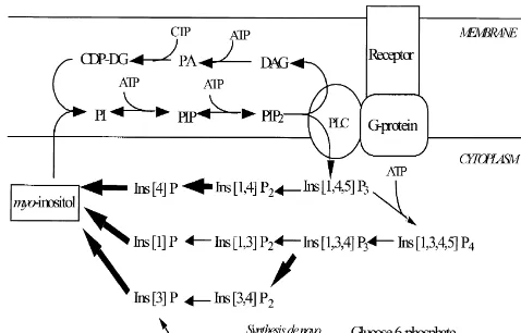
1. Introduction lithium’s mechanism of action is the inositol-depletion hypothesis [3] outlined in Fig. 1. This hypothesis proposes Bipolar disorder is a serious psychiatric illness affecting that uncompetitive inhibition of inositol monophosphatase at least 1% of the population and is characterized by (IMPase) by lithium [17,36] leads to an accumulation of fluctuations between manic and depressed mood states inositol monophosphates and a corresponding depletion of [11]. While the physiological basis of this condition myo-inositol, particularly in overactive cells. Regarding
remains to be elucidated, the search for common mecha- lithium’s effects on the PI-cycle, animal studies have nisms of action between mood stabilizers may provide provided support for this hypothesis [1,19,28,41]. A num-insight into the causes of bipolar disorder. ber of in vitro cell studies have also found changes Although lithium has remained the primary treatment of consistent with the inositol depletion hypothesis following bipolar disorder for decades, other medications like sodium stimulation of the PI cycle via receptor linked activation by valproate are now commonly used as mood stabilizers a suitable agonist [15,18,42,46]. In contrast to lithium, [29]. One widely accepted hypothesis used to explain valproate does not appear to inhibit IMPase [47] thus leading to the conclusion that this drug should not affect concentration levels of myo-inositol or inositol
mono-*Corresponding author. Tel.:11-780-407-6576;11-780-407-6672.
E-mail address: [email protected] (P.H. Silverstone). phosphates.
and depressed bipolar patients [21,23,24] and decreased PME peak ratios during the euthymic phase of the illness compared to healthy controls [21,24]. However, these effects have been observed in both medicated and unmedi-cated bipolar patients [9,10,21] and thus it is difficult to isolate drug effects from the effects of the illness.
In the present study, we examined the effects of chronic lithium and sodium valproate administration on the con-centrations of myo-inositol and inositol monophosphates in whole rat brain. Acute D-amphetamine was administered
and tested as a possible in vivo stimulant of the PI-cycle. Also examined were the effects of these two drugs on glycine, the other brain neurochemical contributing to the
1
human in vivo H myo-inositol resonances, and
glucose-6-Fig. 1. Phosphoinositol cycle (PI-cycle). Lithium blocks the enzyme,
phosphate (G6P) and phosphocholine (PC), two
com-inositol monophosphatase preventing the conversion of com-inositol-1-phos- 31
pounds which contribute to the human in vivo P PME.
phate, (Ins[1]P), inositol-1-phosphate (Ins[3]P), and inositol-4-phosphate
(Ins[4]P) to myo-inositol. It has been hypothesized that this leads to an High resolution in vitro NMR was used to resolve these
accumulation of inositol monophosphates and a depletion of myo-inositol peaks into their component parts, which cannot yet be (steps blocked by lithium are shown by bold arrows). Reduced
myo-accomplished in humans in vivo. Finally, we wished to
inositol levels prevent the efficient formation of phosphoinositol (PI)
examine the effects of lithium and valproate on the
from myo-inositol and cytidine diphosphodiacylglycerol (CDP-DG). 1
commonly used in vivo H reference peaks of creatine1 Phosphatidylinositol-4,5-bisphosphate (PIP ), which is cleaved into two2
second messengers, inositol-1,4,5-triphosphate (Ins[1,4,5]P ) and dia-3 phosphocreatine (Cr1PCr) and N-acetyl aspartate (NAA)
cylglycerol (DAG) is subsequently decreased. An overall dampening of which are often used in the expression of ratio data where PI-cycle functioning is thus postulated to occur. Of note, de novo
absolute quantification methods are not employed.
synthesis of myo-inositol is also prevented in the presence of lithium as the metabolism of glucose-6-phosphate to myo-inositol passes through Ins[3]P.
As observed in Fig. 1, the generation of two important 2. Materials and methods
intracellular second messengers, inositol 1,4,5-triphosphate
(Ins[1,4,5]P ) and sn-1,2-diacylglycerol (DAG), occurs3 This study was approved by the local ethics committee within the PI-cycle. Their formation is thus dependent on and all procedures were carried out in accordance with the the efficient breakdown of inositol phosphates and sub- guidelines of the Canadian Council on Animal Care. Adult sequent formation of inositol. A dampening of the PI-cycle male Sprague–Dawley rats (Ellerslie Biosciences), weigh-by lithium has a number of implications for cell function ing 250–350 g were housed in Plexiglas cages. The rats based on the roles of Ins[1,4,5]P and DAG in mediating3 were given free access to food and water and were
21
Ca release from the endoplasmic reticulum and protein maintained on an alternating 12-h light / 12-h dark cycle. kinase C (PKC) activity, respectively. Injections of lithium, sodium valproate, and saline were In vivo magnetic resonance spectroscopy (MRS) is started 4 days after the rats arrived giving them an increasingly being used to examine the neurochemical opportunity to adjust to their new environment.
basis of psychiatric illness, and the effects of psychiatric Each rat received a twice daily (b.i.d) intraperitoneal medications. MRS has gained popularity in the study of (IP) injection of either 2.0 mmol / kg lithium chloride bipolar disorder because myo-inositol concentrations can (n518) (Fisher Scientific, Fair Lawn, NJ, USA), 300
1 1
be measured using H MRS. Quantitative in vivo H MRS mg / kg sodium valproate (n518) (Sigma Chemical Com-studies of bipolar patients treated with or without lithium pany, St Louis, MO, USA), or saline (n518) at 07.30 h have revealed that myo-inositol decreases following and 16.00 h for 14 days. Sodium valproate was initially lithium treatment [34,35] and that bipolar patients may administered in a dose of 400 mg / kg b.i.d but this was have elevated pre-treatment levels of myo-inositol [51]. decreased after 2 days when the rats exhibited symptoms
31
Human P MRS has also been used extensively in of excessive sedation. All injections were administered in studies of bipolar disorder, since the inositol monophos- volumes of 2 ml / kg and the doses used in this study have phates are contained in the phosphomonoester (PME) peak been previously shown to produce therapeutic serum levels
31
of P spectra. In one study yielding results consistent with of lithium [14] and valproate [6] in rats.
the inositol depletion hypothesis, an increase in the PME On day 15, the rats were administered their morning peak was found in lithium-treated healthy controls as injection followed by an acute injection of 3.0 mg / kg of compared to placebo-treated controls following amphet- D-amphetamine or saline (n59 for all six treatment groups)
in ice-cold isopentane, and then maintained at2808C until (saline, amphetamine) as the two factors. Results were extract preparation and NMR analysis. deemed significant at P#0.05.
Samples were prepared using a modified version of the total lipid extraction method as described by Bligh and
Dyer [5]. Whole rat brains were homogenized in 4 volumes 3. Results
of methanol / chloroform (2:1,v / v; Fisher Scientific, Fair
1
Lawn, NJ, USA). This was followed by the subsequent 3.1. H NMR spectroscopy
additions of one part of chloroform with homogenization,
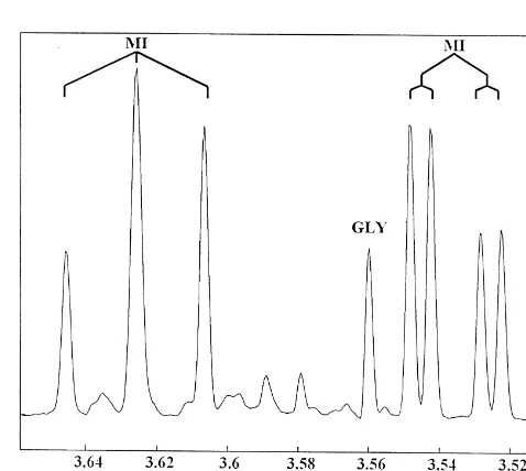
and one part of water with homogenization. Ten milliliters Myo-inositol has been previously shown to give
multi-1
of homogenate was transferred to a test tube and cen- plet signals at 3.28, 3.54, 3.62, and 4.06 ppm in H NMR trifuged at 1000 g for 15 min in a bench top centrifuge spectra of rat brain extracts [2]. Because the signals at 3.54 (Sorvall GLC-2B, Dupont, Wilmington, DE, USA). Fol- and 3.62 ppm are well resolved and relatively free of lowing centrifugation, 3.5 ml of the water / methanol layer overlap from other metabolite signals, an average area of was transferred to a 100313 mm screw cap culture tube these two multiplets was used to quantify myo-inositol and maintained at2208C overnight. The next day, samples concentrations in the brain extracts. Fig. 2 shows a cropped
1
were dried using vacuum centrifugation (Speed Vac, 500 MHz H NMR spectrum of the brain extracts focused Savant) and then reconstituted in 0.83 ml of deuterated on the region where these two peaks are located. Con-water (D 0; Aldrich Chemical Company, Milwaukee, WI,2 centrations for NAA, glycine, and creatine were calculated
1
USA) containing the H NMR standard, sodium 3-tri- using the singlet peaks at approximately 2.02, 3.56, and 2
methylsilyl [2,2,3,3, - H] propionate (TSP; Canadian 3.93 ppm, respectively [2]. 31
Isotopes, Pointe-Claire, Que., CA), and the P NMR Whole brain concentrations (mmol / g wet weight) of standard, methylenediphosphonic acid trisodium salt tetra- myo-inositol after chronic lithium, sodium valproate, or
hydrate (DSS; Sigma Chemical Company, St Louis, MO, saline administration are illustrated graphically in Fig. 3 USA), at concentrations of 1.56 mM and 0.98 mM, and numerically in Table 1. Significant drug effects of respectively. In addition to acting as internal chemical shift lithium and sodium valproate were observed (F523.559, references, these two compounds made exact quantification d.f.52, P,0.001). Tukey post-hoc results showed that of metabolite concentrations possible. both lithium- (P,0.001) and valproate- (P,0.001) treated
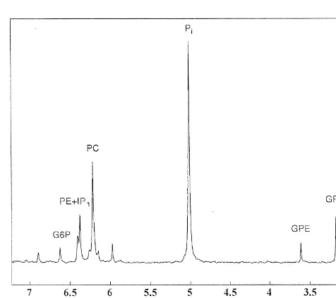
31
P NMR spectra of the extracts were recorded at 11.75 rats exhibited significantly decreased whole brain myo-T on a Varian Unity 500 NMR spectrometer using 45 inositol concentrations compared to saline-treated rats. D
-degree (10 ms) pulses and an acquisition time of 1.6 s amphetamine had no effect on myo-inositol concentrations 31
spanning 20 kHz. Typical P spectra were the sum of (F51.274, d.f.51, P,0.27). 1
3000–4000 scans each with H broad band decoupling As shown in Figs. 4 and 5, significant drug effects of 1
using a standard Varian broad band 5 mm NMR probe. H lithium and sodium valproate were also observed with NMR spectra of the extracts were obtained on the same Cr1PCr (F516.284, d.f.52, P,0.001) and NAA (F5
spectrometer using 40 degree (4 ms) pulses, 32 acquisi-tions / spectrum, and an acquisition time of 3 s spanning 8 kHz.
Spectral analysis was carried out blind to the drug treatment received by the animals. After noise filtering and baseline correction, peak areas in extract spectra were calculated using a Gaussian total line shape analysis using the Peak Research (PERCH) spectrum analysis software package (distributed by PERCH project, Department of Chemistry, University of Kuopio, Kuopio, Finland). Con-centrations were determined for myo-inositol, glycine, Cr1
1
PCr, and NAA using H NMR while concentrations for inositol monophosphates1phosphoethanolamine (IP11
PE), G6P, PC, glycerophosphoethanolamine (GPE), and 31 glycerophosphocholine (GPC) were determined using P NMR. All metabolite concentrations were calculated by comparing peak areas to that of the added internal chemi-cal shift standards of known concentration. To determine significant changes in concentration, two-way analysis of variance (ANOVA) with Tukey post-hoc analysis was
1
employed (SPSS for Windows, Release 7.5.1) with chronic Fig. 2. 500 MHz H NMR spectrum from 3.51 to 3.66 ppm of rat brain
Fig. 3. Whole rat brain concentrations of myo-inositol following chronic Fig. 5. Whole rat brain concentrations of NAA following chronic saline, saline, lithium or sodium valproate treatment and acute treatment of either lithium or sodium valproate treatment and acute treatment of either saline saline orD-amphetamine. *significantly different from rats treated chroni- orD-amphetamine. *significantly different from rats treated chronically cally with saline, P,0.001.D-amphetamine had no significant effects. with saline, P,0.001.D-amphetamine had no significant effects.
Table 1
0.09). Whole brain concentrations of glycine following
a Concentration of metabolites in rat brain following chronic treatment
chronic saline or drug administration are illustrated
nu-Metabolite Saline Lithium Valproate
merically in Table 1. D-amphetamine had no effect on
(mmol / g) (mmol / g) (mmol / g)
whole brain glycine concentrations (F52.110, d.f.51, P, Cr1PCr 10.5660.40 9.6560.38* 9.8460.37* 0.16).
Myo-inositol 7.2860.31 6.6060.33* 6.6360.17*
NAA 8.2060.40 7.4760.40* 7.2660.24* 31
3.2. P NMR spectroscopy Glycine 1.1660.14 1.1260.06 1.1660.08
IP11PE 1.2060.07 1.3960.16* 1.3960.12*
31
G6P 0.22660.021 0.21760.013 0.22260.019 Fig. 6 shows a typical 202.3 MHz P NMR spectrum of GPC 0.63160.241 0.52560.065 0.50260.049 brain extract. It should be noted that the inositol mono-GPE 0.26160.052 0.22460.027 0.23760.012
phosphates (IP )1 are co-resonant with
phos-PC 2.5560.11 2.6060.18 2.656.0.11
phoethanolamine (PE) in the region around 6.4 ppm [39].
Myo-inositol /(Cr1PCr) 0.69960.021 0.68860.037 0.68060.027
Because it is not possible to resolve the IP signal from the
a 1
Values represent the mean6S.D. Concentrations were calculated using
1 PE signal, a combined concentration for these two com-the internal standards, TSP, in high resolution (500 MHz) H NMR
31 pounds was calculated from the observed peak area. Fig. 7 spectra and DSS, in high resolution (202.3 MHz) P NMR spectra.
*
Significantly different (P,0.05) from saline-treated rats. and Table 1 illustrate the brain concentrations of IP11PE calculated from the observed peak following chronic lithium or sodium valproate administration. As with myo-24.061, d.f.52, P,0.001). Tukey post-hoc results showed
inositol, there was a significant group effect with IP1PE
that both lithium- (P,0.001) and valproate- (P,0.001) 1
treated rats exhibited significantly decreased whole brain creatine and NAA concentrations compared to saline-treated rats. Whole brain concentrations of Cr1PCr and NAA following chronic saline or drug administration are illustrated in Table 1. D-amphetamine had no effect on
Cr1PCr (F50.100, d.f.51, P,0.76) or NAA (F50.206, d.f.51, P,0.66) concentrations.
No significant drug effects were observed with whole brain concentrations of glycine (F52.607, d.f.52, P,
31
with sodium valproate was shown to lead to an accumula-tion of Ins[1,4,5]P , but not inositol monophosphates [12].3 Another study found a significant attenuation of striatal agonist-stimulated inositol phosphate formation following chronic sodium valproate treatment suggesting effects of chronic sodium valproate on PI-cycle functioning [26].
Both lithium and sodium valproate have been shown to have effects on the protein kinase C (PKC) family of enzymes closely associated with PI-cycle functioning.
Fig. 7. Whole rat brain concentrations of IP11PE following chronic
PKC isozymes are activated when they are translocated
saline, lithium or sodium valproate treatment and acute treatment of either
from the membrane to the cytosol in the presence of DAG,
saline orD-amphetamine. *significantly different from rats treated
chroni-one of the PI-cycle’s two second messengers [7,30,32].
cally with saline, P,0.001.D-amphetamine had no significant effects.
PKC isozymes a and e have been found to be down-regulated in brain cells of rats following chronic adminis-due to chronic drug treatment (F517.315, d.f.52, P, tration of both lithium and sodium valproate [25,31]. 0.001). Tukey post-hoc results showed that both lithium- Extending the effect of these two drugs on PKC activity, (P,0.001) and valproate- (P,0.001) treated rats exhibited there have also been reported changes in PKC substrates significantly increased IP11PE whole brain concentrations following drug administration. Myristoylated alanine-rich-compared to saline-treated rats. D-amphetamine had no C-kinase substrate (MARCKS), associated with signal
effect on IP11PE concentrations (F50.880, d.f.51, P, transduction and neurotransmitter release, is inactivated by
0.36). PKC phosphorylation and has been found to be reduced in
No significant changes were found with G6P (F50.642, hippocampal cells following chronic lithium [49] or so-d.f.52, P,0.53), PC (F52.322, d.f.52, P,0.62), GPE dium valproate [48] administration. Interestingly, the dow-(F51.910, d.f.52, P,0.16), or GPC (F52.491, d.f.52, n-regulation of PKC isozymes and MARCKS by lithium is
P,0.10). Whole brain concentrations of these compounds prevented in the presence of excess inositol [7,48]. The following chronic saline or drug administration are illus- effect of excess inositol in the presence of sodium val-trated in Table 1. D-amphetamine had no effect on PC proate has not yet been studied. Finally, an increase in the
(F50.263, d.f.51, P,0.11), GPE (F50.014, d.f.51, P, binding activity of AP-1, another PKC substrate and a 0.91), or GPC (F50.089, d.f.51, P,0.77) concentrations major transcription factor in the CNS known to regulate but did have a significant effect on G6P (F55.035, d.f.51, the activity of many genes, has been observed following
P,0.030). the incubation of brain cells with both valproate [8] and
lithium [38,50]. Thus it is possible that both lithium and sodium valproate have common effects on the PI-cycle
4. Discussion second messenger system that are also reflected in actions on ‘downstream’ systems such as PKC isozyme activity. 4.1. Effects of lithium and sodium valproate on the
PI-cycle 4.2. Effects of lithium and sodium valproate on creatine
and NAA
The inositol depletion hypothesis was originally
pro-1
posed to explain lithium’s clinical effectiveness following A number of in vivo H MRS studies have used a ratio evidence of IMPase inhibition by lithium. For the first time method of data expression where the Cr1PCr peak acts as we have demonstrated that therapeutic doses of either a reference against which all compounds of interest are lithium or sodium valproate increase the concentration of compared. Ratio data expression is usually employed inositol monophosphates and decrease the concentration of where technology is not available to quantify metabolites
1
myo-inositol following chronic administration in rats. using in vivo H MRS. However, we have demonstrated However, it is unlikely that both of these drugs act directly that this method of data expression is inappropriate when via IMPase, since only lithium has been shown to have an lithium or sodium valproate are administered. Both drugs effect on this enzyme [17,41,47]. Thus, at present, the caused a significant decrease in Cr1PCr concentrations in mechanism of valproate remains unknown even though whole rat brain following chronic administration. There-lithium and valproate appear to be having similar effects fore, the Cr1PCr peak is likely not an adequate internal
1
on the PI-cycle. In contrast to our hypothesis regarding the standard for in vivo H MRS research where lithium or stimulation of the PI-cycle,D-amphetamine did not poten- sodium valproate is administered to patients or controls
numerator are of the same magnitude as those in the resonate at 3.54 ppm in in vivo spectra at field strengths denominator. This is illustrated in Table 1 where ratios of currently employed. Since no concentration changes in
1
myo-inositol /(Cr1PCr) from the present study are shown glycine were detected using high resolution H NMR
to be non-significant. following chronic lithium or sodium valproate
administra-1
In human in vivo H MRS research, NAA is another tion in rat brain, this suggests that any observed significant commonly used internal standard. Because it is found changes in the 3.54 ppm peak observed in in vivo studies primarily in neurons, NAA is often used as a neuronal of the effects of lithium are most likely due to changes in marker in MRS research [4]. Also there is evidence that a myo-inositol.
31
decrease in NAA correlates well with a decrease in The PME peak observed in in vivo P spectra has been neuronal density [13,37]. However, it is not known reported to be abnormal in both medicated and unmedi-whether NAA concentrations may change in response to cated bipolar patients [9,11,14,21,22,24]. This peak is acute or chronic drug effects. In the present study, chronic made up of contributions from the inositol monophos-lithium and sodium valproate both caused NAA con- phates, PC, PE, and sugar phosphates like G6P [16] with centrations to significantly decrease in rat brain. These the signal from the inositol monophosphates forming results suggest that caution must be exercised when approximately 10% of the peak [43]. Therefore, it is interpreting decreases in NAA as cell death when the difficult to know which of these contributing compounds is possibility exists that neurons can have varying levels of responsible for the changes in the PME peak observed in NAA caused by an inherent dysfunction or the presence of bipolar patients. Also, it is not known what the effects of drugs like lithium or sodium valproate. Further research is lithium and sodium valproate are on the other contributors required to identify the mechanism by which neuronal to the PME peak (i.e. PC, PE, sugar phosphates). However, NAA is decreasing in the presence of lithium or valproate. one animal study used in vitro NMR of brain extracts to
31
In terms of MRS research and its use as an internal show that changes in the in vivo P PME peak following standard, like creatine, NAA is likely not an adequate lithium treatment were correlated with increases in internal standard where patients or controls have been inositol-1-phosphate measured quantitatively [40].
31 medicated with lithium or sodium valproate. These find- In the present study, high resolution 202.3 MHz P
1
ings with creatine and NAA suggest that future in vivo H NMR allowed us to isolate PC and G6P from the inositol 31 MRS research of lithium- and sodium valproate-treated monophosphates of the PME peak found in in vivo P patients must employ quantification methods versus ratio MRS spectra. No significant changes in PC or G6P data expression if the results obtained are to be reliable. concentrations were found in the high resolution NMR extracts of whole brain following chronic drug treatment 4.3. Implications for human in vivo MRS research of with lithium or valproate. Therefore it is unlikely that
31
lithium- or valproate-medicated subjects changes in the PME peak of in vivo P MRS spectra of medicated subjects is due to changes in PC or G6P Since magnetic field strengths of 4 tesla (T) or less are concentrations. Also, because the concentrations of these
1
normally used on in vivo human brain, the resolution of H two compounds did not change, it is very likely that 31
NMR spectra tends to be poor and the myo-inositol peaks changes in the PME peak of in vivo P MRS spectra are co-resonant with signals from glycine and the inositol represent changes in inositol monophosphate concentra-monophosphates. At field strengths of 3 T or greater, two tions.
myo-inositol peaks can be adequately resolved for quantifi- The current study has the limitations of being carried out cation at 3.54 and 3.66 ppm. However, the peak at 3.54 with rat brain, and therefore may not be directly compar-ppm also contains contributions from glycine and the able to human studies. However, since it is not yet possible inositol monophosphates, whereas the myo-inositol peak at to do in vivo studies of human brain at the resolution 3.66 ppm is only contaminated with signals from the necessary to examine each compound of interest indi-inositol monophosphates, but is usually of poorer quality vidually, we must rely on animal data. Furthermore, the
than that at 3.54 ppm. current study was conducted using post-mortem brain
Previous studies have suggested that both lithium and tissue, which again may not be directly comparable to sodium valproate may alter glycine levels in rat brain human tissue in vivo. However, there were consistent [20,27,33]. However, in the present study we have found changes in the lithium- and valproate-treated rats, which that neither lithium or sodium valproate cause changes in were not observed in the saline-treated rats, which suggests rat brain glycine concentrations. The inconsistency in these that there were no systematic post-mortem changes which findings makes it difficult to draw conclusions regarding could account for the data.
concen-[13] T. Ebisu, W.D. Rooney, S.H. Graham, M.W. Weiner, A.A. Maudsley,
trations. While the precise mechanism underlying these
N-acetylaspartate as an in vivo marker of neuronal viability in
changes is not known at the present time, this finding 1
kainate-induced status epilepticus: H magnetic resonance
spectro-suggests that lithium and sodium valproate may act scopic imaging, J. Cereb. Blood Flow Metab. 14 (1994) 373–382. through a common cellular mechanism involving the PI- [14] D. Ghoshdastidar, R.N. Dutta, M.K. Poddar, In vivo distribution of
cycle. We have also shown that both lithium sodium lithium in plasma and brain, Ind. J. Exp. Biol. 27 (1989) 950–954. [15] P.P. Godfrey, S.J. McCkye, A.M. White, A.J. Wood, D.G.
Grahame-valproate decreased the concentrations of both Cr1PCr
Smith, Subacute and chronic in vivo lithium treatment inhibits
and NAA, two common internal references used in human
agonist and fluoride-stimulated inositol phosphate production in rat
1
in vivo H MRS studies. This finding highlights the need cortex, J. Neurochem. 52 (1989) 498–506.
for caution in the interpretation of in vivo MRS studies [16] L. Gyulai, L. Bolinger, J.S.Jr. Leigh, C. Barlow, B. Chance,
using ratio data. Phosphorylethanolamine – the major constituent of the
phos-31
phomonoester peak observed by P-NMR on developing dog brain, FEBS Lett. 178 (1984) 137–142.
[17] L.M. Hallcher, W.R. Shermann, The effects of lithium ion and other
Acknowledgements agents on the activity of myo-inositol-1-phosphatase from bovine brain, J. Biol. Chem. 255 (1980) 10896–10901.
[18] M.R. Hirvonen, Cerebral lithium, inositol and inositol
monophos-Grant support provided by the Medical Research
Coun-phates, Pharmacol. Toxicol. 69 (1991) 22–27.
cil of Canada, Canadian Psychiatric Research Foundation,
[19] M.R. Hirvonen, H. Lihtamo, K. Savolainen, A gas chromatographic
Alberta Heritage Foundation for Medical Research. We method for the determination of inositol monophosphates in rat also wish to thank Dr Glen Baker and Mrs Gail Rauw of brain, Neurochem. Res. 13 (1988) 957–962.
the Neurochemical Research Unit, Department of Psychi- [20] R.S. Jope, J.M. Miller, T.N. Ferraro, T.A. Hare, Chronic lithium treatment and status epilepticus induced by lithium and pilocarpine
atry, University of Alberta, for exceptional technical
cause selective changes of amino acid concentrations in rat brain
support and advice on the assay development, and Mr
regions, Neurochem. Res. 14 (1989) 829–834.
Richard Strel for technical assistance. [21] T. Kato, T. Shioriri, S. Takahashi, T. Inubushi, Measurement of
brain phosphoinositide metabolism in bipolar patients using in vivo 31
P MRS, J. Affect. Disord. 22 (1991) 185–190.
[22] T. Kato, S. Takahashi, T. Shioiri, T. Inubushi, Brain phosphorus
References metabolism in depressive disorders detected by phosphorus-31
magnetic resonance spectroscopy, J. Affect. Disord. 26 (1992) [1] J.H. Allison, M.A. Stewart, Reduced brain inositol in lithium-treated 223–230.
rats, Nature New Biol. 233 (1971) 267–268. [23] T. Kato, S. Takahashi, T. Shioiri, T. Inubushi, Alterations in brain
1 31
[2] K.L. Behar, T. Ogino, Assignment of resonance in the H spectrum phosphorus metabolism in bipolar disorder detected by in vivo P 7
of rat brain by two-dimensional shift correlated and J-resolved NMR and Li magnetic resonance spectroscopy, J. Affect. Disord. 27 spectroscopy, Mag. Reson. Med. 17 (1991) 285–303. (1993) 53–59.
[3] M.J. Berridge, C.P. Downes, M.R. Hanley, Lithium amplifies [24] T. Kato, S. Takahashi, T. Shioiri, J. Murashita, H. Hamakawa, T. agonist-dependent phosphatidylinositol responses in brain and saliv- Inubushi, Reduction of brain phosphocreatine in bipolar II disorder ary glands, Biochem. J. 206 (1982) 587–595. detected by phosphorus-31 magnetic resonance spectroscopy, J. [4] D.L. Birken, W.H. Oldendorf, N-acetyl-L-aspartic acid: a literature Affect. Disord. 31 (1994) 125–133.
review of a compound prominent in 1H-NMR spectroscopic studies [25] R.H. Lenox, D.G. Watson, J. Patel, J. Ellis, Chronic lithium of brain, Neurosci. Biobehav. Rev. 13 (1989) 23–31. administration alters a prominent PKC substrate in rat hippocampus, [5] E.G. Bligh, W.J. Dyer, A rapid method of total lipid extraction and Brain Res. 570 (1992) 333–340.
purification, Can. J. Biochem. Physiol. 37 (1959) 911–917. [26] R. Li, L.L. Wing, R.J. Wyatt, D.G. Kirch, Effects of haloperidol, [6] G. Chen, L.D. Huang, Y.M. Jiang, H.K. Manji, The mood-stabilizing lithium, and valproate on phosphoinositide turnover in rat brain,
agent valproate inhibits the activity of glycogen synthase kinase-3, J. Pharmacol. Biochem. Behav. 46 (1993) 323–329.
¨ ¨
Neurochem. 72 (1999) 879–882. [27] W. Loscher, D. Horstermann, Differential effects of vigabatrin, [7] G. Chen, H.K. Manji, D.B. Hawver, C.B. Wright, W.Z. Potter, gamma-acetylenic GABA, aminooxyacetic acid, and valproate on Chronic sodium valproate selectively decreases protein kinase C levels of various amino acids on rat brain regions and plasma, alpha and epsilon in vitro, J. Neurochem. 63 (1994) 2361–2364. Naunyn-Schmiedeberg’s Arch. Pharmac. 349 (1994) 270–280. [8] G. Chen, P. Yuan, D.B. Hawver, W.Z. Potter, H.K. Manji, Increase in [28] B. Lubrich, Y. Patishi, O. Kofman, G. Agam, M. Berger, R.H.
AP-1 transcription factor DNA binding activity by valproic acid, Belmaker, D. van Calker, Lithium-induced inositol depletion in rat Neuropsychopharmacology 16 (1997) 238–245. brain after chronic treatment is restricted to the hypothalamus, Mol. [9] R.F. Deicken, G. Fein, M.W. Weiner, Abnormal frontal lobe phos- Psychiat. 2 (1997) 407–412.
phorus metabolism in bipolar disorder, Am. J. Psychiat. 152 (1995) [29] H.K. Manji, J.M. Bebchuk, G.J. Moore, D. Glitz, K.A. Hasanat, G.
915–918. Chen, Modulation of CNS signal transduction pathways and gene
[10] R.F. Deicken, M.W. Weiner, G. Fein, Decreased temporal lobe expression by mood-stabilizing agents: therapeutic implications, J. phosphomonoesters in bipolar disorder, J. Affect. Disord. 33 (1995) Clin. Psychiat. 60 (Suppl. 2) (1999) 27–39.
198–199. [30] H.K. Manji, G. Chen, J.K. Hsiao, E.D. Risby, M.I. Masana, W.Z. [11] Diagnostic and Statistical Manual of Mental Disorders, 4th Edition, Potter, Regulation of signal transduction pathways by mood-stabiliz-American Psychiatric Association, Washington, D.C, 1994. ing agents: implications for the delayed onset of therapeutic efficacy, [12] J.F. Dixon, L.E. Hokin, The antibipolar drug valproate mimics J. Clin. Psychiat. 57 (Suppl. 13) (1996) 34–46.
lithium in stimulating glutamate release and inositol 1,4,5-trisphos- [31] H.K. Manji, R.H. Lenox, Long-term action of lithium: a role for phate accumulation in brain cortex slices but not accumulation of transcriptional and posttranscriptional factors regulated by protein inositol monophosphates and bisphosphates, Proc. Natl. Acad. Sci. kinase C, Synapse 16 (1994) 11–28.
Molecular targets for lithium’s actions, Arch. Gen. Psychiat. 52 [42] W.R. Sherman, L.Y. Munsell, B.G. Gish, M.P. Honchar, Effects of (1995) 531–543. systematically administered lithium on phosphoinositide metabolism [33] A. Martin-Gallard, P. Rodriguez, M. Lopet, J. Benavides, M. Ugarte, in rat brain, kidney, and testis, J. Neurochem. 44 (1985) 798–807. Effects of dipropylacetate on the glycine clearance enzyme system [43] P.H. Silverstone, C.C. Hanstock, J. Fabian, R. Staab, P.S. Allen, and glycine levels, Biochem. Pharmac. 34 (1985) 2877–2882. Chronic lithium does not alter human myo-inositol or
phos-1 31 [34] G.J. Moore, J.M. Bebchuk, M.W. Faulk, I.B. Wilds, N. Seraji- phomonoester concentrations as measured by H and P MRS,
Bozorgzad, C.L. Arfken, J. Strahl-Bevacqua, K. Hasanat, L. Jol- Biol. Psychiat. 40 (1996) 235–246.
kovsky, H.K. Manji, Lithium and bipolar disorder: CNS neuro- [44] P.H. Silverstone, S. Rotzinger, A. Pukhovsky, C.C. Hanstock, chemical changes associated with mood stabilization, in: Int. Soc. Effects of lithium and amphetamine on inositol metabolism in the
1 31
Magn. Res. Med., 7th annual meeting, Philadelphia, PA, 1999, human brain as measured by H and P MRS, Biol. Psychiat. 46
Abstract 1409. (1999) 1634–1641.
[35] G.J. Moore, J.M. Bebchuk, H.K. Manji, Quantitative proton MRS in [45] A.L. Stoll, W.E. Severus, Mood stabilizers: shared mechanisms of bipolar affective disorder: monitoring of lithium induced modulation action at postsynaptic signal-transduction and kindling processes, of brain myo-inositol, in: Int. Soc. Magn. Res. Med., 5th Annual Harvard Rev. Psychiat. 4 (1996) 77–89.
Meeting, Vancouver, BC, 1997, Abstract 1214. [46] G.Y. Sun, M. Navidi, F.G. Yoa, T.N. Lin, O.E. Orth, E.B. Stubbs Jr., [36] S.R. Nahorski, C.I. Ragan, R.A.J. Challiss, Lithium and the phos- R.A. MacQuarrie, Lithium effects on inositol phospholipids and phoinositide cycle: an example of uncompetitive inhibition and its inositol phosphates: evaluation of an in vivo model for assessing pharmacological consequences, Trends Pharmacol. Sci. 12 (1991) polyphosphoinositide turnover in brain, J. Neurochem. 58 (1992)
297–303. 290–297.
[37] M. Nakano, H. Ueda, J.Y. Li, M. Matsumoto, T. Yanagihara, [47] R. Vadnal, R. Parthasarathy, Myo-inositol monophosphatase: diverse Measurement of regional N-acetylaspartate after transient global effects of lithium, carbamazepine, and valproate, Neuropsychophar-ischemia in gerbils with or without ischemic tolerance: an index of macology 12 (1995) 277–285.
neuronal survival, Ann. Neurol. 44 (1998) 334–340. [48] D.G. Watson, R.H. Lenox, Chronic lithium-induced down-regulation [38] N. Ozaki, D.-M. Chuang, Lithium increases transcription factor of MARCKS in immortalized hippocampal cells: potentiation by binding to AP-1 and cyclic AMP-responsive element in cultured muscarinic receptor activation, J. Neurochem. 67 (1996) 767–777. neurons and rat brain, J. Neurochem. 69 (1997) 2336–2344. [49] D.G. Watson, J.M. Watterson, R.H. Lenox, Sodium valproate down-[39] N.E. Preece, D.G. Gadian, J. Houseman, S.R. Williams, Lithium- regulates the myristoylated alanine-rich C kinase substrate induced modulation of cerebral inositol phosphate metabolism in the (MARCKS) in immortalized hippocampal cells: a property of rat: a multinuclear magnetic resonance study in vivo, Lithium 3 protein kinase C-mediated mood stabilizers, J. Pharmacol. Exp.
(1992) 287–297. Ther. 285 (1998) 307–316.
[40] P.F. Renshaw, J.J. Summers, C.E. Renshaw, K.G. Hines, J.S.Jr. [50] P.X. Yuan, G. Chen, L.D. Huang, H.K. Manji, Lithium stimulates 31
Leigh, Changes in the P-NMR spectra of cats receiving lithium gene expression through the AP-1 transcription factor pathway, Mol. chloride systemically, Biol. Psychiat. 21 (1986) 694–698. Brain Res. 58 (1998) 225–230.
1 [41] W.R. Sherman, A.L. Leavitt, M.P. Honchar, L.M. Hallcher, B.E. [51] K. Yue, P. Davanzo, M. Strober, M.A. Thomas, Frontal lobe H MR
Phillips, Evidence that lithium alters phosphoinositide metabolism: spectroscopy of children with bipolar disorder, in: Int. Soc. Magn. chronic administration elevates primarily D-myo-inositol-1-phos- Res. Med., 7th Annual meeting. Philadelphia, PA, 1999, Abstract phate in cerebral cortex of the rat, J. Neurochem. 36 (1981) 1471.