1
TESTICULAR TERATOMA
Henny Syahrini Lubis, Savita Handayani, Dairion Gatot, Jarmila Elmaco Hematology and Oncology Division, Departement of Internal Medicine Medical Faculty of North Sumatera University/Adam Malik General Hospital
TESTICULAR TERATOMA Abstract
Testicular teratoma is a sub-type of Non-Seminomatous Germ Cell Tumour (NSGCT) and often
occurs in two distinct age groups. Adult testicular teratomas are often mixed and are malignant.
Teratoma can be divided histologically into mature and immature. Pure mature teratoma of the testis
is rare. Intratubular Germ Cell Neoplasia (ITGCN) is a common feature associated with teratoma.
Teratoma is frequently chemoresistant and clinical management of these tumours includes radical
inguinal orchidectomy followed by Retroperitoneal Lymph Node (RPLND) dissection if indicated.
Long-term oncological outcomes for mature and immature testicular teratoma are equivocal.
A man 43 years old present with abdominal bloating since 2 years. Originally a small lump
scrotum left one year, over time the size of quail eggs like a chicken egg. Testicles left bluish red.
Bumps gradually enlarged the size of a baseball was followed by a ball at the foot of the middle
abdomen. Suffocating in the gut encountered. Pain was found, is intermittent. Weight loss ± 15 kg in
two years. Decreased sexual desire, even sex is not performed since last 1 year. Decreased appetite
encountered. History of parents and family members suffering from cancer pain can not be found.
Status praesens found the general state of being, a state of underweight malnutrition and disease
state is bad. On physical examination symmetrical enlarged abdomen encountered inspection,
palpation abdominal distension, liver/spleen/renal difficult to assess, and percussion different. Genital
erythema and hematoma of the left testis, palpable mass the size of 2x2 cm immobile and painful.
Laboratory : AFP : 0.61 ng/mL; LDH 276 U/L; Blood β HCG : 0 mIU/mL. USG conclusion
looks intra-abdominal mass. CT Scan Whole Abdomen with IV contrast conclusion: Cystic mass
density, solid fat and the pelvic cavity to the abdominal size +/- 20-30 cm. The protesters urged the
intestines to the lateral and cranial. Nothing seemed overview of hydronephrosis. Very probably a
teratoma. Histopathology conclusions : Immature teratoma with liposarcoma components.
Patients underwent radical orchiectomy. After radical orchiectomy, patients planned
chemotherapy. But the patient's condition worsened and the patient died.
2
1. INTRODUCTION
Testicular tumours are uncommon but constitute an important group of malignancies in
young men. Worldwide, it is estimated that there were more than 48.500 new cases and 8.900
deaths from the disease in 2002.1 The vast majority are primary germ cell tumours (GCTs) and the incidence has doubled in the past 30 years (with most of the increase in seminomas).2 While most patients present with early-stage and highly curable disease, the continued rise in
the incidence of these tumours presents a major challenge. At diagnosis, 1-2% of cases are
bilateral and the predominant histology is germ cell tumour (90 -95% of cases).3 Peak incidence is in the third decade of life for non-seminoma, and in the fourth decade for pure
seminoma.
Testicular teratoma is a sub-type of Non-Seminomatous Germ Cell Tumour (NSGCT)
and often occurs in two distinct age groups. Adult testicular teratomas are often mixed and
are malignant. Teratoma can be divided histologically into mature and immature. Pure mature
teratoma of the testis is rare. Intratubular Germ Cell Neoplasia (ITGCN) is a common feature
associated with teratoma. Teratoma is frequently chemoresistant and clinical management of
these tumours includes radical inguinal orchidectomy followed by Retroperitoneal Lymph
Node (RPLND) dissection if indicated. Long-term oncological outcomes for mature and
immature testicular teratoma are equivocal.4
Teratoma is a GCT that predominantly occurs in the gonads: the testis and ovaries.
They contain well-differentiated or incompletely differentiated elements of at least two germ
cells layers (endoderm, ectoderm and/or mesoderm). Mature teratomas are well differentiated
relative to the germ cell layers. Immature teratomas are incompletely differentiated and are
similar to foetal or embryonic tissue.5 This article discusses and summaries the pertinent features of mature and immature teratomas of the testis and their significance.
2. CASE REPORT
A man 43 years old present with abdominal bloating since 2 years. Originally a small
lump scrotum left one year, over time the size of quail eggs like a chicken egg. Testicles left
bluish red. Bumps gradually enlarged the size of a baseball was followed by a ball at the foot
of the middle abdomen. Suffocating in the gut encountered. Pain was found, is intermittent.
Weight loss ± 15 kg in two years. Decreased sexual desire, even sex is not performed since
last 1 year. Decreased appetite encountered. History of parents and family members suffering
3
Status praesens found the general state of being, a state of underweight malnutrition
and disease state is bad. On physical examination symmetrical enlarged abdomen
encountered inspection, palpation abdominal distension, liver/spleen/renal difficult to assess,
and percussion different. Genital erythema and hematoma of the left testis, palpable mass the
size of 2x2 cm immobile and painful.
Laboratory : Hb : 11.00 g/dl; Leukosit : 6.110/mm3; Trombosit : 351.000/mm3. AFP : 0.61 ng/mL; LDH 276 U/L; Blood β HCG : 0 mIU/mL; CEA 2.5 ng/mL. Thorax X-Ray and
ECG is normal. USG abdomen : liver : regular surface, sharp edge, normal size, parenchym
homogeneous. Ascites (-). Portal venous blood vessels difficult to assess. Hepatic veins
difficult to assess. Spleen : normal size. Splenic vein difficult to assess. Gall bladder : size
difficult to assess. Pancreatic : difficult to assess. Normal kidneys. Conclusion Sonogram:
Looks intra-abdominal mass. CT Scan Whole Abdomen with IV contrast conclusion: Cystic
mass density, solid fat and the pelvic cavity to the abdominal size +/- 20-30 cm. The
protesters urged the intestines to the lateral and cranial. Nothing seemed overview of
hydronephrosis. Very probably a teratoma. Histopathology conclusions : Immature teratoma
with liposarcoma components.
Patients underwent radical orchiectomy. After radical orchiectomy, patients planned
chemotherapy. But the patient's condition worsened and the patient died.
3. DISCUSSION
Two distinctive groups of testicular teratomas occur according to age: pre and
post-pubertal. Pure teratomas (nonmixed) are common in the paediatric sub-group, however these
are rare in adults. A mixed variant neoplasm is more commonly seen in adults. Mature
pre-pubertal teratomas are benign and represent approximately 30% of testicular germ cell
tumours in children. Post-pubertal (adult) testicular teratomas are malignant. Malignant
testicular teratomas have a higher metastasis rate of 20% as opposed to their ovarian
counterparts. Pure teratoma in the testis is rare accounting for 4% of GCT in this organ
compared to pure teratoma in 95% of GCTs found in the ovary. As previously mentioned,
teratomatous features are more commonly found in mixed GCTs in the testis, rather than pure
teratoma, and are apparent in approximately 50% of these tumour.4
4
Clinical presentation of teratoma is similar to other neoplasms of the testis. Teratoma,
both mature and immature, often present clinically as a painful testicular mass.6 The presenting symptom of testicular pain can be attributed to haemorrhage and haematoma
formation and thus the pressure applied to the testicle during tumour formation.7 Teratomas are renowned for their rapid growth and highly vasculitic nature compared to seminoma.
These tumours are often discovered incidentally or indirectly during the assessment of
testicular trauma as an initial presenting complaint. They can be present for some time prior
to detection, as is the case with most testicular masses.
Both mature and immature teratomas are generally associated with normal testicular
tumour markers: Beta Human Chorionic Gonadotropin (B-hCG) and Alpha Feto-Protein
(aFP). Occasionally some exhibit mildly elevated aFP levels. On sonographic evaluation,
teratomas typically demonstrate the appearance of cystic areas with intervening septa and
solid areas. The presence of calcifications in the tumour is another helpful sonographic
finding associated with teratomas, however, diagnosis can only be confirmed by pathologic
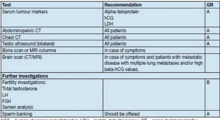
evaluation.8 To evaluate for metastatic disease a staging CT thorax and abdomen is recommended in all cases of testicular cancer.6 Up to 10% of patients present with subpleural nodes, which are not detectable by conventional chest radiograph. There is no evidence for
fluorodeoxygenase PET (FDG-PET) imaging in the staging of testicular cancer.9
Table 1. Recommended tests for staging at diagnosis10
5
Pre-pubertal teratomas are diploid, often lack chromosomal imbalances and do not
exhibit isochromosome formation. In contrast, adult teratomas, most of which are mixed type,
are hypotriploid and are associated with chromosomal abnormalities. A pertinent genetic
feature of testicular GCTs is the acquisition and over representation of 12p sequences. Gain
of one or more of these genes on 12p is crucial in the development of testicular GCTs. Other
genetic changes found in a quarter of these tumours include: partial loss of chromosome 13
(particularly q31) and gain of chromosome 7 (particularly q11), chromosome 8 and the X
chromosome.4
3.3 Macroscopic and microscopic features
Mature post-pubertal teratomas have a macroscopic appearance of solid testicular
tumours. Microscopically mature teratomas have a disordered arrangement and demonstrate
cytological atypia. Adjacent seminiferous tubules often display carcinoma in situ or
intratubular germ cell neoplasia (ITGCN) in up to 90%, which is associated with malignant
potential. Pre-pubertal teratomas, which are benign tumours, are seldom associated with
ITGCN. There is frequently widespread testicular atrophy and absent spermatogenesis. As
teratomas are germ cell tumours they can host a variety of non-tumour native tissues. For
instance, in mature ovarian teratomas choroid plexus, thyroid and pituitary tissue can be
present, with the latter manifesting systemically as prolactinomas. In mature testicular
tumours however, this non-tumour native tissue is seen less frequently.4
Pure adult testicular teratomas are rare and around a third are mixed GCTs. The
occurrence of mixed teratoma GCTs can be attributed to the pathogenesis whereby malignant
germ cells (ITGCN) differentiate into non-teratomatous cells prior to the formation of
teratomatous elements. This accounts for the association of ITGCN seen in pure teratomas, as
well as metastases of non-teratomatous cell types. The diagnosis of immaturity for a teratoma
requires tissue resembling embryonic origin, in particular the appearance of neuroepithelium.
However, the presence of this tissue or ‘immaturity’ of a post-pubertal testicular teratoma is not significant. Some pathologists believe that the teratomas should not be classified as
mature or immature due to they having the same genetic changes and biological potential, in
both pre and post-pubertal populations. As mentioned previously, the pathogenesis of
testicular teratoma is that malignant transformation occurs prior to teratomatous
differentiation. Hence the further immature differentiation of teratomatous tissue is irrelevant,
6
Primitive Neuro ectodermal Tumour (PNET) is diagnosed in the presence of an
overgrowth of immature neural elements in teratomas. Extra testicular PNETs from GCTs as
a result of metastatic spread are frequently resistant to chemotherapy and are associated with
a high rate of mortality. Despite testicular teratomas having malignant behaviour in the
absence of excess neural tissue (i.e. PNET), the presence of this tissue does have a negative
prognostic value.
3.4. Associated tumours: dermoid and epidermoid cysts
Testicular dermoid cysts are rare lesions and whether these should be classed as a
variant of mature teratoma is still under discussion. Epidermoid cysts represent 1% of all
testicular tumours but whether they are true neoplasms still remains a topic of debate.
Testicular dermoid cysts are synonymous with the features of ovarian dermoid cysts. Their
macroscopic appearance is of a cystic tumour often containing grossly identifiable hair. In
contrast to mature postpubertal teratomas, these lesions often occur in a testis with normal
spermatogenesis and lack of atrophy. They lack adjacent ITGCN and cytological atypia,
which are microscopic features of mature postpubertal teratomas. Lipogranuloatous reaction
in the testicular parenchyma and pilosebaceous cyst arrangement is diagnostically
characteristic of testicular dermoid cysts. Epidermoid cysts are similarly not associated with
ITGCN and lack atypia and mitotic activity.4
Figure.1 Immature teratoma (H&E 200x) showing immature neuroepithelium (left side)10
4. TREATMENT AND OUTCOMES
7
Only 2 to 6% of NSCGT consist of pure teratoma in adults. As previously discussed,
mature pre-pubertal teratoma has a benign clinical course, however in adults metastases for
both mature and immature teratoma have been reported in 13 to 60% of cases at initial
presentation. The standard treatment for all testicular tumours (GCTs) in adults is radical
inguinal orchidectomy. Retroperitoneal metastasis has been identified in approximately 20 to
30% of patients with clinical stage I pure testicular teratoma treated with primary
Retroperitoneal Lymph Node Dissection (RPLND). However, RPLND is still controversial in
patients with clinical stage I pure teratoma. The adjuvant treatment options remain RPLND
or surveillance, however as previously stated around a quarter of these patients will have
retroperitoneal metastasis and increased to 75% for patients with clinical stage IIA disease.
The teratomatous component of GCTs is particularly resistant to chemotherapy, which is
often an adjuvant treatment option used following RPLND for other viable NSGCT
elements.4
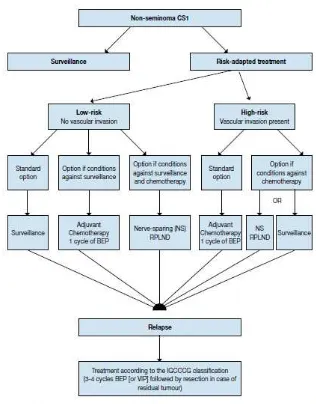
Table 2.Guidelines for the treatment of NSGCT stage I.10
8
Figure 2. Risk-adapted treatment in patients with clinical stage 1 non-seminoma NSGCT CS1.10
Advanced stage (Stage IIA/B)
Almost two thirds of men with NSGCT’s, which include pure teratoma, present with
advanced metastatic disease. This particular group of men often has regression of other
NSGCT elements in the testis. Tumour markers may be elevated in this sub group and the
presence of other GCT’s such as yolk sac or embryonal sac carcinoma are often present in metastasised sites. Treatment is entirely dependent upon histology of primary tumour and the
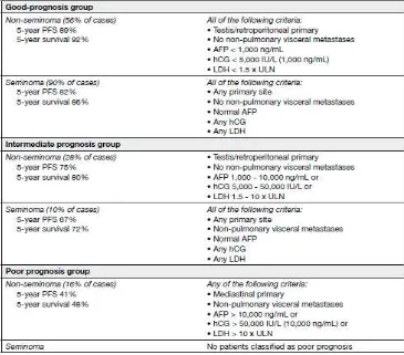
prognosis as defined as per the International Germ Cell Consensus Classification guidelines
(IGCCCG), which is based on 5202 non-seminomatous cases. There is a general consensus
that patients with advanced stage NSCGT can be managed with chemotherapy except for
9
treated with RPLND or surveillance. If surveillance is chosen, patients should be scheduled
for review six weeks postdiagnosis specifically looking for disease progression. Growth of
the lesion without a respective raise in tumour markers suggests teratoma or other malignant
transformation and RPLND is highly recommended. If tumour markers are also raised, then
chemotherapy is the initial indication; with PEB (cisplatin, etoposide, bleomycin) being the
chemotherapy of choice as per the IGCCCG guidelines. Patients refusing to undergo primary
chemotherapy may have RPLND with adjuvant PEB (2 cycles) in the case of metastatic
disease. The outcomes post RPLND and chemotherapy are equivocal with 98% cure rate. The
side effects of either treatment are different, thus extensive discussion needs to be taken
between patient and clinician with regards to treatment options. Seminomas are radiosensitive
hence Radiotherapy (RT) is an effective treatment for stage I and IIA-B disease. However,
RT is not recommended for the treatment of NSGCTs. These tumours, including mature and
immature teratoma, are often mixed cell type therefore chemotherapy is required to kill the
chemosensitive components in combination with surgical excision of residual mass (e.g.
teratoma).
10 Advanced stage (Stage IIC and III)
Around 37% of patients with pure teratoma present with advanced disease. All
advanced (stage III) NSGCTs, including mature and immature teratoma, should be assessed
and stratified on a clinical basis into either favourable-risk or intermediate/poor risk group.
Recommended treatment for the favourable risk group is at least three cycles of adjuvant
chemotherapy (PEB). These patients should all undergo restaging following primary
chemotherapy and if there is complete remission resection is not indicated. The extent of
resection of residual masses (e.g. RPLND or wedge lung resection) should also take into
account individual patient and quality of life factors. The histological diagnosis should not
influence the treatment course in stage III disease and in general all advanced stage NSGCTs,
including pure teratoma, should be treated according to the same pathway as described
above.4
The Impact of teratoma in Mixed NSGCTs
Teratoma in adults frequently presents as a mixed type tumour, with yolk sac or
embryonal sac tumour in 50% of cases. Theimpact of teratomatous elements in orchidectomy
specimens has beeninvestigated to detail. Currently, evidence shows a correlation between
teratoma within the orchidectomy specimen with an increased likelihood of teratoma in the
post chemotherapy RPLND specimenwith a current rate of 20% of teratomatous elements
found in primaryRPLND and 40% of teratomatous elements in patients undergoingRPLND
post chemotherapy. On the contrary, the absence ofteratoma in the retroperitoneum is not
suggested if teratoma is notpresent in the orchidectomy specimen.Mature teratoma is found
in approximately 35 to 40% of primarychemotherapy RPLND (PC-RPLND) specimens for
advanced NSGCT. One study by Steyerberg et al. analysed 556 RPLND specimens
post-chemotherapy for NSGCTs and found mature teratoma in 42%and that a teratoma negative
primary tumour was a strong predictor of necrosis in the PC-RPLND specimen. Currently
there is noimaging modality that accurately distinguish between necrosis, viable cancer or
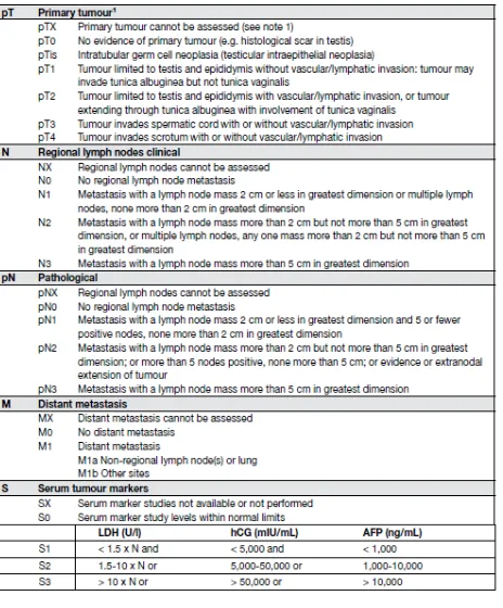
12 Table 5. Stage Grouping TNM classification 2009.10
Stage IA
patients have primary tumours limited to the testis and epididymis, with no evidence of microscopic vascular or lymphatic invasion by tumour cells on microscopy, no sign of metastases on clinical examination or imaging, and post-orchiectomy serum tumour marker levels within normal limits. Marker decline in patients with clinical stage I disease should be assessed until normalisation.
Stage IB
patients have a more locally invasive primary tumour, but no sign of metastatic disease. Stage IS
patients have persistently elevated (and usually increasing) serum tumour marker levels after orchiectomy, indicating subclinical metastatic disease (or possibly a second germ cell tumour in the remaining testis).
Retroperitoneal teratoma in NSCGT treated with primary chemotherapy (<1 cm)
Serologic and radiographic complete response to first line chemotherapy (defined as a
residual transverse axial lesion on CT less than 1 cm) is achieved in 26 to 64% of patients
with advanced NSCGT. These patients are considered to be in a low–risk group, however,
some centers still recommend RPLND following primary chemotherapy from a rationale that
radiographic estimation of the size of residual nodal tissue following primary chemotherapy
is unreliable. This remains a controversial topic and in particular the lack of imaging criteria
and consensus on nodal size criteria. A study by Oldenburg et al. of 87 RPLND patients
post-chemotherapy showed that 30% with lymph nodes <1 cm had viable tumour present (20%
teratoma and 10% malignant tumour).11 The European Germ Cell Cancer Consensus group and recent literature recommends that patients who achieve remission, defined as residual
13
Retroperitoneal teratoma in NSCGT treated with primary chemotherapy (>1 cm)
Retroperitoneal lymph-node dissection should be performed for radiographic masses
>1cm post-chemotherapy for NSGCTs. Loehrer et al. reported on 51 patients who had
surgical resections of teratoma after cisplatin-based chemotherapy. In this study twenty
patients (39%) experienced relapse with either histologically proven teratoma (10 patients) or
viable GCT (10 patients).16
Sonneveld et al. reported on 51 patients with retroperitoneal teratoma after
chemotherapy for NSGCT. In their series, nine patients experienced a relapse, with growing
mature teratoma in 56%, teratoma with malignant transformation in 33%, and viable GCT in
11%.17
Another contemporary cohort of 210 patients by Carver et al. with only pure
teratomatous elements at post chemotherapy RPLND revealed mature teratoma in 178
patients (85%), immature teratoma in 15 patients (7%) and teratoma with malignant
transformation in 17 patients (8%).18 Of the 193 patients with mature or immature teratoma, 24 patients (12%) experienced relapse after post chemotherapy RPLND and two patients
experienced relapse with teratoma with malignant transformation. For men relapsing with
only teratomatous histology, the most common sites of relapse were retro-crural and
pulmonary in seven and four patients respectively. Other recurrence sites includeliver, pelvis,
neck and para-aortic. Patients with teratoma with malignant transformation are known to
have a poorer prognosis and attempted resection of the primary site of recurrence remains the
treatment of choice due to the poor sensitivity of these tumours to chemotherapy.18
Oncological outcomes
As previously discussed, the oncological outcomes for teratoma pre and post
chemotherapy RPLND are determined by the IGCCCG staging and grading of severity.
Significant predictors for increased risk of disease recurrence include higher pre and post
chemotherapy nodal size, intermediate or poor IGCCG risk classification and evidence of
malignant transformation.18 Of particular note, histological findings of immature teratoma, when compared to mature teratoma did not increase the risk of disease recurrence. However,
mature teratoma in the PC-RPLND specimen is commonly associated with late relapse.
Overall, the oncological outcomes for immature and mature teratoma remain equivocal as per
14
5. PROGNOSIS
Table 6. Risk factors for occult metastatic disease in stage I testicular cancer.10
Table 7. Prognostic-based staging system for metastatic germ cell cancer.10
6. CONCLUSION
In adults mixed GCTs with teratomatous elements are more common than pure
teratoma. Mature and immature teratomas in the post-pubertal setting and often exhibit
malignant behavior. Distinction between mature and immature teratoma is made
histologically, with the latter closely resembling foetal or embryonic tissue. The biological
behaviour of immature teratoma is identical to that of mature teratoma. RPLND for patients
15
teratomas is complex but is often primary chemotherapy followed by RPLND or surveillance
and is guided clinically. However, teratomas are resistant and therefore often respond poorly
to chemotherapy. Long-term oncological outcomes are equivocal for both mature and
16 REFERENCE
1. International Agency for Reseach on Cancer. World Health Organization. Available
at: http://wwwdep. iarc.fr/. Accessed March 9, 2010.
2. McGlynn KA, Devesa SS, Sigurdson AJ, et al. Trends in the incidence of testicular
germ cell tumour s in the United States. Cancer 2003;97:63-70.
3. La Vecchia C, Bosetti C, Lucchini F, et al. Cancer Mortality in Europe, 2000-2004,
and an overview of trends since 1995. Ann Oncol 2010 Jun;21(6):1323-60.
4. David Wetherell, Mahesha Weerakoon, David Williams, Bhawanie Koonj Beharry,
Ania Sliwinski, Darren Ow, et al. Mature and Immature Teratoma: A Review of
Pathological Characteristics and Treatment Options. Australia ; Med Surg Urol 2014,
3:1.
5. McDougal WS, Wein AJ, Kavoussi LR, Novick AC, Partin AW, et al. (2012)
Campbell-Walsh Urology. (10thedn). Elsevier Saunders, USA.
6. Albers P, Albrecht W, Algaba F, Bokemeyer C, Cohn-Cedermark G, et al. (2011)
EAU guidelines on testicular cancer: 2011 update. Eur Urol 60: 304-319.
7. See WA, Hoxie L (1993) Chest staging in testis cancer patients: imaging modality
selection based upon risk assessment as determined by abdominal computerized
tomography scan results. J Urol 150: 874-878.
8. Simmonds PD, Lee AH, Theaker JM, Tung K, Smart CJ, et al. (1996) Primary pure
teratoma of the testis. J Urol 155: 939-942.
9. de Wit M, Brenner W, Hartmann M, Kotzerke J, Hellwig D, et al. (2008) [18F]-
FDG-PET in clinical stage I/II non-seminomatous germ cell tumours: results of the
German multicentre trial. Ann Oncol 19: 1619-1623.
10.P. Albers (Chair), W. Albrecht, F. Algaba, C Bokemeyer, G. Cohn-Cedermark, K.
Fizazi, et al. Guidelines on Testicular Cancer. European Association of Urology 2015.
Page 5-52.
11.Oldenburg J, Alfsen GC, Lien HH, Aass N, Waehre H, et al. (2003)
Postchemotherapy retroperitoneal surgery remains necessary in patients with
nonseminomatous testicular cancer and minimal residual tumor masses. J Clin Oncol
21: 3310-3317.
12.Ehrlich Y, Brames MJ, Beck SD, Foster RS, Einhorn LH (2010) Long-term follow-up
17
nonseminomatous germ cell tumors: is a postchemotherapy retroperitoneal lymph
node dissection needed after complete remission? J Clinical Oncol 28: 531-536.
13.Krege S, Beyer J, Souchon R, Albers P, Albrecht W, et al. (2008) European consensus
conference on diagnosis and treatment of germ cell cancer: a report of the second
meeting of the European Germ Cell Cancer Consensus group (EGCCCG): part I. Eur
Urol 53: 478-496.
14.Kollmannsberger C, Daneshmand S, So A, Chi KN, Murray N, et al. (2010)
Management of disseminated nonseminomatous germ cell tumors with riskbased
chemotherapy followed by response-guided postchemotherapy surgery. J Clin Oncol
28: 537-542.
15.Zuniga A, Kakiashvili D, Jewett MA (2009) Surveillance in stage I nonseminomatous
germ cell tumours of the testis. BJU Int 104: 1351-1356.
16.Loehrer PJ Sr, Hui S, Clark S, Seal M, Einhorn LH, et al. (1986) Teratoma following
cisplatin-based combination chemotherapy for nonseminomatous germ cell tumors: a
clinicopathological correlation. J Urol 135: 1183-1189.
17.Sonneveld DJ, Sleijfer DT, Koops HS, Keemers-Gels ME, Molenaar WM, et al.
(1998) Mature teratoma identified after postchemotherapy surgery in patients with
disseminated nonseminomatous testicular germ cell tumors: a plea for an aggressive
surgical approach. Cancer 82: 1343-1351.
18.Carver BS, Shayegan B, Serio A, Motzer RJ, Bosl GJ, et al. (2007) Long-term clinical
outcome after postchemotherapy retroperitoneal lymph node dissection in men with