ANTIBACTERIAL EFFECTS OF ETHANOLIC LEAF EXTRACTS OF
BACHANG (
MANGIFERA FOETIDA
LOUR.) ON
STREPTOCOCCUS
MUTANS
Lilik Koernia Wahidah1, Ahmad Rokiban1, Subur Widodo1, Dewi Mut Mainah1, Yulianty2 and Mohammad Kanedi2*
1
Department of Pharmacy, Faculty of Mathematics and Sciences, Tulang Bawang University,
Bandar Lampung, Indonesia.
2
Department of Biology, Faculty of Mathematics and Sciences, University of Lampung,
Bandar Lampung, Indonesia.
ABSTRACT
Bachang (Mangifera foetida Lour.) get less attention in scientific
studies, so that it is difficult to find literature about the benefits of this
mango in the folk medicine practice and contemporary pharmacology.
The study was carried out to find out if the leaf extract of M. foetida is
potent as antibacterial against Streptococcus mutans, the dental
caries-causing bacteria. Seven different solution were prepared for treatments
namely aquadest (as negative control), erythromycin (as positive
control) and five levels concentration of bachang leaves extract (v/v),
ie 20%, 40%, 60%, 80% and 100%. Susceptiblity of the bacteria was
assayed by disc diffusion technique with blood agar base. Minimum
inhibitory concentration(MIC) and minimum bactericidal
concentration (MBC) were determined by a serial dilution technique with nutrient broth
media were used. The results showed bachang leaf extracts significantly inhibit the growth of
S.mutans with inhibition zones ranging from 13.74 mm to 19.22 mm. At the maximum
concentration the effect even the same as erythromycin. With the MIC value of 14%, the
extract exhibits bactericidal properties by MBC test.
INTRODUCTION
Bachang (Mangifera foetida Lour.) is one of the mango species native to Indonesia that
possesses particularities on its fruit. Unripe bachang fruit contain an irritant juice which
decrease at maturity and left the irritant sap restricted to the peel that make the fruit flesh can
be eaten fresh. In Indonesia, this mango generally intercropped with other crops in
plantations. In addition, in spite of its turpentine smell and the taste but it is not generally
valued as a table fruit and less marketable.[1]
Due to less commercial, presumably, causing bachang get less attention in scientific studies,
so that it is difficult to find literature about the benefits of this mango in the folk medicine
practice and contemporary pharmacology. In contrast, the scientific works on various aspects
of agronomic, biochemical, pharmacological, medicinal and health benefits of Indian mango
(Mangifera indica L.), the most valuable manggo in the world, are very abundant. Indian
mango, the common mango, in each home land of the plant is known to have numerous
medicinal uses namely to treat diarrhea, cough, anemia, hypotension, itch, diuretic,
rheumatism, gingivitis, dysentery, syphilis, emetic, gastric disorders, hepatic disorders,
bleeding hemorrhoids, lunghemorrhage, diabetes, menorrhagia, jaundice, liver obstruction,
wounds, mouth sores, toothache, chest pain, anemia, skin diseases and dental caries.[2]
Today M. indica suggested as an important source of many pharmacologically and
medicinally important chemicals such as mangiferin, mangiferonic acid hydroxymangiferin,
polyphenols and carotenes. Mangiferin, named based on the generic name of the plants,
exhibits many different pharmacological activities, including antioxidant, radioprotective,
immunomodulatory, anti-allergic, anti-inflammatory, antitumor, antidiabetic, lipolytic,
antiboneresorption, monoamine oxidase-inhibiting, antimicrobial and antiparasitic.[3] In M. indica, the mangiferin can be isolated from leaves, stem bark, fruit peels and root, peels of
raw and ripe fruits, fruit peel and pulp.[4]
Among the few studies on Mangifera foetida, known as wild mango, show that this mango
fruits containing several volatiles substances such as, esters and oxigenated monoterpenes
were dominant, with ethyl butanoate the most abundant.[5] A study on antioxidant properties of fresh, powder and fiber products of M. foetida fruit showed that bachang fruit containing
showed that the ethanol fraction of the wild mango’s leaf extract contained phenols,
coumarins, flavonoids, tannins, alkaloids and quinones.[7]
The existence of flavonoids in M. foetida can be used as a basis to expect that the crude
extract of this mango will exhibit antibacterial properties. There are lots of research reports
on the pharmacological effects of a plant that points flavonoids as active substance.
Flavonoids isolated from leaves of Indian mango (M. indica L.) is known to inhibit the
growth of Lactobacillus sp., Escherichia coli, Azospirillium lipoferum and Bacillus sp.[8] Bioactive substances isolated from leaves and stems of Chromolaena squalida that evidently
shown antibacterial activities on both Gram-positive and Gram-negative bacteria known to
contain flavonoids.[9]
In spite of common mango plant (M. indica) has been used for treating dental caries and the
mango plant extract in fact revealed antibacterial activities, yet there is no report regarding
the effects of the mango plant extract on the bacteria causing dental caries. For that reason,
the study was carried out to find out if the leaf extract of M. foetida is potent to inhibit the
growth of Streptococcus mutans or even possesses bactericidal effects against the bacteria..
MATERIALS AND METHODS Bachang Leaves Extract
Bachang mango leaves used in the study were collected from Karangsari village, sub-district
of Air Naningan, the district of Tanggamus, Lampung province, Indonesia. The leaves that
were chosen as a sample is the third, fourth, fifth, sixth and seventh leaf from the shoot end of
a twig. Once washed with tap water and rinsed with distilled water, the fresh leaves were
chopped into small pieces and then sun-dried after being covered with a black cloth. The
simplicia (100 g) were macerated using 70% ethanol for 24 hours, repeated three times. The
macerate then evaporated using a rotary evaporator until concentrated liquid extracts were
obtained and labelled as stock solution. The stock solution was diluted with distilled water in
accordance with the treatment concentrations designed for the experiment.
Experimental Design
By using a completely randomized design, seven different solution were prepared for
treatments in the study namely aquadest (as negative control), erythromycin (as positive
control) and five levels concentration of bachang leaves extract (v/v), ie 20%, 40%, 60%,
Bacterial Inhibition Test
The disc diffusion assay technique was used, 100 μl of microbes cultures aged 18-24 h were
add to Petri disc and blood agar base (BAB) were poured. After media were solidified, the
disc (with a diameter of 5 mm) that have been previously soaked in treatment solution were
placed. The inoculated agar plates incubated at 37oC for 24 h. The zones of inhibition were then recorded in millimeters.
Determination of MIC and MBC
The minimum inhibitory concentrations(MIC) of the bachang leaves extracts performed by a
serial dilution technique with nutrient broth (NB) media were used. The serial dilution
concentrations of the stock were 1%, 2%, 3%, 4%, 5%, 6%, 7%, 8%, 9%, 10%, 11% , 12%,
13%, 145, 15%, 16%, 17%, 18% and 19% along with three control solution, ie media, plant
extract and inocula. The media control contained 2ml NB; extract control containing 1.8 ml
NB and 0.2ml plant extract; while inocula control containing 1.9ml NB and 0.1ml bacterial
suspension. The minimum inhibitory concentration was defined as the lowest concentration
able to inhibit any visible bacterial growth after being incubated for 24 hours. To determine
the minimum bactericidal concentration (MBC), bacterial suspension in the extract solution
set as MIC were re-cultured on Mueller-Hinton Agar and incubated for 24 hours. The highest
dilution that yielded no single bacterial colony was taken as the minimum bactericidal
concentration.
Statistical Analysis
Both descriptive and inferential statistics were used. Statistical differences between groups
were analyzed by one-way analysis of variance (ANOVA) followed by LSD test. The data
were presented as the mean±SD and the statistical significance was established at p < 0.05.
RESULTS
The susceptibility and resistance of S. mutans against the aquadest (as negative control),
bachang leaf extracts and erythromycin (positive control) treatments are presented in Table 1.
One-way ANOVA statistics applied for the mean value of the inhibition zone diameters
resulted in F-value of 183.527 (F-crit of 2.848) and P-value < 0.001. The data clearly
indicated that ethanolic leaf extracts of the bachang mango (M. foetida Lour.) of all
concentration levels are significantly differ from aquadest (the negative control) and at the
Table 1. The results of disc diffusion test against the effects of aquadest (as negative control), bachang leaf extracts and erythromycin (positive control) on the growth of S. mutans. Mean±SD values followed by the same superscript are not
statistically different at α=0.05, based on the LSD test result.
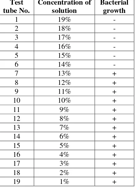
The result of MIC test against S.mutans inocula using a series of dilution concentration of
extract along with the control solution, are tabultaed in Table 2. It is clearly indicated by the
data that there is no bacterial growth visible in the test tube contained leaf extracts of bachang
19% -14%. It is suggested, therefore, that 14% is the minimum inhibitory concentration of
ethanolic leaf extracts of bacang mango against the growth of S.mutans.
20 Media control - 21 Extract control - 22 Bacterial control +
(-): no bacterial growth visible (+): bacterial growth occurs
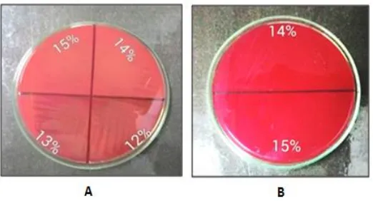
The results of re-culturing test on the growth of S.mutans suspension taken from MIC
solution series (12% - 15%) on BAB agar are depicted by photographs in Fig.1. The
photographs showed that bacterial inocula taken from MIC solution of 14% and 15% yielded
no bacterial colony at all. It can be assumed that the ethanolic leaf extracts of M. foetida
possess bactericidal properties, instead of bacteriostatic, against S.mutans.
Figure 1. Photographs depicted the growth of S.mutans inocula taken from solution used in MIC test. (A) Comparison between the concentration in the MIC test where bacterial growth was visible (12% and 13%) and that was not (14% and 15%); (B) Growth of S. mutans taken only from the dilution concentration that does not show bacterial growth.
DISCUSSION
As explained in the introduction, this plant contains active ingredients such as esters and
monoterpenes, coumarins, tannins, alkaloids, flavonoid, carotenoid, ascorbic acid, phenols,
and quinones. Some of these chemicals have been reported to have anti-bacterial properties.
Monoterpenes with its active components such as (+)menthol, thymol and linalyl acetate was
suggested to result in alterations of membrane permeability and in leakage of intracellular
materials of bacterial cells.[10]
Coumarins and the derivatives such as osthenol showed effective antibacterial activity against
Bacillus cereus, Escherichia coli, Pseudomonas aeruginosa and Staphylococcus aureus with
osthenol was suspected to accoun for antibacterial activities of this molecule against these
strains.[11] Next, tannins. Purified tannin extracts from tannin-containing plants such as oak, locust, skunk bush and plum exhibited a range of antimicrobial activity against Escherichia
coli, Klebsiella pneumoniae and Staphylococcus aureus.[12]
Among bioactive extracts from M. foetida mentioned above, flavonoid is the most suggested
as the highly potent antibacterial. Methanolic leaf extracts from guava plant (Psidium
guajava), which known to contain flavonoids, were suggested as antibacterial due to cause
significant release of RNA in gram-negatives and gram-positives bacteria. The guava extracts
was allegedly disrupt the integrity of the microorganism lipopolysaccharide (LPS) layer.[13] The disruption of bacterial membrane by flavonoid treatments has also been confirmed by
more recent studies. Dzoyem et al.[14] found that flavonoids from Dorstenia species lead to depolarization of membrane and inhibition of DNA, RNA and proteins synthesis in S.aureus,
causing the decrease of cell density and lysis of the bacteria. Quercetin, one of flavonoid
derivatives, attributed to inhibition of DNA gyrase, while the other derivates such as
sophoraflavone G and (−)-epigallocatechin gallate inhibit cytoplasmic membrane function,
and that licochalcones A and C inhibit energy metabolism.[15] Lipophilic flavonoids found to disrupt microbial membranes; catechin inhibit isolated bacterial glucosyltransferases in S.
mutans; robinetin, myricetin and (−)-epigallocatechin are known to inhibit DNA synthesis in
Proteus vulgaris.[16]
The last, due to Mangifera species were the main sources of mangiferin, it is appropriate to
assume that the antibacterial effects of M.foetida might related to mangiferin. In Fusarium
wilt of safflower, mangiferin caused lysis of the hyphal cells and reduced growth and
presumably, also altered the metabolism of the fungus.[17] Mangiferin was considered as an antimicrobial agent upon gram-positive, gram-negative bacteria and yeast Candida albicans
and it was a helping agent for up-regulating the multidrug transporter of
ABCB1/Pglycoprotein.[18]
CONCLUSSION
Ethanolic leaf extracts of bachang mango (Mangifera foetida Lour.) possess antibacterial and
REFERENCES
1. Orwa C, A Mutua, Kindt R, Jamnadass R, S Anthony. 2009. Agroforestree Database:a
tree reference and selection guide version 4.0.
http://www.worldagroforestry.org/sites/treedbs/treedatabases.asp.
2. Wauthoz N., Balde A., Balde E.S. Van Damme M. and Duez P. 2007.
Ethnopharmacology of Mangifera indica L. Bark and Pharmacological Studies of its Main
C-Glucosylxanthone, Mangiferin. International Journal of Biomedical and
Pharmaceutical Sciences, 1(2): 112-119.
3. Sharma S. 2014. Mangiferaindica: Ethnopharmacology of Mangiferin from its Leaf
Extract. International Journal of Science and Research (IJSR). June 2014; 3(6):
1992-1994.
4. Jyotshna, Khare P. and Shanker K. 2016. Mangiferin: A review of sources and
interventions for biological activities. BioFactors 42(5:504–514. Version of Record
online: 23 SEP 2016.
5. Wong K.C. and Ong C.H. 1993. Volatile components of the fruits of bachang (Mangifera
foetida Lour.) and kuini (Mangifera odorata Griff.). Flavour and Fragrance Journal, 8(3):
147–151 May/June 1993.
6. Tyug T.S., Johar M.H. and Ismail A. 2010. Antioxidant Properties of Fresh, Powder, and
Fiber Products of Mango (Mangifera Foetida) Fruit. Journal International Journal of Food
Properties, 2010; 13(4): 682-69.
7. Hillary J. and Nuringtyas T.R. 2016. Inhibitory effect of wild mango (Mangifera foetida
L.) extract on seed germination of Cynodons dactylon (L.) Pers. AIP Conference
Proceedings, 1744(1): 020029-1- 020029-5. doi: 10.1063/1.4953503.
8. Kanwal Q., Hussain I., Siddiqui H.L. and Javaid A. 2009. Flavonoids from mango leaves
with antibacterial activity. J. Serb. Chem. Soc. 2009; 74(12): 1389–1399.
9. Taleb-Contini S.H., Salvador M.J., Watanabe E., Ito I.Y. and de Oliveira D.C.R. 2003.
Antimicrobial activity of flavonoids and steroids isolated from two Chromolaena species.
Revista Brasileira de Ciências Farmacêuticas—Brazilian Journal of Pharmaceutical
Sciences, 2003; 39(4): 403-408.
10.Trombetta D., Castelli F., Sarpietro M.G., Venuti V., Cristani M., Daniele C., Saija A.,
Mazzanti G. and Bisignano G. 2005. Mechanisms of Antibacterial Action of Three
Monoterpenes. Antimicrob Agents Chemother. 2005 Jun; 49(6): 2474–2478.
11.de Souzaa S.M., Monacheb F.D. and Smaˆnia Jr. A. 2005. Antibacterial Activity of
12.Min B.R., Pinchak W.E., Merkel R., Walker S., Tomita G. and Anderson R.C. 2008.
Comparative antimicrobial activity of tannin extracts from perennial plants on mastitis
pathogens. Scientific Research and Essay, February 2008; 3(2): 066-073.
13.Henie E.F.P., Zaiton H. and Suhaila M. 2009. Bacterial membrane disruption in food
pathogens by Psidium guajava leaf extracts. International Food Research Journal, 2009;
16: 297 -311.
14.Dzoyem J.P., Hamamoto H., Ngameni B., Ngadjui B.T. and Sekimizu K. 2013.
Antimicrobial action mechanism of flavonoids from Dorstenia species. Drug Discov
Ther. 2013 Apr; 7(2): 66-72.
15.Cushnie T.P.T. and Lamb A.J. 20015. Antimicrobial activity of flavonoids. International
Journal of Antimicrobial Agents, 2005; 26: 343–356
16.Kumar S. and Pandey A.K. 2013. Chemistry and Biological Activities of Flavonoids: An
Overview. The Scientific World Journal Volume 2013 (2013): 16 pages.
17.Ghosal S., Biswas K., Chakrabarti D.K. and Basu Chaudhary K.C. 1977. Control of
Fusarium wilt of safflower by mangiferin. Phytopathology 67: 548-550.
18.Wei Z.Q., Deng J.G. and Yan L. 2011. Pharmacological Effects of Mangiferin. Chinese