Applied Animal Endocrinology
E. James Squires
Department of Animal and Poultry Science, University of Guelph,
Guelph, Ontario, Canada
CAB International 875 Massachusetts Avenue
Wallingford 7th Floor
Oxon OX10 8DE Cambridge, MA 02139
UK USA
Tel: +44 (0)1491 832111 Tel: +1 617 395 4056
Fax: +44 (0)1491 833508 Fax: +1 617 354 6875
E-mail: [email protected] E-mail: [email protected] Website: www.cabi-publishing.org
©CAB International 2003. All rights reserved. No part of this publication may be reproduced in any form or by any means, electronically,
mechanically, by photocopying, recording or otherwise, without the prior permission of the copyright owners.
A catalogue record for this book is available from the British Library, London, UK.
Library of Congress Cataloging-in-Publication Data Squires, E. James
Applied animal endocrinology/E. James Squires. p. cm
Includes bibliographical references (p. ). ISBN 0-85199-594-2 (alk. paper)
1. Veterinary endocrinology. 2. Livestock–Reproduction. I. Title. SF768.3.S68 2003
636.089´64–dc21
2003003596
ISBN 0 85199 594 2
Typeset by MRM Graphics Ltd, Winslow, Bucks
Contents
Preface X
Dedication xii
Acknowledgements xii
Abbreviations xiii
1 Hormone and Receptor Structure and Function 1
1.1 Introduction 1
What is a hormone? 1
Why are hormones necessary? 1
How do hormones function? 2
What effects are due to hormones? 3
How is hormone action selective? 4
Types of hormones 5
Location of endocrine glands 5
1.2 Synthesis, Release and Metabolism of Hormones 5
Synthesis of protein hormones 5
Synthesis of steroid hormones 7
Synthesis of eicosanoids 9
Synthesis of thyroid hormones 11
Hormone release 11
Metabolism and excretion of hormones 12
1.3 Receptors and Hormone Action 13
Extracellular receptors 13
Second messenger systems 14
Adenylate cyclase–cAMP–protein kinase A pathway 14 Guanyl cyclase–cGMP-dependent protein kinase pathway 15
Genomic actions of cAMP 16
Calcium-dependent phospholipase C–protein kinase C system 16 Interaction of cAMP and Ca2+pathways 18
Tyrosine kinase receptors: catalytic receptors 18
Cytokine receptors 20
Receptor serine kinase 20
Termination of hormone action 20
Intracellular receptors 20
Structural and functional domains of nuclear receptors 22 Binding sites of the hormone–receptor complex on DNA 22 Organization of nuclear chromatin and the nuclear matrix 23
Chromatin 23
Nuclear matrix 24
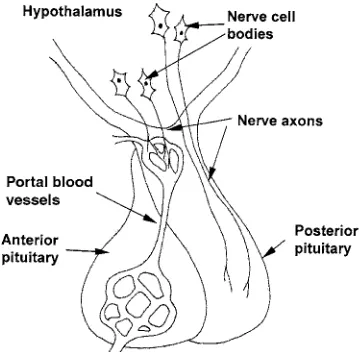
Identification of DNA regulatory sequences 25 Identification of DNA-binding proteins 26 Integration of peptide and steroid hormone actions 26 1.4 Pituitary–Hypothalamic Integration of Hormone Action 27 Structure–function relationship of pituitary and hypothalamus 27
Posterior pituitary hormones 28
Anterior pituitary hormones 28
Hypothalamic control of pituitary hormone secretion 29 Hypothalamic releasing and release-inhibiting hormones 30
Control of hormone release 31
Questions for Study and Discussion 32
Further Reading 33
2 Endocrine Methodologies 35
2.1 Methods for Studying Endocrine Function 35
Model systems 35
Whole animal model 35
Isolated organs or tissues 37
In vitromodels 38
Use of inhibitors and agonists 39
Use of antibodies 40
Immune response 40
Detection and purification of antibodies 41
Monoclonal antibodies 41
Use of antibodies to identify the site of hormone synthesis or
target tissue 42
2.2 Measurement of Hormones and Receptors 42
Assay of hormones 42
Types of hormone assays 43
Bioassays 43
Chemical assays 44
Liquid chromatography 45
Gas chromatography 46
Electrophoresis 47
Competitive binding assays 47
Assay requirements 47
Measurements of hormone–receptor binding 49
Competition binding 50
2.3 Methods for the Production of Hormones 51
Steroids and non-protein hormones 51
Protein and peptide hormones 52
Determination of amino-acid sequence 53
Peptide and protein synthesis 54
Non-peptide mimics of peptides 55
Production of recombinant proteins 56
2.4 Manipulation of Endocrine Function 57
Hormone delivery methods 57
Types of sustained-release devices 58
Pulsatile release of hormone 59
Immunomodulation of hormone action 61
Types of immunoglobulins 61
Transgenic animals 62
Uses for transgenic animals 62
Production of transgenic animals 63
Questions for Study and Discussion 64
Further Reading 65
3 Manipulation of Growth and Carcass Composition 66
3.1 Overview 66
Effects on growth, feed efficiency and lean yield 67
3.2 Anabolic Steroids and Analogues 68
Mechanism of action 70
Direct effects 71
Indirect effects 72
Delivery systems 72
Safety issues 74
3.3 Use of Intact (Uncastrated) Male Pigs 75
Advantages and problems of intact male pigs 75
Effects of sex steroid hormones 76
Description of boar taint 77
Measurement of boar taint 78
Use of tainted meat in processed products 79
Metabolism of androstenone and skatole 80
Factors affecting boar taint 81
Diet and management 82
Genetic factors 82
Immunological methods to control boar taint 83
3.4 Somatotrophin 83
Control of ST release 85
Mechanism of action of ST 88
Direct effects 88
ST receptors 88
Metabolic effects 88
Indirect effects 89
Delivery/dose effects 89
Safety/quality aspects 90
3.5 β-Adrenergic Agonists 90
Mechanism of action 91
β-AA structures 91
β-AA receptors 91
Physiological responses to β-AA 93
Delivery/dose 94
Safety aspects 94
Alternative approaches for using growth promoters 95
3.6 Thyroid Hormones 96
Synthesis and metabolism 96
Metabolic effects 97
Effects on growth and development 98
3.7 Dietary Polyunsaturated Fatty Acids 99
Mechanism of action 99
Linoleic acid, linolenic acid and γ-linolenic acid 100
Applications 102
Conjugated linoleic acid 102
Metabolic effects of CLA isomers 103
Mechanism of action of CLA 104
CLA preparations 104
3.8 Leptin 105
Leptin receptors 105
Involvement in energy metabolism and reproduction 106
Direct effects 106
Applications 107
3.9 Cholecystokinin and Appetite 107
Mechanism of action 107
Applications 108
3.10 Antibiotics, Antimicrobials and Other Factors 108
3.11 Dietary Chromium and Insulin 109
Insulin 109
Glucagon 110
Mechanisms of action 110
Physiological effects 111
Dose 111
Safety issues 112
3.12 Effects of Stress on Meat Quality 112
Pale, soft, exudative and dark, firm, dry meat 112
Porcine stress syndrome 114
Testing for PSS 115
Endocrine factors that affect PSS pigs and PSE pork 117 Manipulations to reduce the incidence of PSE 118
Summary and Conclusions 118
Questions for Study and Discussion 119
Further Reading 120
4 Endocrine Effects on Animal Products 124
4.1 Mammary Gland Development and Milk Production 124
Introduction 124
Mammary gland development 124
Involution and the dry period 126
Model systems for studying mammary gland development and function 126
In vitrocell culture systems 126
Whole animal studies 126
Hormones and mammary gland development (mammogenesis) 127
Hormones and initiation of lactogenesis 128
Maintenance of lactation (galactopoiesis) 129
Hormonal effects 129
Milk removal 130
Effect of bST 130
Mechanism of action 131
Delivery 131
Safety concerns of bST use 132
Factors affecting milk composition 132
Milk protein 132
Milk fat 133
Ketosis 134
Milk fever 134
Hormones involved 134
Predisposing factors 135
Treatment and prevention 135
4.2 Egg Production 136
Sexual development 136
Hormonal effects 137
Genetic effects 137
Regulation of follicular development and egg production 138
Application 140
Eggshell formation 141
Shell matrix 142
Calcium metabolism 143
Applications 145
4.3 Wool Production and Endocrine Defleecing 145
Introduction 145
Defleecing methods 145
Model systems used to study function of follicles 147 Growth factor effects on hair and wool follicles 147
Insulin-like growth factors 147
Fibroblast growth factors 148
Transforming growth factor-β 149
EGF family of growth factors 149
EGF receptor 150
Effects of EGF on follicles 150
Other effects of EGF 151
Summary of growth factors affecting fibre growth 151
Questions for Study and Discussion 151
Further Reading 152
5 Endocrine Manipulation of Reproduction 154
5.1 Manipulation of Reproduction in Mammals 154
Sexual differentiation and maturation 154
Differentiation of the gonads and ducts 155
Differentiation of the brain 155
Sex differentiation in cattle, sheep and pigs 156
Sex-determining genes 157
Regulation of meiosis in germ cells 157
Regulation of the oestrous cycle 158
Overview of the oestrous cycle 159
Follicular development 159
Oestrus and ovulation 161
Luteal phase 161
Pregnancy 162
Parturition 162
Puberty and seasonality 162
Regulation of LH production 163
Regulation of steroidogenesis 164
Manipulation of the oestrous cycle 164
Hormone preparations for manipulating reproduction 165 Use of hormone agonists to control fertility 170
Methods for the detection of oestrus 171
Oestrus behaviour 171
Milk progesterone 172
Induction and synchronization of oestrus 172 Strategies for synchronizing oestrus 173 Prostaglandin F2α-based systems 173 GnRH and the Ovsynch®protocol 173
Progestin-based systems 174
Superovulation and embryo transfer 175
In vitroproduction of embryos 176
Maintenance of pregnancy 177
Induction of abortion/parturition 177
Postpartum interval 178
Cystic ovarian disease 178
Effects of nutrition 179
Effects of stress 179
Inducing puberty 180
Advancing cyclicity in seasonal breeders 180
Immunological control of reproduction 181
5.2 Endocrine Manipulations in Aquaculture Fish 182
Control of reproduction 182
Sex reversal 182
Hormonal treatments for sex reversal 183
Indirect methods 183
Induction of spawning 185
Effects on growth and nutrient utilization 186
Applications 187
Stress and effects on the immune system 188
Applications 189
Questions for Study and Discussion 189
Further Reading 189
6 Effects on Animal Behaviour, Health and Welfare 192
6.1 Control of Broodiness in Poultry 192
Applications 193
6.2 Applications of Pheromones 194
Types of pheromones 194
Chemistry of pheromones 194
Pheromone production and release 195
Detection of pheromones 196
Vertebrate pheromones 196
Rodents 197
Pigs 198
Cattle 198
Sheep and goats 199
Fish 199
Other 199
Insect pheromones 200
Applications 202
Pest control 202
Population monitoring 202
Mass trapping 203
Insect management 204
Reproduction control in mammals 204
6.3 Effects of Stress 204
What is stress? 204
Hormonal responses to stress 205
SA system 205
HPA axis 206
CRH and CRH receptors 207
Role of various hormones in stress responses 208
Assessment of stress 210
Behavioural and physiological measures 210
Hormonal measures 210
Issues related to sampling 211
Effects of stress on the immune system and disease resistance 212
Overview 212
Stress effects on the immune system 214
Effects on reproduction 215
Effects on growth performance 217
Summary 218
6.4 Endocrine Applications in Toxicology 218
Endocrine disruptors or modulators 218
Assessment of endocrine disruptor activity 219
In vitroassays 220
In vivoassays 220
Sources of endocrine disruptors 221
Plant-derived endocrine modulators 221
Xenobiotic endocrine modulators 223
Indirect mechanisms of action 224
Effects on hormone metabolism 224
Effects on thyroid function 225
Effects on adrenal function 225
Effects on CNS function and behaviour 225
Summary 226
Questions for Study and Discussion 227
Further Reading 227
Index 230
The purpose of this book is to provide information on a number of different endocrine systems that affect animal production, and to describe how these systems can be manipulated or mon-itored to advantage. A number of excellent endocrinology texts are already available that describe the function of various endocrine systems, but these texts, for the most part, deal with human or comparative endocrinology. This book focuses on commercial animals, and endocrine systems that can affect growth and carcass composition, the production of animal products (milk, eggs and wool), reproduction efficiency and animal health, behaviour and welfare are described. Detailed information on the mechanism of action of the endocrine sys-tems is covered, and an attempt is made to integrate knowledge from similar topics by focus-ing on common mechanisms and themes (for example, see the discussion of dietary polyunsaturated fatty acids (PUFA) in Chapter 3, Section 3.7). This information is used to understand potential methods for altering these systems and, hopefully, to stimulate ideas for the development of new methods.
The first two chapters cover the essential background information in endocrinology, my version of ‘Endocrinology 101’, but also include information on the production of hormones and the methods for manipulating endocrine systems. In the remaining chapters, endocrine systems that affect some aspect of animal production are described. Each chapter includes an overview of the problem or application, followed by a description of the endocrine systems affecting the problem and a discussion of how these systems can be manipulated or monitored to advantage.
In Chapter 1, the structure and function of hormones and receptors are covered. The main concepts of endocrinology are reviewed in sufficient depth to provide the necessary back-ground material for the rest of the book. An attempt was made to avoid excessive detail and to introduce some potential applications to heighten interest. An initial overview of hormones and endocrinology is followed by a discussion of the synthesis, release and metabolism of hor-mones, the intracellular and extracellular mechanisms of hormone action and the integration of hormone action at the level of the hypothalamus and pituitary.
Chapter 2 covers various endocrine methodologies. The methods that are used to study how endocrine systems function are described, followed by methods for measuring hormones and receptors. Methods used for the production of hormones are then described and, finally, a number of methods for manipulating hormone function are covered.
In Chapter 3, endocrine systems that affect growth rate, feed efficiency and carcass com-position are described. This includes anabolic steroids and analogues, use of uncastrated (intact) male pigs and the problem of boar taint, somatotrophin, β-adrenergic agonists, thyroid hormones, dietary PUFA (linoleic, linolenic, γ-linolenic acid (GLA) and conjugated linoleic acid (CLA)), leptin, control of appetite by cholecystokinin (CCK), antibiotics, antimicrobials and other factors, dietary chromium and insulin, and the effects of stress on meat quality.
In Chapter 4, the endocrine effects on animal products other than meat are covered. These include endocrine effects on mammary gland development and milk production (including the regulation of mammogenesis, lactogenesis and galactopoiesis), the effects of bovine soma-totrophin (bST), the factors affecting milk composition, and metabolic diseases related to lac-tation. This is followed by a discussion of endocrine effects on egg production, including those on ovary sexual development in chickens, and the regulation of follicular development and eggshell formation. Finally, wool production and endocrine defleecing are covered.
Chapter 5 is concerned with the endocrine manipulation of reproduction. In the first sec-tion, sexual differentiation and maturation of mammals are covered, followed by the regula-tion of the oestrous cycle, pregnancy and parturiregula-tion. Methods for manipulating reproducregula-tion are then discussed, including manipulation of the oestrous cycle, pregnancy, the postpartum interval, inducing puberty and advancing cycling in seasonal breeders. The next section cov-ers endocrine manipulations in aquaculture, including control of reproduction, effects on growth and nutrient utilization and the effects of stress.
In Chapter 6, the applications of endocrinology in monitoring and manipulating animal behaviour, health, performance and welfare are described. The control of broodiness in turkeys and applications of pheromones in vertebrates and insects are discussed first. This is followed by a section on the effects of stress, including the hormonal responses to stress and the effects of stress on immune function, reproduction and growth performance. The endocrine applications in toxicology are covered in the final section, which illustrates that changes in endocrine function can be used as indicators of endocrine disruptors in the envi-ronment and food chain.
This text is aimed at senior undergraduate and graduate students in animal science and veterinary medicine, although others in academia and industry who are interested in applica-tions of endocrinology in animal production systems should also find it useful. It is based on a course, ‘Applied Endocrinology’, that has been taught at the Department of Animal and Poultry Science, University of Guelph, for the past 15 years. It is my hope that it will help to integrate knowledge of endocrine function in commercial animals and stimulate new ideas and applications for improving animal production, health and welfare. Constructive criticism and comments will be most appreciated.
Acknowledgements
Abbreviations
AA arachidonic acid
β-AA β-adrenergic agonists
Ab antibody
ACTH adrenocorticotrophic hormone ADG average daily gain
ADH antidiuretic hormone
ADI acceptable daily human intake
Ag antigen
AI artificial insemination
AIS androgen insensitivity syndrome ALA α-linolenic acid
AMH anti-Müllerian hormone AP-1 activating protein-1
AR androgen receptor
ARA androgen receptor co-activator ATP adenosine triphosphate
BCIP/NBT 5-bromo-4-chloro-3-indoyl phosphate/nitro blue tetrazolium BMP bone morphogenic protein
BMR basal metabolic rate bST bovine somatotrophin
BW body weight
cAMP cyclic adenosine monophosphate CAD cation–anion difference
CAP 6-chloro-8-dehydro-17-acetoxy-progesterone CBG corticosteroid binding globulin
CBP corticoid-binding protein
CCK cholecystokinin
cDNA complementary DNA
CFTR cystic fibrosis transmembrane conductance regulator CIDRs controlled internal drug-releasing devices
CL corpus luteum
CLA conjugated linoleic acid CNS central nervous system
ConA concanavalin A
COX cyclooxygenase
CRC calcium release channel
CREB cAMP-responsive-element binding protein
CRF corticotrophin releasing factor CRH corticotrophin releasing hormone
CRHR corticotrophin releasing hormone receptor CrNic chromium nicotinate
CRP C-reactive protein CrPic chromium picolinate cST chicken somatotrophin CV coefficient of variation
DAG diacylglycerol
DDT dichlorodiphenyltrichloroethane DES diethylstilbestrol
DFD dark, firm, dry (meat) DGLA dihomo-γ-linolenic acid DHA docosahexaenoic acid DHEA dehydroepiandrosterone
DHEAS dehydroepiandrosterone sulphate DHT 5α-dihydrotestosterone
E2 oestradiol
EBI ergosterol biosynthesis inhibiting (fungicides) eCG equine chorionic gonadotrophin
ECM extracellular matrix ECP oestradiol cypionate
EDC endocrine disruptor chemical EGF epidermal growth factor
EGFR epidermal growth factor receptor ELISA (or EIA) enzyme-linked immunosorbent assay
EPA eicosapentaenoic acid OR Environmental Protection Agency
ER oestrogen receptor
EROD 7-ethoxyresorufin-O-deethylase
FA fatty acid
FAS fatty acid synthase
FEBP fetoneonatal oestrogen binding protein FFA free fatty acid
FGA fluorogestone acetate FGF fibroblast growth factor
FGFR fibroblast growth factor receptor FIA fluorescence immunoassay FID flame ionization detector FSH follicle stimulating hormone
GC gas chromatography
GDF-9 growth differentiation factor 9 GDP guanosine diphosphate GFP green fluorescent protein GH growth hormone, somatotrophin GHBP growth hormone binding protein GHRH growth hormone releasing hormone GH-RIH growth hormone release-inhibiting hormone GHRP growth hormone releasing peptide
GHS growth hormone secretagogue
GHS-R growth hormone secretagogue receptor
GI gastrointestinal
GLA γ-linolenic acid GLUT4 glucose transporter 4
GnRH gonadotrophin releasing hormone
GnRH-A GnRH analogue
GR glucocorticoid receptor
GRIPI glucocorticoid receptor interacting protein I GTF glucose tolerance factor
GTP guanosine triphosphate
HAT hypoxanthine–aminopterin–thymidine (medium) HB-EGF heparin-binding epidermal growth factor hCG human chorionic gonadotrophin
9-HDA 9-hydroxydec-2-enoic acid HDL high-density lipoprotein
H-FABP fatty acid binding protein isolated from heart HGF hepatocyte growth factor
HGPRT hypoxanthine guanine phosphoribosyl transferase HIT histidine triad
HMG high-mobility group (proteins) HMI 3-hydroxy-3-methylindolenine HMOI 3-hydroxy-3-methyloxindole HNF hepatic nuclear factor hnRNA heterogeneous nuclear RNA HOB methyl p-hydroxybenzoate HPA hypothalamic–pituitary–adrenal HPI hypothalamic–pituitary–interrenal (axis) HPLC high performance liquid chromatography HREs hormone-responsive elements
17β-HSD 17β-hydroxysteroid dehydrogenase
hST human somatotrophin
HVA 4-hydroxy-3-methoxyphenylethanol IBMX isobutylmethylxanthine
IFN interferon
IGF-I insulin-like growth factor-I
IGFBP insulin-like growth factor binding protein
IL interleukin
IP3 inositol-1,4,5-phosphate
IP4 inositol-1,3,4,5-tetrakisphosphate IPM integrated pest management IRM integrated resource management IVF in vitrofertilization
IVM in vitromaturation
JAK Janus kinase
KGF keratinocyte growth factor KLH keyhole limpet haemocyanin
LA linoleic acid
LDL low-density lipoprotein
LH luteinizing hormone
LHRH luteinizing hormone releasing hormone
LMWCr low molecular weight chromium-binding substance β-LPH β-lipotrophin
LPS lipopolysaccharide
LT leukotriene
LUC-NE nerve fibres of the locus ceruleus that secrete noradrenaline MALDI-TOF matrix-assisted laser desorption ionization–time of flight mass
spectrometry
MAP 6-methyl-17-acetoxy-progesterone MAS meiosis activating substance MCH melanin concentrating hormone MCSF macrophage colony stimulating factor MDGI mammary-derived growth inhibitor MGA melengestrol acetate
mGnRH mammalian GnRH
MH malignant hyperthermia
MHC major histocompatibility complex MIF melanotrophin release-inhibiting factor MIH MSH inhibiting hormone
MIS Müllerian inhibiting substance
MOET multiple ovulation and embryo transfer 3MOI 3-methyloxindole
MPS meiosis preventing substance MR mineralocorticoid receptor MRF MSH releasing factor MRI magnetic resonance imaging MRL maximum residue level
MS mass spectrometry
MSH melanocyte stimulating hormone MUP major urinary protein
NDF Neu differentiation factors NEFA non-esterified fatty acids NF-κB nuclear factor-κB NGF nerve growth factor NOEL no observed effects limit NOS nitric oxide synthase
NPY neuropeptide Y
NSAIDs non-steroidal anti-inflammatory drugs
OC17 ovocleidin-17
9-ODA 9-oxodec-2-enoic acid ODP odorant-binding protein
OECD Organization for Economic Cooperation and Development 1,25(OH)2D3 1,25-dihydroxyvitamin D3
OPN osteopontin
OR odour receptor
oST ovine somatotrophin PCBs polychlorinated biphenyls PCPA p-chlorophenylalanine PCR polymerase chain reaction PDGF platelet-derived growth factor PDI potential daily intake
PKCI protein kinase C inhibitor
PLC phospholipase C
PMA phorbol 12-myristate 13-acetate PMSG pregnant mare serum gonadotrophin
POA preoptic area
POMC pro-opiomelanocortin
PPARγ peroxisome proliferator-activated receptor γ PPRE peroxisome proliferator responsive element PR progesterone receptor
PRF prolactin releasing factor PRH prolactin releasing hormone
PRID progesterone-releasing intrauterine device
PRL prolactin
PSE pale, soft, exudative (meat) PSS porcine stress syndrome pST porcine somatotrophin PST phenol sulphotransferase
PTH parathyroid hormone
PTHrP parathyroid hormone related protein PTK phosphotyrosine kinase
PTU 6-propyl-2-thiouracil PUFA polyunsaturated fatty acid
PYY peptide YY
QMP queen mandibular pheromone QTL quantitative trait locus RAR retinoic acid receptor
REM rapid eye movement
RER rough endoplasmic reticulum
RFLP restriction fragment length polymorphism RFN reddish-pink, firm, non-exudative (meat)
RIA radioimmunoassay
RSE reddish-pink, soft, exudative (meat)
rT3 reverse T3
RXR 9-cisretenoic acid receptor
SA system sympathetic nervous system activation of the adrenal medulla SARMs selective androgen receptor modulators
SCD stearoyl-CoA desaturase SDS sodium dodecyl sulphate SF-1 steroidogenic factor 1 sGnRH-A salmon GnRH analogue SH2 src homology region 2 SHBG sex hormone binding globulin SLA swine lymphocyte antigen SPF specific-pathogen-free SR sarcoplasmic reticulum
SREBP sterol regulatory elements binding protein
SS somatostatin
ST somatotrophin, growth hormone StAR steroid acute regulatory protein
STATs signal transducers and activators of transcription 3,3’-T2 diiodotyrosine
T3 triiodothyronine
T4 thyroxine
TAT transactivator protein TBA trenbolone acetate
TBT tributyltin
TCD thermal conductivity detector TCDD tetrachlorodibenzo-p-dioxin TDF testis-determining factor TF transcription factor Tfm testicular feminization TGFβ transforming growth factor-β
Th helper T cells
TMB 3,3’,5,5’-tetramethylbenzidine TNF-α tumour necrosis factor-α TP-1 trophoblast protein-1 TPA tetradecanoylphorbol acetate TR thyroid hormone receptor TRH thyrotrophin releasing hormone
TSH thyroid stimulating hormone (thyrotrophin) TXA2 thromboxane A2
UCP uncoupling protein
VDR 1,25-hydroxy vitamin D3receptor VEGF vascular endothelial growth factor VIP vasoactive intestinal peptide
VNO vomeronasal organ
1.1 Introduction
What is a hormone?
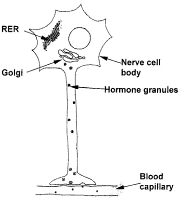
A hormone is a chemical messenger that coordinates the activities of different cells in a multicellular organism. Bayliss and Sterling first used the term in 1904 to de-scribe the actions of secretin, which is a hormone produced by the duodenum to stimulate the flow of pancreatic juice. The classical definition of a hormone is: a chemical substance that is synthesized by particular endocrine glands and then enters the bloodstream to be carried to a target tissue, which has specific recep-torsthat bind it. Other mechanisms of hor-mone delivery also exist (Fig. 1.1). Neuroendocrine hormones are synthe-sized by nervous tissue and carried in the blood to the target tissue; for example, the various releasing factors that are pro-duced in the hypothalamus, which travel to the anterior pituitary via the hypo-thalamus–pituitary blood portal system. Neurocrine hormones are released into the synaptic cleft by neurones that are in contact with the target cells. Paracrine hor-mones diffuse to neighbouring cells, while autocrinehormones feed back on the cell of origin in a form of self-regulation. At the other extreme, pheromones are produced by one animal and released into the environ-ment to be received by other animals (see Section 6.2).
Why are hormones necessary?
Hormones are involved in maintaining homeostasis – consistency of the internal environment that is maintained for the bene-fit of the whole organism. Homeostasis was first recognized by Claude Bernard in the 19th century, who noted that the internal environment (i.e. the fluid bathing cells) had to be regulated independently of external environment. Being able to regulate and maintain its internal environment gives the animal freedom from changes in the external
1 ©CAB International 2003.Applied Animal Endocrinology(E.J. Squires)
1
Hormone and Receptor
Structure and Function
Fig. 1.1.Mechanisms of hormone delivery.
Endocrine/neuroendocrine
Paracrine
Autocrine Blood stream
Target tissue Endocrine
gland
environment, allowing it to live in changing or harsh environments. However, there are metabolic costs associated with maintaining homeostasis. For example, maintenance of a constant body temperature allows animals to function in cold environments, while cold-blooded animals (poikilotherms) only func-tion during warm temperatures. The added energy costs of maintaining deep body tem-perature above that of the environment means that warm-blooded animals have a higher energy requirement for maintenance than do poikilotherms.
Homeostasis is maintained by negative feedback.For example, an endocrine tissue produces a hormone that affects the produc-tion of a metabolite by the target tissue. The metabolite then interacts with the endocrine gland to reduce the production of the hor-mone. This forms a cyclic system in which the levels of the metabolite are maintained at a
particular level. The set point of the system can be altered to affect the levels of the metabolite by altering the sensitivity of the target tissue to the hormone or the sensitivity of the endocrine gland to negative feedback from the metabolite (Fig. 1.2).
In addition to maintaining homeostasis, hormones can also be used to drive change in an organism. In this case, levels of hormone increase to some peak, and this occurs by positive feedback.Positive feedback ampli-fies the response, so the tissue must be desen-sitized or turned over to stop the response. An example of this response is the surge of luteinizing hormone (LH) that leads to ovula-tion (see Secovula-tion 5.1). LH produced by the pituitary gland stimulates the developing ovarian follicle to produce oestrogen, which stimulates the hypothalamus to produce gonadotrophin releasing hormone and increase LH production by the pituitary. This produces a surge of LH, which decreases only after the follicle ovulates (Fig. 1.3).
How do hormones function?
Hormones cause a trigger effect to modulate the activity of the target tissue. The effects of hormones are seen long after levels of the hormone return to basal values. In contrast, nervous signals are short lasting and more immediate. However, nervous signals can regulate hormone production as well. Hormones are present in trace amounts in plasma, usually ranging from 10–9 to 10–6g ml–1. They are present at all times in order to maintain receptors in the target tis-sue and keep the tistis-sue primed for a Fig. 1.2.Feedback system to regulate hormone
production.
Fig. 1.3.Positive feedback system leading to the LH surge and ovulation.
Hormone
Metabolite Endocrine
gland
Target tissue
LH
Ovulation
Hypothalamus
Oestrogen LH (pituitary)
response. Hormones are secreted in variable amounts according to need, and there is a constant turnover by inactivation and excre-tion of the hormone.
The combined effects of more than one hormone on a biological response can occur in a number of different ways (Fig. 1.4). The actions of different hormones are concertedor additive if they cause the same response and the combined effect of the hormones is simply the sum of the separate actions of the individ-ual hormones. This suggests that the two hor-mones act by different mechanisms. In some cases, two hormones can cause the same effect but the effects due to the different hormones are non-additive. This implies that the two hormones may act by the same common mech-anism. The effects of two different hormones aresynergisticwhen the combined effect of the two hormones together is more than the sum of the separate effects of the individual hor-mones. Some hormones, for example steroid hormones and thyroid hormones, can have a
permissiveaction and have no effect on their own but must be present for another hormone to have an effect. A permissive hormone could act by increasing the number of receptors or affecting the activity of the second messenger system for the second hormone. For example, oestradiol has a permissive action for terone by inducing the expression of proges-terone receptors in the oviduct.
What effects are due to hormones?
Hormones cause changes in cellular metabo-lism, but they do not make a cell do some-thing it was not previously capable of. Hormones do not directly cause changes in gene structure but can activate genes to influ-encegene expressionand ultimately protein synthesis. Hormones can alter catalytic rates of enzymes, by mechanisms such as the phosphorylation and dephosphorylation of proteins. Hormones can also alter membrane permeability to affect transport processes Hormone and Receptor Structure and Function 3
Fig. 1.4.Concerted, non-additive, synergistic and permissive action of hormones.
Concerted R e sp o n se s R e sp o n se s R e sp o n se s R e sp o n se s Synergistic Permissive Non-additive
A B A & B A B A & B
A B A & B
A B A & B
and ion movements, alter muscle contraction, exocrine secretion and water permeability.
These general mechanisms can cause a variety of effects in the animal. Hormones can:
• cause morphological changes, such as the differences in body shape between males and females;
• act as mitogens to accelerate cell division or alter gene expression to trigger differen-tiation of cells;
• stimulate the overall rate of protein syn-thesis or the synsyn-thesis of specific proteins; • be involved in stimulating smooth muscle
contraction; for example, oxytocin stimu-lates contraction of the myoepithelium in the mammary gland for milk ejection; • affect exocrine secretions; for example,
secretin, a peptide hormone from intestinal mucosa, stimulates pancreatic secretions; • control endocrine secretions, and a number
of trophic hormones from the anterior pituitary can stimulate or inhibit hormone secretion from target organs;
• regulate ion movements across mem-branes and control permeability to water; for example, antidiuretic hormone (ADH, vasopressin) increases water reabsorption by the kidney; and
• have a dramatic effect on behaviour, such as sex-related behavioural characteristics, maternal behaviour, nesting activity and broodiness (see Chapter 6).
How is hormone action selective?
The method of hormone delivery to the target cells and the presence of specific receptors in
the target cells determine the selectivity of hormone action. For example, the hypophy-seal–portal system linking the hypothalamus to the pituitary delivers releasing hormones from the hypothalamus directly to the target cells in the anterior pituitary. Smaller quanti-ties of hormone are needed since there is less dilution of the hormone in selective delivery systems compared to hormones that reach their target via the peripheral circulation. Many hormones are linked to carrier proteins in the blood, which stabilize the hormone and increase its half-life in the circulation. For example, sex hormone binding globulin is synthesized in the liver and binds testos-terone and oestradiol in the circulation with a high affinity. However, the main factor that determines the sensitivity of a particular tis-sue to a hormone is whether or not the tistis-sue contains the specific receptor for the hormone – the tissue will not respond to the hormone unless it has enough of the specific receptor.
Receptorsare specific proteins present in target cells that bind a particular hormone and initiate a response. Receptors are nor-mally present in small numbers (10,000 mol-ecules per cell). There are two general types, cell-surface receptors (Fig. 1.5) and intracellu-lar receptors (Fig. 1.6). Peptide and protein hormones generally do not enter the cell, but interact with cell-surface receptors. For some cell-surface receptors, a second messenger system is needed to transmit the hormone response signal from the outside to the inside of the cell. This involves the activation of a protein kinase, which phosphorylates spe-cific proteins within the cell to alter their function. Steroid hormones and thyroid
hor-Fig. 1.5.Action of hormones via cell-surface receptors.
Cell membrane
Hormone
Protein substrate phosphorylated
Cellular response Active
mones enter the cell to interact with intracel-lular receptors and regulate gene expression.
Types of hormones
The major structural groups of hormones are:
• steroids;
• proteins, polypeptides and glycoproteins; • amino-acid derivatives (especially
deriva-tives of tyrosine); and
• fatty acids and derivatives, such as prostaglandins.
The structures of some non-protein hor-mones are given in Fig. 1.7.
Location of endocrine glands
The location of the key endocrine glands is given in Fig. 1.8. Table 1.1 lists the hormones produced by these glands and their func-tions. Applications involving many of these hormones are covered in this text and the rel-evant sections are listed in Table 1.1.
1.2 Synthesis, Release and
Metabolism of Hormones
Synthesis of protein hormones
Peptide and protein hormonesconsist of a linear chain of amino acids. As with any pro-tein, the specific sequence of the different amino acids determines the primary
struc-ture and nastruc-ture of the protein. The amino-acid sequence information for a protein is contained in the sequence of bases (A,C,G,T) in the coding region of the gene that codes for the protein. A three-base sequence codes for one amino acid; this is known as the genetic code. This code is copied from DNA into mRNA by transcription and the mRNA is used to direct protein synthesis by the process of translation (Fig. 1.9).
Signal peptides are short sequences of 15–30 hydrophobic amino acids located at the amino-terminal (beginning) of proteins. The presence of a signal sequence (S) directs the newly synthesized protein into the endoplas-mic reticulum and then to export from the cell. Other proteins enter the cytosol and from there are directed to the mitochondria (M) or nucleus (N), or to other sites within the cell. Proteins move between the various compartments by vesicular transport. The uptake of proteins by particular vesicles is controlled by the sorting signal sequences in the proteins (Fig. 1.10). A program (SignalP) for identifying signal peptides and their cleavage sites has been described by Nielsen
et al. (1997) and can be accessed online (http://www.cbs.dtu.dk/).
Newly synthesized protein hormones containing signal sequences are known as prehormones. Some peptide hormones are synthesized as part of a larger precursor, called a prohormone. Examples of prohor-Hormone and Receptor Structure and Function 5
Fig. 1.6.Action of hormones via intracellular receptors.
Protein synthesis
Nuclear
membrane DNA
responsive gene
Cellular response Hormone
mRNA Cell
membrane
Fig. 1.7.Structures of representative non-protein hormones.
mones include proparathyroid hormone, the precursor of parathyroid hormone and pro-insulin, which is the precursor of insulin. Pro-opiocortin is the precursor of several trophic peptide hormones produced in the anterior pituitary. The newly synthesized prohor-mone with a signal peptide is known as a preprohormone(Fig. 1.11).
A number of hormones, including thy-roid stimulating hormone (TSH), follicle stimulating hormone (FSH) and LH, have
sugar units attached to the amino-acid side-chains, and are known as glycoproteins. After protein synthesis, the preprohormone moves from the endoplasmic reticulum to the Golgi apparatus, where sugar residues are attached to asparagine, serine and other amino-acid side-chains in a process called glycosylation. These sugar units can form complex branched chains. From the Golgi apparatus, the proteins are packaged into secretory vesicles and the active hormone is generated by cleavage of the prohormone sequences. The secretory granules fuse with the plasma membrane to release their con-tents by exocytosis when the cell is stimulat-ed. For more information on the mechanisms of protein secretion, see the review by Blázquez and Shennan (2000).
Synthesis of steroid hormones
Steroid hormones are produced in the gonads and the adrenal gland. The gonadal Hormone and Receptor Structure and Function 7
Fig. 1.9.Transcription of DNA to RNA and transla-tion of RNA into protein.
Fig. 1.8.The location of key endocrine glands in cattle: 1, pineal; 2, hypothalamus; 3, pituitary; 4, thyroid; 5, parathyroid; 6, pancreas; 7, adrenal; 8, kidney; 9, ovary (testis in males).
Fig. 1.10.Role of signal peptides in directing the movement of proteins within cells. A typical signal sequence (S) is: M-M-S-F-V-S-L-L-L-V-G-I-L-F-W-A-T-E-A-E-Q-L-T-K-C-E-V-F-Q- (a patch of hydropho-bic amino acids is underlined). The signal (M) for importing into the mitochondria is: M-L-S-L-R-Q-S-I-R-F-F-K-R-A-T-R-T-L-C-S-S-R-Y-L-L-. The signal (N) for importing into the nucleus is: P-P-K-K-K-R-K-V-.
Endoplasmic reticulum
Cell surface
steroids include the progestins, oestrogens andandrogens. Progesterone is a major prog-estin, which prepares the lining of the uterus for implanting of the ovum and is involved in the maintenance of pregnancy. Oestrogens, such as oestradiol, are involved in the devel-opment of female secondary sex characteris-tics and in the ovarian cycle. The androgens are involved in the development of male sec-ondary sex characteristics. Testosterone is a major androgen. The adrenal cortex produces glucocorticoids and mineralocorticoids. Cortisol, a major glucocorticoid, promotes
gluconeogenesis and fat and protein degra-dation. Aldosterone, a major mineralocorti-coid, increases absorption of sodium, chloride and bicarbonate by the kidney to increase blood volume and blood pressure.
The synthesis of steroid hormones occurs on the smooth endoplasmic reticulum and in the adrenal mitochondria. Cholesterol is the precursor of all steroid hormones and is present as low-density lipoprotein (LDL) in plasma. Many of the steps in the biosynthesis of steroids involve an electron transport chain in which cytochrome P450 is the termi-Table 1.1.Summary of hormones produced by various endocrine glands and their functions.
Relevant book Endocrine glands Hormones produced Physiological response sections
Hypothalamus TRH TSH and PRL by anterior pituitary See Section 1.4 for
GnRH LH and FSH interaction between
CRH ACTH, β-endorphin, stress hypothalamus and
GHRH and GH-RIH GH pituitary
PRF and PIF PRL MRF and MIF MSH
Anterior pituitary GH Somatomedin by liver Section 3.4 (adenohypophysis) PRL Mammary gland/lactogenesis Section 4.1
TSH Thyroid hormone Section 3.6
FSH E2, follicular growth/spermatogenesis Sections 4.2, 5.1 LH E2and P4, ovulation/androgen Section 5.1 ACTH Adrenal steroids Sections 3.12, 6.3
MSH Melanogenesis
β-endorphin Analgesic
Posterior pituitary Oxytocin Milk ejection Section 1.4 (neurohypophysis) Vasopressin Antidiuretic hormone
Pineal Melatonin Seasonality, gonad function Section 5.1 Parathyroid PTH Calcium and phosphorus metabolism Sections 4.1, 4.2 Thyroid T4and T3 Metabolic rate Section 3.6 Adrenal cortex Cortisol, corticosterone, Carbohydrate metabolism, Sections 3.12, 6.3 (outer part) aldosterone sodium retention
Adrenal medulla (Nor)Epinephrine Alarm reactions Sections 3.5, 6.3 Gonads Androgens, oestrogens Sexual development/behaviour Sections 3.2, 3.3,
Progestins Pregnancy 4.1, 5.1, 5.2
Inhibin FSH
Relaxin/oxytocin Parturition
Pancreas Insulin, glucagon Blood glucose Section 3.11 Gastrointestinal Gastrin, GIP, secretin HCl and bicarbonate Section 3.9
tract CCK Pancreatic enzymes
Motilin, neurotensin Gastric activity
VIP Blood flow
Kidney Erythropoietin Blood cell formation
nal electron acceptor and carries out hydrox-ylation reactions. The overall scheme is shown in Fig. 1.12.
The conversion of cholesterol to preg-nenolone (Fig. 1.13) involves removal of the C6side chain from cholesterol by hydroxyla-tion at C20and C22and cleavage of this bond by desmolase (P450 side-chain cleavage). This step occurs in adrenal mitochondria and is stimulated by adrenocorticotrophic hor-mone (ACTH).
Pregnenolone is then converted to prog-esterone by oxidation of the 3-hydroxy to a 3-keto group and isomerization of the ∆5 double bond to a ∆4 double bond. Progesterone is converted to cortisol by hydroxylation at C17, C21 and C11. Progesterone is converted to aldosterone
by hydroxylation at C21and C11, and oxida-tion of the C18 methyl to an aldehyde (Fig. 1.14).
Progesterone is converted into andro-gens by hydroxylation at C17and cleavage of the side-chain to form androstenedione (an androgen). The 17-keto group is reduced to a hydroxyl to form testosterone. Androgens are converted into oestrogens by loss of the C19 methyl group and aromatization of the A ring. The formation of oestrogens from androgens is catalysed by the aromatase enzyme CYParom (CYP19) (Fig. 1.15).
Synthesis of eicosanoids
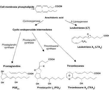
The eicosanoid hormones include prostaglandins, prostacyclins, thrombox-anesandleukotrienes. They are locally pro-duced within cell membranes and have autocrine and paracrine effects. They stimu-late inflammation, regustimu-late blood flow and blood pressure, affect ion transport and mod-ulate synaptic transmission. They are synthe-sized from 20 carbon fatty acids, such as Hormone and Receptor Structure and Function 9
Fig. 1.11.Structure of insulin illustrating the sig-nal peptide and pro-sequence.
Fig. 1.13.Conversion of cholesterol to pregnenolone.
arachidonic acid (20:4) derived from mem-brane lipids (Fig. 1.16).
The enzyme cyclooxygenase (COX) catalyses the first step in the conversion of
arachidonate to prostaglandins and throm-boxanes. Non-steroidal anti-inflammatory drugs (NSAIDs), such as aspirin, ibuprofen and acetaminophen, inhibit COX and reduce Fig. 1.14.Metabolism of pregnenolone to aldosterone.
the production of prostaglandins and throm-boxanes. Prostaglandin E2 (PGE2) and F2α (PGF2α) control vascular smooth muscle activity. Prostaglandin I2 (PGI2) is produced by the blood vessel wall and is the most potent natural inhibitor of blood platelet aggregation. Thromboxanes such as TXA2are produced by thrombocytes (platelets) and are involved in the formation of blood clots and the regulation of blood flow to the clot. Leukotrienes are made by leukocytes and are extremely potent in causing vasocontraction and inducing vascular permeability.
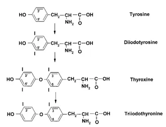
Synthesis of thyroid hormones
Synthesis of thyroid hormonesoccurs in the thyroid gland. The synthesis is stimulated by thyrotrophin (TSH) released from the anteri-or pituitary. Thyrotrophin is released in response to thyrotrophin releasing hormone produced from the hypothalamus. Thyroid
hormones are synthesized by iodination of tyrosine residues in the thyroglobulin pro-tein. Proteases in lysosomes degrade thy-roglobulin to release triiodothyronine (T3) and thyroxine (T4) (Fig. 1.17).
Hormone release
[image:30.530.83.449.99.403.2]Steroids are not stored but released immedi-ately to diffuse out of the cell. Protein and peptide hormones are stored in granules within the gland and are released in response to various stimuli. Trophic hormones can stimulate hormone release: for example TSH, which stimulates the release of thyroxine. The trophic hormones FSH and LH stimulate the synthesis and release of gonadal steroids, while ACTH stimulates the synthesis and release of adrenal steroids (see Section 1.4). Hormones can be released in response to nervous stimuli from environmental cues such as light, smell, sound and temperature. Hormone and Receptor Structure and Function 11
Fig. 1.16.Synthesis of eicosanoids.
Arachidonic acid
Thisneuroendocrine transductionillustrates the integration of the nervous and endocrine systems. Hormones are also released in response to levels of various metabolites. For example, intracellular glucose levels control glucagon and insulin secretion, amino acids stimulate somatotrophin release and increase uptake of amino acids, while extracellular Ca2+regulates parathyroid hormone and cal-citonin secreting cells. These effects are all examples of stimulus–response coupling.
Metabolism and excretion of hormones
Hormones must be metabolized rapidly and removed so that feedback mechanisms can operate and hormones can regulate cellular functions. Removal or inactivation follows exponential decay kinetics (Fig. 1.18). The half-life of the hormones in the circulation is a measure of the longevity of hormone action. Many synthetic hormones and hor-mone analogues are designed to have a longer half-life and thus be effective for longer periods of time than natural hor-mones.
Peptide hormones are degraded by
[image:31.530.101.430.101.352.2]pep-tidases, such as the cathepsins in lysosomes, which split the peptide bonds in the mol-ecule. Exopeptidases degrade protein from the carboxy-terminal end or the amino-termi-nal end. Endopeptidases,such as trypsin and chymotrypsin, degrade the protein at internal sites with some specificity. Trypsin hydro-lyses peptide bonds where the carboxyl group is from lysine or arginine, while for chymotrypsin the carboxyl group in the pep-tide bond comes from phenylalanine, trypto-Fig. 1.17.Synthesis of thyroid hormones.
Fig. 1.18.Inactivation of hormones follows expo-nential decay.
H
o
rmo
n
e
co
n
ce
n
tra
ti
o
[image:31.530.272.448.524.661.2]phan or tyrosine. Deamination or reduction of disulphide bonds (e.g. insulin) can also inactivate proteins. This occurs in kidney, liver and in target cell lysosomes.
Steroid hormones are bound to protein carriers in blood, such as serum albumin or steroid-binding globulin, which are necessary since steroids are lipophilic. Binding to protein carriers also increases the half-life of steroids. Physiologically, only 5–10% of the hormone is present in the free or unbound form.
Steroids are degraded by a two-phase process in the liver and in the kidney. This process inactivates the steroids and makes them more water soluble for excretion. In phase one, enzymes such as cytochrome P450 (CYP) add functional groups such as hydroxyl groups. These metabolites are then conjugated to glucuronic acid or sulphates by transferase enzymes (Fig. 1.19). These more water-soluble metabolites are excreted by the kidney in the urine or by the liver in the bile salts.
1.3 Receptors and Hormone Action
Hormones interact with receptors located either on the cell surface or inside the cell to initiate their effects on the target tissue. Binding of hormones to cell-surface receptors activates intracellular enzyme systems to alter cell function. Hormones that cross the cell membrane act by binding to intracellular receptors. The hormone–receptor complex then interacts with DNA to affect expression of specific genes.
Extracellular receptors
Extracellular receptorsare large macromol-ecules located on the outer surface of the
plasma membrane in target tissues. For example, the insulin receptor has a molecular mass of 200–400 kDa, consisting of two α-subunits of 130 kDa and two β-subunits of 90 kDa linked by disulphide bonds. Usually, there are separate receptors for each hormone and the function of the cell (i.e. the cell type) dictates whether a particular receptor will be present on a cell and the number of receptors present. Experimental evidence that a hor-mone receptor is located on the cell surface includes:
1.Demonstrating that antibodies against the receptor can block hormone action;
2.Limited proteolysis of intact cells would be expected to destroy the receptor and remove the hormone response;
3.Coupling the hormone to a large molecule that cannot enter the cell and demonstrating that the effect of the hormone is still present; and
4.Demonstrating that the receptor is present in a plasma membrane preparation produced by subcellular fractionation (100,000 g pellet).
Hydrophobic regions on the receptor protein interact with lipid in the membrane. The receptor can be solubilized with detergents and purified by affinity chromatography using the hormone bound to a column matrix. Receptors can be glycoproteins and contain carbohydrate residues.Experimental toolsto demonstrate this are:
1.Treat the receptor preparation with neur-aminidase or β-galactosidase to remove the sugar residues. This inhibits binding of the hormone.
2.Concanavalin A (ConA; a protein from jack bean that binds to a D-glucosyl residues) can Hormone and Receptor Structure and Function 13
be used to inhibit hormone binding. In addi-tion, ConA can be used for affinity chro-matography of glycoproteins (see Section 2.2, Chemical assays).
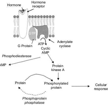
Second messenger systems
Some hormones interact with a cell-surface receptorand stimulate the synthesis of intra-cellular second messengercompounds. The hormone does not enter the cell to elicit a response but stimulates one of two main pathways:
1. The adenylate cyclase–cAMP–protein kinase A pathway or the related guanylate cyclase–cGMP-dependent protein kinase pathway; and
2. The calcium-dependent phospholipase C–protein kinase C pathway.
In the first system, hormone binding to the receptor activates the enzyme adenylate cyclase or guanylate cyclase, which synthe-size either cAMP or cGMP. These second messengers activate protein kinase A. In the second system, binding of the hormone to the receptor activates phospholipase C, which splits phosphatidylinositol in the cell mem-brane to inositol phosphate and diacylgly-cerol. The inositol phosphate increases levels
of intracellular calcium, which, together with the diacylglycerol, activates protein kinase C.
Both protein kinase A and protein kinase C can phosphorylate and activate various intracellular proteins to alter cellular metabo-lism. These proteins are inactivated by removing the phosphate using the enzyme phosphoprotein phosphatase.
Adenylate cyclase–cAMP–protein kinase A pathway
[image:33.530.173.395.458.673.2]The enzyme adenylate cyclase catalyses the formation of cAMP from ATP. cAMP acti-vates protein kinases which phosphorylate intracellular proteins to alter their activity (Fig. 1.20). The formation of cAMP is an amplification step that increases the effective hormone concentration, since one adenylate cyclase enzyme catalyses the formation of many cAMP molecules. The enzyme phos-phodiesterase degrades cAMP to AMP by the following reaction:
ATP → cAMP → AMP
Adenylate Phosphodiesterase cyclase
Several properties of cAMP make it suitable as a second messenger. It is derived from ATP
Fig. 1.20.Cyclic AMP second messenger system.
Adenylate cyclase
Protein kinase A AMP
Protein G Protein
Cellular response Phosphorylated
protein
Phosphoprotein phosphatase Phosphodiesterase
ATP Cyclic
AMP Hormone Hormonereceptor
but is chemically stable. ATP is ubiquitous and cAMP is formed from it in a single reac-tion. Since cAMP is not a metabolic precursor, but an allosteric regulator, it can be controlled independently of metabolism. cAMP is a small and easily diffusable molecule and it has a number of functional groups that allow specific binding to regulatory subunits of protein kinases.
Experimentally, the involvement of cAMP as a second messenger can be deter-mined if physiological levels of hormone increase cAMP in cells and cAMP production precedes the physiological effect. The hor-mone should also stimulate adenylate cyclase activity in broken cells. Treatment with exogenous cAMP or its analogues, such as dibutyryl cAMP and 8-bromo cAMP (Fig. 1.21) should produce the hormone response. Phosphodiesterase inhibitors, such as theo-phylline, caffeine or isobutylmethylxanthine (IBMX), will decrease cAMP clearance and potentiate the response. Adenylate cyclase can be activated by treatment with the diter-pene, forskolin (Fig. 1.22), which binds direct-ly with the catadirect-lytic subunit to permanentdirect-ly activate it.
A number of different hormones act via the cAMP second messenger system (Table 1.2). The substrates for cyclic AMP-depend-ent protein kinases include triglyceride lipase, which is involved in the regulation of lipolysis, phosphorylase bkinase in the regu-lation of glycogenolysis, cholesterol ester hydrolase in the regulation of steroidogenesis and fructose 1,6-diphosphatase in the regula-tion of gluconeogenesis. These latter enzymes are all activated by phosphoryla-tion. Enzymes that are inactivated by phos-phorylation include pyruvate kinase in the regulation of glycolysis and gluconeogenesis, glycogen synthase in the regulation of glyco-gen synthesis, and 3-hydroxy-3-methylglu-taryl-CoA reductase in the regulation of cholesterol biosynthesis.
Guanyl cyclase–cGMP-dependent protein kinase pathway
The cGMP system is similar to the cAMP sys-tem, but may act in opposition to cAMP. This is known as the yin–yang hypothesis. For example, activation of the cAMP-dependent kinases results in smooth muscle relaxation, Hormone and Receptor Structure and Function 15
while activation of the cGMP-dependent kinases results in smooth muscle contraction. Levels of cGMP are normally 10–50 times lower than those of cAMP.
GENOMIC ACTIONS OF CAMP. Activating pro-tein kinase A and subsequent phosphoryla-tion of intracellular proteins can cause immediate cellular responses, such as modifi-cation of metabolic pathways and regulation of ion flows and muscle contraction. However, cAMP can also have effects on gene transcription by protein kinase A activation of the cAMP-responsive-element binding protein (CREB), or modification of the struc-tural proteins in chromatin. Activated CREB binds to specific cAMP-responsive elements in the regulatory regions of certain genes to activate gene expression.
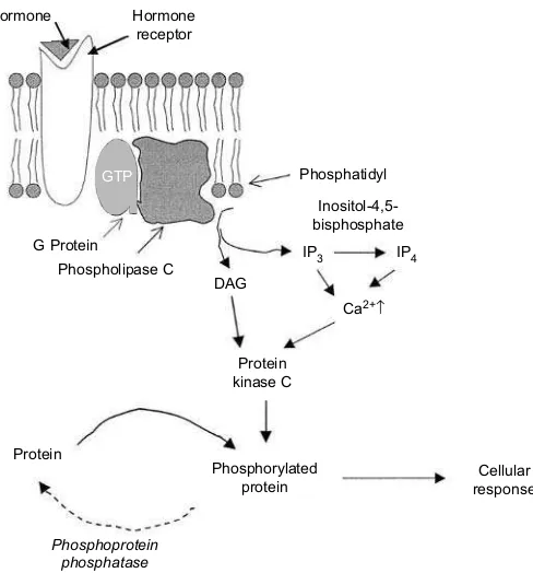
Calcium-dependent phospholipase C–protein kinase C system
The primary intracellular effector in this pathway is calcium, which activates a calci-um-dependent protein kinase C. Hormone binding activates phospholipase C, which catalyses the hydrolysis of
phosphatidylinos-itol-4,5-bisphosphate to produces inositol-1,4,5-phosphate (IP3) and diacylglycerol (DAG) (Fig. 1.23). Inositol-1,4,5-phosphate and its metabolite, inositol-1,3,4,5-tetra kisphosphate (IP4), increase intracellular Ca2+ by activating calcium channels at the endo-plasmic reticulum and cell surface. Diacylglycerol activates protein kinase C by increasing its affinity for Ca2+, and protein kinase C then phosphorylates cellular pro-teins to regulate their activity (Fig. 1.24).
In both of these systems, the receptor is discrete from the enzyme system that it acti-vates; both ‘float’ in the cell membrane lipid. The receptor interacts with the adenylate cyclase or phospholipase C via an intermedi-ate G-protein. The G-protein is activintermedi-ated by binding GTP and inactivated when the GTP-ase activity converts the GTP to GDP. G-protein function can be studied using a non-hydrolysable form of GTP or cholera toxin, both of which inhibit the GTP-ase activity. The Ras oncogene codes for a permanently active G-protein, which might explain its role in the development of cancer. G-proteins act to couple cell-surface receptors for hormones, neurotransmitters, odorants and photons of light to effector molecules such as ion chan-nels or enzymes that generate second mes-senger molecules.
An example of the stimulus response coupling due to this system is the effect of gonadotrophin releasing hormone (GnRH). GnRH is produced by the hypothalamus and causes LH and FSH release by the anterior pituitary. GnRH increases cellular Ca2+levels and affects inositol metabolism. LH release is Ca2+dependent and an increase in intracellu-lar Ca2+causes a release of LH.
Calcium also binds to calmodulin to form an active complex. Calmodulin is a heat-stable globular protein of molecular Fig. 1.22.Forskolin, an activator of adenylate
cyclase.
Table 1.2.Hormones that act via the adenylate cyclase–cAMP–protein kinase A pathway.
Glucagon Chorionic gonadotrophin Vasopressin Parathyroid hormone Thyrotrophin (TSH) Calcitonin
mass 16 kDa and is a calcium-dependent reg-ulatory protein found in all eukaryotic cells. It controls intracellular Ca2+ and binds four Ca2+ions per molecule to form an active com-plex. This complex acts as an allosteric regula-tor of protein kinase C and other enzymes. It also controls the activity of cellular filamentous organelles (via actin and myosin) responsible for cell motility, exoplasmic flow (hormone
secretion) and chromosome movement. Experimental tools used to determine the involvement of the calcium-dependent phopholipase C–protein kinase C system are:
1. Increase intracellular Ca2+ levels using Ca2+-selective ionophores (A23187) or lipo-somes loaded with Ca2+.
2. Decrease intracellular Ca2+ by chelating Hormone and Receptor Structure and Function 17
[image:36.530.174.418.392.654.2]Fig. 1.24.Calcium-dependent protein kinase C second messenger system. Fig. 1.23.Action of phospholipase C.
Hormone
Protein
Ca2+↑
Cellular response Phosphorylated
protein
Phosphoprotein phosphatase
Protein kinase C G Protein
Phospholipase C DAG Hormone
receptor
Phosphatidyl
Inositol-4,5-bisphosphate
with EGTA, using Ca2+-channel blockers, or inorganic Ca2+antagonist (La3+).
3. Use phorbol esters (tetradecanoylphorbol acetate (TPA)), which resemble diacylglycerol (Fig. 1.25, see structure inside dashed lines), to activate protein kinase C.
4.Inhibit phospholipase C with U73122; the inactive analogue, U73343, is used as a posi-tive control.
INTERACTION OF CAMP AND CA2+ PATHWAYS. There is a considerable amount of ‘cross-talk’ between the different secondary messenger systems. Ca2+ binds to calmodulin and this complex can bind to phosphodiesterase to activate it and decrease cAMP levels. Protein kinase A, which is activated by cAMP, can phosphorylate some Ca2+ channels and pumps and alter their activity to affect intra-cellular calcium levels. Protein kinase C can be phosphorylated by protein kinase A to change its activity. Protein kinase C and pro-tein kinase A can phosphorylate different sites on the same protein, so that its activity is regulated by both cAMP and Ca2+.
Tyrosine kinase receptors: catalytic receptors
The tyrosine kinase receptors do not use a second messenger system to activate a sepa-rate protein kinase, but have a kinase domain
as part of the receptor structure. The activat-ed receptor phosphorylates tyrosine residues in its kinase domain and can then phospho-rylate other proteins, and is thus called a tyrosine kinase receptor. The receptor con-sists of a transmembrane domain, an extra-cellular domain for hormone recognition and a cytoplasmic domain that transmits the reg-ulatory signals and contains ATP binding sites. The cytoplasmic domain has a C-termi-nal tail with autophosphorylation sites. The phosphorylated receptor acts as a kinase enzyme and phosphorylates substrates. These phosphorylated substrates transmit several regulatory signals into the cell.
There are three main classes of tyrosine kinase receptors (Fig. 1.26). The class I receptor (e.g. epidermal growth factor (EGF) receptor) is a monomeric transmembrane protein with intracellular and extracellular domains on the same molecule. The extracellular domain con-tains two cysteine-rich repeat regions. The class II receptor (e.g. insulin receptor) is a heterotetrameric receptor in which the two α-subunits and the two β-subunits are linked by disulphide bonds. The class III receptor (e.g. receptors for platelet-derived growth fac-tor (PDGF) or nerve growth facfac-tor (NGF)) is a monomeric protein with cysteine residues over the extracellular domain. The intracellu-lar domain has unique amino acid inserts in the middle of the kinase domain.
[image:37.530.79.221.80.262.2]Hormone binding causes dimerization of the monomeric receptors (Fig. 1.27). The kinase domains in the monomers are then phosphorylated and activated by their part-ner. The kinases can then phosphorylate and activate other proteins. The receptors also interact with proteins containing SH2 domains that bind to phosphotyrosines. The amino-acid sequence next to the phospho-tyrosines specifies which protein, containing an SH2domain, can bind to receptor. The SH2 domain protein can be attached to a different enzymatic domain or be a linker molecule that binds to other enzyme molecules that could not normally bind to the receptor. In this way, linker molecules can bind a number of different specific molecules to bring them together to produce the desired biological effect. For example, the appropriate kinase and phosphatase can be held in position so Fig. 1.25.Tetradecanoylphorbol acetate (TPA) or
that a protein (receptor, ion channel, etc.) is activated by the kinase in the presence of the appropriate signalling molecule and deacti-vated by the phosphatase when the
signalling molecule is absent. The role of SH2 domain proteins is described in a popular press article by Scott and Pawson (2000).
Hormone and Receptor Structure and Function 19
Fig. 1.26.Types of tyrosine kinase receptors.
CYTOKINE RECEPTORS. Receptors for cytokines (growth hormone (GH), prolactin, erythro-poietin, interferons and interleukins) do not have intrinsic kinase activity (Fig. 1.28). The GH receptor exists as a monomer when it is not bound to hormone. The binding of hor-mone causes dimerization of the receptor and binding of Janus kinase (JAK) tyrosine kinase, which phosphorylates the receptor to activate it. Phosphotyrosines act as docking sites for intracellular signalling molecules, such as STATs (signal transducers and activa-tors of transcription), which activate various genes.
RECEPTOR SERINE KINASE. Receptor serine kinases include receptors for transforming growth factor-β (TGFβ), activin and inhibin. These peptide growth factors are involved in the control of cell proliferation and differenti-ation. The existence of these receptors was demonstrated by chemical cross-linking of radiolabelled hormone to cell-surface pro-teins on responsive cells.
The binding of hormone results in for-mation of a heterodimer of receptors I and II. Serine residues on RI are phosphorylated by RII to activate the complex (Fig. 1.29).
Termination of hormone action
After hormones interact with receptors, they cluster together, and this triggers vesicu-larization of membrane and endocytosis. The receptors may then be degraded by lyso-mal enzymes or the receptor can be recycled. The hormone at the cell surface can be degraded by serum enzymes. The cyclic nucleotides are degraded by phosphodi-esterases and the phosphorylated proteins are dephosphorylated by phosphoprotein phosphorylase.
Intracellular receptors
Steroid hormone receptors move between the nucleus and cytoplasm and, in the absence of hormone, are bound to the 90 kDa heat-shock protein complex (Hsp90). (TR, RAR and VDR do not bind Hsp90.) Binding of the hormone to the recep-tor results in release of the Hsp90 com-plex and translocation of the hormone– receptor complex to the nucleus. A dimer of the hormone–receptor complex then interacts
with hormone-responsive elements on specific genes to affect DNA transcrip-tion. This exposes template sites on DNA, either directly or by influencing pre-existing repressor molecules, to increase the initiation sites for RNA polymerase and increase tran-scription (Fig. 1.30). These actions of steroid hormones occur over a much longer time frame (hours) compared to peptide hor-mones.
Hormone and Receptor Structure and Function 21
Fig. 1.29.Mechanism of action of receptor serine kinases.
Fig. 1.30.Mechanism of action of steroid hormones.
Cytoplasm Nucleus
DNA
Acceptor site Uptake
Hormone-dependent
genes Biological
response
Heat-shock protein
Hormone-dependent RNA
Protein synthesis
Translocation Binding and
Steroid hormones can also affect the extent of mRNA degradation. The effects of steroid hormones can be determined experi-mentallyby:
1.Measuring steady-state levels of mRNA by Northern blotting. Levels would increase with increased synthesis or decreased degra-dation of mRNA.
2.In order to separate synthesis from degra-dation, a nuclear run-on assay can be used to measure the rate of mRNA synthesis. 3.Actinomycin D (an inhibitor of RNA tran-scription) and puromycin (an inhibitor of protein synthesis) can be used to inhibit the effects of steroid hormones.
Steroid hormones can affect the response to other hormones. This can be through synthe-sis of receptors or protein kinases to increase hormone response or phosphoprotein phos-phatases, which are antagonistic to cyclic nucleotide actions.
Structural and functional domains of nuclear receptors
Nuclear receptors have common structural domains involved in DNA binding and
hor-mone binding (Fig. 1.31). Other regions are involved in dimerization of the receptor and translocation of the receptor to the nucleus.
The ligand-binding domain is the region where the hormone binds