Cognitive Functioning, Cortisol Release, and Symptom
Severity in Patients with Schizophrenia
Deborah J. Walder, Elaine F. Walker, and Richard J. Lewine
Background:
There is substantial evidence of
dysregula-tion of cortisol secredysregula-tion, hippocampal abnormalities, and
memory deficits in schizophrenia and other psychotic
disorders. Research also suggests that cortisol secretion
augments dopaminergic activity, which may result in
increased symptom expression in this clinical population.
Methods:
We examined the relations among cortisol
release, cognitive performance, and psychotic
symptom-atology. Subjects were 18 adults with schizophrenia or
schizoaffective disorder, seven with a nonpsychotic
psy-chiatric disorder, and 15 normal control subjects. Tests of
memory and executive function were administered.
Corti-sol was assayed from multiple saliva samples.
Results:
Findings indicated the following: 1) patients with
psychotic disorders scored below the comparison groups
on the cognitive measures; 2) for the entire sample,
cortisol levels were inversely correlated with performance
on memory and frontal tasks; and 3) among patients,
cortisol levels were positively correlated with ratings of
positive, disorganized, and overall symptom severity, but
not with negative symptoms.
Conclusions:
The present results suggest that
abnormal-ities in the hypothalamic–pituitary–adrenal axis and
hip-pocampal systems play a role in observed cognitive
deficits across populations. Among psychotic patients,
elevated cortisol secretion is linked with greater symptom
severity.
Biol Psychiatry 2000;48:1121–1132 ©
2000
Society of Biological Psychiatry
Key Words:
Cortisol, hippocampus, memory, cognitive,
symptoms, schizophrenia
Introduction
P
atients with psychotic disorders manifest deficits in
short-term verbal and visuospatial memory functions
(Gold et al 1992a, 1992b; Goldberg et al 1990; Hoff et al
1992; Saykin et al 1994; Schmand et al 1992). Paralleling
these findings, research has shown that schizophrenia is
associated with hippocampal volumetric reductions (e.g.,
Bogerts et al 1985; Breier et al 1992; Jeste and Lohr 1989;
Suddath et al 1990; Waldo et al 1994) and cellular
abnormalities (e.g., Akbarian et al 1993; Altshuler et al
1990; Benes et al 1991; Bogerts et al 1990; Jeste and Lohr
1989; Kovelman and Scheibel 1984). In recent studies,
reductions in hippocampal volume have been found in
first-episode schizophrenia patients, suggesting that the
abnormality is not a treatment artifact (Hirayasu et al
1998; Velakoulis et al 1999). This also extends to patients
with other psychotic disorders (Velakoulis et al 1999).
A link between hippocampal morphology and memory
has been established in studies of nonschizophrenic
sam-ples, such that the volume of the hippocampus is inversely
related with memory test performance (Lencz et al 1992;
Starkman et al 1992). This is consistent with the prevailing
theory that the hippocampal system plays a major role in
declarative (or explicit) memory (Eichenbaum et al 1996;
Squire 1987, 1992). A few studies show no significant
relationship between delayed memory and hippocampal
volume in schizophrenia spectrum patients (Colombo et al
1993; Delisi et al 1991; Nestor et al 1993; Torres et al
1997). In contrast, Goldberg et al (1994) found a strong
association between intrapair differences in a parameter of
verbal memory and intrapair differences in left
hippocam-pal volume in monozygotic twins discordant for
schizo-phrenia. Discrepancies in these findings may be due to
variability in the cognitive measures selected to index
memory. It is also noteworthy that Goldberg et al (1994)
focused on the hippocampal volume difference between
affected twins and their healthy monozygotic cotwins.
Thus, there was some control for genetically determined
differences in volume.
In addition to its role in memory functioning, the
hippocampus is assumed to play a role in the regulation of
the hypothalamic–pituitary–adrenal (HPA) axis.
Specifi-cally, it appears that the stimulation of glucocorticoid
receptors in the hippocampus contributes to a negative
feedback system that dampens HPA activity. Animal
studies suggest that prolonged stress and elevations in
corticosteroids can damage the hippocampus (Sapolsky
and McEwen 1986; Sapolsky et al 1990), which may lead
to dysregulation of the HPA axis via a decrease in
glucocorticoid negative feedback. Thus, studies of human
subjects revealing an inverse correlation between cortisol
From the Departments of Psychology (DJW, EFW) and Psychiatry (RJW), Emory University, Atlanta, Georgia.
Address reprint requests to Deborah J. Walder, M.A., Emory University, Depart-ment of Psychology, 532 North Kilgo Circle, Atlanta GA 30322.
Received March 28, 2000; revised August 28, 2000; accepted September 1, 2000.
and hippocampal volume (Rao et al 1989; Starkman et al
1992) are not surprising.
Consistent with the above findings, there is also an
inverse relation between cortisol level and performance
on measures of hippocampal function, particularly
declar-ative memory, in healthy human subjects. This relation has
been shown in experimental studies in which
glucocorti-coids are manipulated and memory decrements ensue
(Kirschbaum et al 1996; Newcomer et al 1994, 1999),
studies in which stress-induced alterations in cortisol are
associated with memory deficits (Lupien et al 1997), and
longitudinal studies of the relation between age-related
declines in cortisol and memory performance (Seeman et
al 1997). It is noteworthy that Kirschbaum et al (1996)
showed that memory performance deficits follow acute,
relatively low-stress exposure (e.g., Trier Social Stress
Test), or what is presumed to be relatively low-dose
increases in cortisol. In contrast, one more recent study
(Newcomer et al 1999) found that similar cognitive
impairments following exogenous glucocorticoid
treat-ment are high-dose specific (e.g., approximating cortisol
exposure during moderate to maximum stress conditions).
Future research aimed at clarifying the specific effects of
low- versus high-dose and acute versus chronic
glucocor-ticoid exposure is warranted.
The precise nature of memory deficits following acute
elevations in cortisol is also unclear. Some studies suggest
impairments in immediate recall following stress-induced
cortisol secretion (Lupien et al 1997), whereas others
suggest a retrieval-specific impairment following
exoge-nous administration of corticosteroids (De Quervain et al
2000). Overall, this body of literature suggests that cortisol
is linked with memory function in two ways: 1) directly,
by acutely disrupting working memory and short-term
recall, and 2) indirectly, through the effects of persistent
cortisol elevations on hippocampal integrity.
To date, only two studies have explored the relation
between glucocorticoids and memory performance in
schizophrenia. One early report (Newcomer et al 1991)
showed an inverse relation between early morning (8:00
AM
) postdexamethasone cortisol levels and auditory verbal
learning deficits in unmedicated schizophrenic patients. In
a more recent experimental study, Newcomer et al (1998)
examined the effects of dexamethasone versus a placebo
on verbal memory performance in schizophrenic patients
over 4 days. The finding of an association between higher
plasma cortisol concentrations (before dexamethasone
treatment) and reduced memory performance in patients
with schizophrenia extended their earlier findings. There
was no relation, however, between postdexamethasone
cortisol levels and recall performance. It is noteworthy that
although several studies evidence elevated basal levels of
cortisol (Walker and Diforio 1997) and
postdexametha-sone nonsuppression (Arana et al 1983; Sharma et al 1988)
in schizophrenia, these findings are not consistently
ob-served in this population.
Previous studies indicate a relation between HPA
activ-ity and symptomatology in schizophrenia. In some,
corti-sol secretion was primarily associated with more severe
positive symptoms (Kaneko et al 1992; Keshavan et al
1989; Rybakowski et al 1991), whereas in others it was
associated with higher ratings of negative symptoms
(Newcomer et al 1991; Tandon et al 1991). It has been
suggested that the relation between cortisol levels and
symptom severity is due to the augmenting effects of
cortisol on dopamine activity (Walker and Diforio 1997).
Findings from several studies revealing a relation of
pre- or postdexamethasone suppression test cortisol levels
with psychotic symptoms suggest that this association is
not affected by level of depression (Schatzberg and
Roth-schild 1988; Sharma et al 1988). Thus, the association of
cortisol with symptom severity is not attributable to the
presence of mood disorder in some psychotic patients.
This study is based on the assumption that heightened
cortisol release is associated with memory deficits in
normal healthy control subjects. Thus, it was hypothesized
that cortisol level would be inversely correlated with
memory performance in patients with psychotic disorders.
In addition, we examined the association of symptom
severity with both cortisol secretion and memory
perfor-mance. Given the evidence that HPA activation augments
dopamine activity (Schatzberg et al 1985), it was predicted
that elevated cortisol levels would be associated with
increased symptom severity. This study is novel in that it
examines the relations among cognitive performance,
symptom expression, and neuroendocrine measures in a
psychiatric population, in the absence of the stress
typi-cally associated with blood sampling procedures or
exog-enously administered pharmacologic agents.
situation diminishes. These factors were considered when
interpreting the findings in this study.
Methods and Materials
Subjects
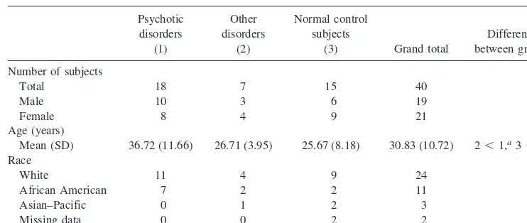
The study samples were 15 normal control (NC) subjects, 18 patients with current or past (in remission) diagnosis of a psychotic disorder (PD), and seven individuals with other psy-chiatric disorders (ODs). Subjects ranged from 18 to 58 years of age, with an overall mean age of 30.8 years (see Table 1 for demographic characteristics). The PD group included patients who met DSM-IV criteria for schizophrenia, including paranoid, disorganized, residual, and undifferentiated type (n 5 12) or schizoaffective disorder (n56). Subjects in the OD group met criteria for affective disorders without psychotic features (e.g., major depressive disorder, history of a major depressive epi-sode), and one subject met criteria for pervasive developmental disorder. Normal control subjects were recruited from the com-munity; all other subjects were recruited from inpatient and outpatient psychiatric services. Fifteen of the 18 subjects with psychotic disorders were being treated with antipsychotics. Some patients refused medication.
Informed consent was obtained from all subjects before their participation in this research study. The study protocol was reviewed and approved by the Human Investigations Committee at Emory University before data collection.
Diagnostic Instruments
DIAGNOSTIC INTERVIEW. The Structured Clinical
Inter-view for DSM-IV, research version (SCID-IV; First et al 1997), the Schedule for Assessment of Positive Symptoms (SAPS; Andreasen 1983), and the Schedule for Assessment of Negative Symptoms (SANS; Andreasen 1981) were administered by trained examiners to all participants at the initial assessment. Diagnostic interviews were videotaped so that consensus diag-noses could be established.
SALIVA SAMPLING FOR THE ASSAY OF CORTISOL. The measurement of salivary cortisol has been purported to be a reliable tool for investigating HPA activity (Kiess et al 1995; Kirschbaum and Hellhammer 1989; Laudat et al 1988). More-over, as pointed out by Weinstein et al (1999), salivary, urinary, and plasma measures of cortisol are highly interrelated, as well as comparable in sensitivity as measures of stress reactivity (Bassett et al 1987; Shipley et al 1992). Finally, the noninvasive nature of saliva sampling yields it a more preferable method for repeated assessments (Baum and Grunberg 1995).
Saliva samples (about 1 mL each) for the assay of cortisol were obtained from subjects three to five times during the assessment. These samples were obtained by asking the subjects to spew saliva into plastic specimen tubes, in which the samples were stored. To maintain uniformity in the portion of the diurnal variation in cortisol excretion represented in the samples, all assessments were conducted at the same time of day. The first sample was obtained at approximately 8:00 AM. Subsequent samples were obtained at approximately 9:00 AM, 10:00 AM, 11:00AM, and 12:00PM. By obtaining multiple measures, it was possible to examine group and individual differences in change, as well as mean levels.
CORTISOL ASSAY. The saliva samples were prepared for
assay according to standard procedures. They were stored at
220°C in a 13– cu ft S/P (Asheville, NC) cryofreezer. In preparation for assay, the samples were rapidly thawed and centrifuged at 300gfor 10 min to remove coagulated protein and other insoluble material. Cortisol was assayed in duplicated 200-mL aliquots of the clear supernatant using materials and procedures provided by Incstar (Stillwater, MN). The assay was performed in tubes coated with an antiserum that shows signif-icant cross-reactivity only with prednisone (83%), 11-deoxycor-tisol (6.4%), cortisone (3.6%), and corticosterone (2.3%). Stan-dards in the range 1–30 ng/mL consisted of the serum stanStan-dards provided with the kit materials diluted with 200 mL of phos-phate-buffered saline. Protein concentrations were equalized in standards and samples by adding cortisol-free serum to the Table 1. Characteristics of Current Sample
Psychotic disorders
(1)
Other disorders
(2)
Normal control subjects
(3) Grand total
Difference between groups
Number of subjects
Total 18 7 15 40
Male 10 3 6 19
Female 8 4 9 21
Age (years)
Mean (SD) 36.72 (11.66) 26.71 (3.95) 25.67 (8.18) 30.83 (10.72) 2,1,a3,1b
Race
White 11 4 9 24
African American 7 2 2 11
Asian–Pacific 0 1 2 3
Missing data 0 0 2 2
samples. For the most recent assays conducted by the lab, the mean coefficients of variation between duplicates and between assays were less than 5%. Relative to the serum standards, the mean recovery of cortisol from saliva has been indistinguishable from 100%.
Neuropsychological Assessment
The neuropsychological measures were selected to yield a comprehensive profile of hippocampal functioning and atten-tional functioning.
HIPPOCAMPAL FUNCTIONING. California Verbal
Learn-ing Test (CVLT; Delis et al 1983)The CVLT is typically used to assess memory deficits associated with hippocampal functioning. It measures recall and recognition of lists containing related words, across several trials.
The composite score derived for this test was an average of the standardized scores on the seven subtests of this measure, including two measures of immediate recall, two measures of short-delay recall (free and cued), two measures of long-delay recall (free and cued), and one measure of long-delay recogni-tion. Higher scores indicate better performance.
Wechsler Memory Scale—Revised (WMS-R; Wechsler 1987)
Subtests of the WMS-R are also frequently used to assess memory deficits associated with hippocampal functioning. The WMS-R scales require recall of stories and paired word associ-ates. Patients with schizophrenia have significant memory im-pairments on the original Wechsler Memory Scale (Goldberg et al 1990) and the WMS-R (Gold et al 1992b). Research findings suggest that memory impairment based on this measure is not solely attributable to general cognitive deterioration in schizo-phrenia (Gold et al 1992b). The four subtests administered from this measure (Logical Memory I, Logical Memory II, Verbal Paired Associates I, and Verbal Paired Associates II) focus on short-term learning, recall, and delayed recall of verbal material. The composite score derived for the WMS-R was an average of the standardized scores on the four subtests, immediate and delayed recall of a story, and immediate and delayed recall of verbal paired associates. Higher scores indicate better performance.
Modified Wisconsin Card Sorting Test (MCST; Nelson 1976)
This test is a simplified version of the Wisconsin Card Sorting Task, revised to reduce ambiguity concerning the sorting princi-ple active at any point in the test. Poor performance on the MCST has been demonstrated in schizophrenic patients (Nelson 1976). The MCST is a measure traditionally used to assess frontal lobe (or executive) functioning. There is evidence, however, suggest-ing that volume reductions in the anterior hippocampal formation may predict neuropsychological deficits in executive functions (Bilder et al 1995). Therefore, given the hypothesized connec-tivity between the frontal and hippocampal brain regions, the MCST was selected as an additional potential indicator of hippocampal-related deficits.
In this test the subject is required to sort a deck of stimulus cards into groups corresponding to the color, form, or number
printed on four key cards before him or her. The subject is not told the correct sorting principle but is informed by the examiner whether or not each card was sorted correctly. The subject must deduce the correct sorting rule based upon the information provided.
Indices derived from this test are total number of correct responses, total number of errors, and number of perseverative errors. The composite score was an average of the standardized scores for the total number of errors and number of perseverative errors. Thus, for total number of correct responses, higher scores indicate better performance. For the composite score, however, higher scores indicate poorer performance.
ATTENTION. Continuous Performance Task—Vigil (CPT-Vigil) (ForThought, Nashua NH) CPT-Vigil is a computerized attention measure typically used to assess vigilance, or mainte-nance of attention, over time. It has been shown to be a valid test of individual differences in vigilance performance (Buchsbaum and Sostek 1980). Because memory involves aspects of attention, this measure was administered to a subgroup of 33 subjects to examine the relation between memory performance and atten-tional functioning. Data were not collected on the remaining seven subjects of the total sample because of technical problems with computer equipment.
This computerized task measures sustained concentration and attention by presenting target stimuli (e.g., letters) on the computer screen one at a time. The “AK” version of the task, which requires the subject to press the space bar on the keyboard in response to a complex target stimulus (e.g., the letterKonly when preceded by the letter A), was used. This stimulus is presented among other randomly presented nontarget stimuli (e.g., other letters of the alphabet presented in an order not ascribing to the rule outlined above).
The test apparatus consisted of a desktop computer monitor and keyboard. The signal stimuli were presented on the computer screen for a duration of 85.3 msec and at an interstimulus interval of 910.3 msec. A total of 100 targets out of 480 total stimuli were presented. The stimulus sequence was randomized. A response to a nontarget stimulus was considered an “error of commission,” and a failure to respond in the presence of a target stimulus was considered an “error of omission.” From these measures, an index of sensitivity (d9) was derived and used in the current analyses. Sensivity (d9) has been shown to have moderate reliability (Buchsbaum and Sostek 1980) and is a measure of attentiveness or discriminability.
Procedures and Time Frame
The order of administration of the tests was the same for all subjects: CVLT (immediate recall), MCST, CVLT (20-min delayed recall), subtests of the WMS-R (immediate recall), subtests of the WMS-R (delayed recall), and CPT-Vigil.
Assessments began at approximately 8:00AM, with a descrip-tion of the study goals and measures, and took roughly 3.5– 4.5 hours to complete. The consent form described the procedures for maintaining confidentiality, the study protocol, and the subject’s right to withdraw from participation at any time.
every hour. The diagnostic interview (SCID-IV), from which SANS and SAPS ratings were determined, followed the initial saliva sampling and was followed by the neuropsychological assessment. All interviewers were trained in conducting the SCID-IV before commencement of this study. The neuropsycho-logical assessment was administered by another trained research assistant, who was blind to the subjects’ interview results.
Subjects were asked to refrain from alcoholic beverages for one day before the assessment. They were also instructed to avoid consumption of caffeinated food and beverages beginning the night before the assessment and to eat a light breakfast before their appointment. Any deviations from the instructions were recorded.
Analysis
CORTISOL. Cortisol levels were measured at times 1, 2, and
3 for all subjects. Cortisol levels at times 4 and 5 were only collected for those subjects for whom the duration of the assessment continued for 3– 4 hours. This was more often the case for subjects belonging to the inpatient and outpatient clinical samples, as compared with NC subjects. Because 42.5% of the total sample was missing data for cortisol at time 4 and 82.5% of the total sample was missing data for cortisol at time 5, these measures were excluded from all analyses.
Two additional indices of HPA activity were computed for each subject: 1) the average of cortisol at times 1, 2, and 3 and 2) the slope of cortisol across times 1, 2, and 3 (i.e., the linear slope of the regression of cortisol on time). The linear slope provides an index of the magnitude of the change in cortisol levels during the assessment period for each subject. The slope of cortisol has previously been used as a sensitive measure to index cortisol responsivity to the initial novelty of similar assessment procedures (Weinstein et al 1999). One-sample t tests were conducted comparing the slope for each of the four groups to a test value of zero. Results indicated that the slope value for each of the four groups was significantly different from zero (total sample,t(39)514.143,p5.000; NC group,t(14)57.421,p5
.000; PD group, t(17) 5 9.296,p 5 .000; OD group, t(6)5
9.906,p5.000).
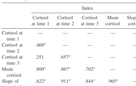
Table 2 lists correlation coefficients among measures of cortisol. The high intercorrelations indicate reliable assay of cortisol levels.
It has been shown that exposure to novelty can heighten cortisol levels (Al’Absi and Lovallo 1993; Hennessy et al 1995). Thus, it is possible that in this study cortisol at time 1 served as an index of sensitivity to the initial novelty of the assessment. In contrast, saliva samples at times 2 and 3 may reflect baseline levels of cortisol. If this is the case, then the slope of cortisol may be an alternative measure of stress sensitivity as well, given that it indexes the magnitude of the linear change in cortisol from the initial level over time.
COGNITIVE MEASURES. Interitem correlations among the
WMS-R, CVLT, and MCST standardized scores were examined and internal consistency (a) was computed. Results indicated high internal consistency among the four WMS-R subtests (a 5
.8804), the seven CVLT subtests (a 5 .9445), and the two MCST indices (a 5.9424). Therefore,zscores for the subtests of each of these measures were averaged together to derive CVLT, WMS-R, and MCST composite scores, respectively. Sensitivity (d9) was calculated according to standard procedures (McNicol and Willson 1971).
The CVLT and WMS-R composite scores were highly corre-lated (a 5.8516). Therefore, they were averaged to compute an index of “explicit memory.” There were high intercorrelations among the CVLT, WMS-R, and MCST total number of correct responses scores (a 5.7947). Therefore, the three scores were averaged to compute an aggregate index of “hippocampal func-tioning.” Scaling was the same for all variables in the composite scores.
SYMPTOM RATINGS. Recent research findings suggest that a three-symptom dimension model may best fit the data on clinical symptoms in patients with schizophrenia (Andreasen et al 1995). Accordingly, positive symptoms are divided into two dimensions, a psychosis dimension (i.e., delusions and halluci-nations) and a disorganized dimension (i.e., disorganized speech, disorganized behavior and inappropriate affect). The third di-mension is composed of negative symptoms (i.e., affective flattening, physical anergia). Using Andreasen et al’s (1995) three-factor solution, we derived Positive, Negative, and Disor-ganized Symptoms scale scores to measure symptomatology.
Internal consistency (Chronbach’sa) was examined for each of the hypothesized scales, excluding items with missing data and/or that had restricted variance across ratings. The reliabilities for the hypothesized Positive Symptoms scale (a 5 .8631), Negative Symptoms scale (a 5.6986), and Disorganized Symp-toms scale (a 5.7100) were high. Therefore, ratings on the items within each scale were averaged to obtain a composite index of severity of positive, negative, and disorganized symptoms, re-spectively. The items comprising each scale and their associated criteria are contained in Appendix 1.
Neuropsychological dysfunction in schizophrenia may be more closely associated with overall symptom severity than specific symptom dimensions (Goldberg and Weinberger 1995). Therefore, ratings across the three symptom scales were aver-aged to create a global index of symptom severity. The reliability Table 2. Correlation Coefficients for Cortisol Measures for
for this scale was low (a 5.3078). This was expected, given that the three scales were presumed to measure different dimensions of symptomatology.
DIAGNOSTIC GROUP DIFFERENCES. Independent-sam-plesttests were employed to test for group differences in age, cognitive performance, cortisol levels, and symptom scale rat-ings. One-tailed tests were used because the hypothesized group differences in the examined variables were directional in nature.
Group Demographics The PD group was significantly older than the OD group [t(23) 5 2.198,p 5 .019] and NC group [t(31)5 20.3090,p5.002] (Table 1).x2tests comparing the
PD, OD, and NC groups on gender and race indicated no group differences in these demographic characteristics.
Cognitive Measures The PD group scored below the OD group on d9 [t(9) 5 21.885, p 5 .038], the global index of hippocampal functioning [t(23) 5 25.000,p5 .000], and the global index of explicit memory [t(23)5 25.018,p5.000]. The PD group also scored below the NC group ond9[t(28)53.241,
p5.002], the global index of hippocampal functioning [t(31)5
6.233, p 5 .000], and the global index of explicit memory [t(31)55.941,p5.000]. The PD group scored higher (poorer performance) on the MCST composite score than the OD group [t(23)52.416,p5.012] and the NC group [t(31) 5 23.373,
p5.001].
These analyses were also conducted with age as a covariate because there were group differences in age. There was no longer a significant difference between the PD and OD groups ond9. This was the only change in the pattern of results.
Symptom Scale Ratings The PD group had significantly higher ratings than the OD group on the Positive Symptoms scale [t(23) 5 1.854, p5 .039], the Disorganized Symptoms scale [t(23) 5 2.526, p 5 .010], and the Overall Symptoms scale [t(23)53.160,p5.002]. The diagnostic groups did not differ on negative symptoms.
Cortisol There were no diagnostic group differences on the measures of cortisol. However, it is possible that psychotropic
medication was masking group differences, given evidence that antipsychotics reduce cortisol levels (Walker and Diforio 1997). In addition, some studies have evidenced hypercortisolemia in depressed patients (Carpenter and Bunney 1971; Deuschle et al 1998). More specifically, Galard et al (1991) and Gotthardt et al (1995) showed significantly higher basal levels of cortisol in depressed patients relative to NC subjects. The inclusion of a large majority of subjects with a history of depression in the OD group might therefore account for the absence of a significant difference in cortisol secretion between the PD and OD groups. Some psychiatric populations (viz., depressed patients) have also been shown to exhibit hypersecretion of cortisol during the afternoon (Christie et al 1986; Seckl et al 1991). Given the diagnostic nature of the OD group subjects, restriction of the cortisol samplings to morning hours in this study may account for the lack of significant group differences in cortisol.
Results
The Relation between Cortisol and Cognitive
Performance
All correlational analyses were conducted using one-tailed
tests because of the directional nature of the hypothesized
relations among the examined variables. Pearson
correla-tion analyses conducted across the total sample (PD group,
OD group, and NC group) indicated that explicit memory
and hippocampal functioning were negatively correlated
with time 3 cortisol (Table 3). Hippocampal functioning
was also negatively correlated with time 2 cortisol. The
pattern of results indicates that higher cortisol was
asso-ciated with poorer performance. Performance on the
MCST, as indexed by the composite score, was not
correlated with any of the cortisol measures.
Correlational analyses examining discrete aspects of
memory performance—namely, immediate, short-term,
and long-term memory—were also conducted (Table 4).
Results indicated that time 1 cortisol was not associated
with any of the three derived subcomponents of memory
in either the total sample or the NC group. Time 2 cortisol
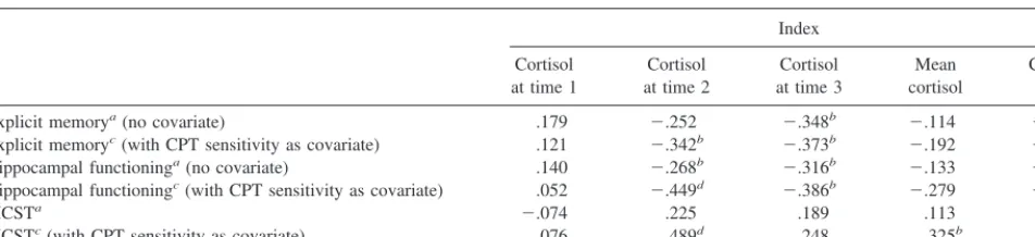
Table 3. Correlation Coefficients and Partial Correlation Coefficients Relating Cortisol with Explicit Memory and Hippocampal Functioning, for Total SampleIndex
Cortisol at time 1
Cortisol at time 2
Cortisol at time 3
Mean cortisol
Cortisol slope
Explicit memorya(no covariate) .179 2.252 2.348b 2.114 2.228
Explicit memoryc(with CPT sensitivity as covariate) .121 2.342b 2.373b 2.192 2.301b
Hippocampal functioninga(no covariate) .140 2.268b 2.316b 2.133 2.233
Hippocampal functioningc(with CPT sensitivity as covariate) .052 2.449d 2.386b 2.279 2.379b
MCSTa 2.074 .225 .189 .113 .173
MCSTc(with CPT sensitivity as covariate) .076 .489d .248 .325b .372b
CPT, Continuous Performance Test; MCST, Modified Wisconsin Card Sorting Test.
an540.
bp,.05, one-tailed test. cn533.
was significantly correlated with all three memory
vari-ables in the NC group, but with only immediate recall in
the total sample. Time 3 cortisol was significantly
corre-lated with all three memory variables in the total sample,
but with only immediate and long-term memory in the NC
group. Mean cortisol and cortisol slope were significantly
correlated with immediate and long-term memory in the
NC group. In the total sample, cortisol slope was
signifi-cantly correlated with immediate memory; no other
cor-relations were significant within this group. There were no
significant findings in the PD and OD groups alone.
Overall, these results suggests that cortisol levels were
associated with immediate, short-term, and long-term
memory performance .
It is possible that attentional problems contribute to the
observed memory deficits in schizophrenia. The
correla-tional analyses were therefore repeated controlling for
attention, as indexed by sensitivity performance on the
CPT-Vigil. All of the significant correlations held after
covarying for attention. In addition, there were significant
negative correlations for cortisol slope with explicit
mem-ory and hippocampal functioning, as well as time 2
cortisol with explicit memory, and significant positive
correlations for time 2 cortisol, mean cortisol, and cortisol
slope with MCST (Table 3). These findings suggest that
cognitive impairment attributable to deficits in attention
did not account for the relation between cortisol and
memory performance. Instead, individual differences in
attention were obscuring the relations of cortisol with
memory and executive functioning.
The above analyses were repeated with the PD group
alone (Table 5). Results indicated no significant
correla-tions for cortisol with explicit memory, hippocampal
functioning, or MCST performance. After covarying for
attention, there was a trend toward a negative correlation
for time 2 cortisol with hippocampal functioning, as well
as a positive correlation for time 2 cortisol and cortisol
slope with MCST performance.
The majority of the findings among the PD group were
consistent with findings among the total sample in that,
although they were of lesser magnitude, they were in a
similar direction. Findings in the total sample, and to a
lesser degree in the PD group, suggest that increased
cortisol secretion, particularly those measures presumed to
reflect baseline cortisol, is associated with impaired
per-formance on cognitive measures indexing hippocampal
functioning.
The Relation between Cortisol and Symptoms
Age was negatively correlated with time 1 cortisol (
r
5
2
.451,
p
5
.030) and mean cortisol (
r
5 2
.410,
p
5
.045)
within the PD group. Therefore, correlational analyses of
Table 4. Correlation Coefficients Relating Cortisol with Immediate, Short-Term, and Long-TermMemory for Total Sample, Psychotic Disorder Group, Other Disorders Group, and Normal Control Group
Memory
Index
Cortisol at time 1
Cortisol at time 2
Cortisol at time 3
Mean cortisol
Cortisol slope
Total samplea
Immediate .055 2.280b 2.347b 2.191 2.277b
Short term .028 2.179 2.312b 2.152 2.221
Long term .052 2.254 2.334b 2.172 2.255
Normal controlc
Immediate 2.206 2.860d 2.758d 2.670d 2.768d
Short term .203 2.504b 2.398 2.222 2.346
Long term .044 2.734d 2.581b 2.446b 2.570b
Psychotic disorderse
Immediate .148 .146 .083 .160 .147
Short term 2.186 2.022 2.222 2.161 2.159
Long term 2.104 .065 .031 2.015 .016
Other disordersf
Immediate .427 .361 2.343 .367 .155
Short term .146 .351 2.369 .116 2.022
Long term .405 .118 2.644 .141 2.200
an540.
bp,.05, one-tailed test. cn515.
dp,.01, one-tailed test. en518.
cortisol with symptom severity in the PD group controlled
for age for these two measures of cortisol (Table 6).
The pattern of results indicated that higher cortisol
levels were associated with more severe symptoms (Table
6). Time 1 cortisol was positively correlated with
disor-ganized and overall symptoms, and mean cortisol was
correlated with positive and overall symptoms. After
covarying for age, these findings held and mean cortisol
was additionally correlated with disorganized symptoms.
In addition, time 2 cortisol was correlated with positive
symptoms and time 3 cortisol was correlated with positive,
disorganized, and overall symptoms. Higher cortisol slope
was linked with higher ratings of positive and overall
symptoms.
The Relation between Cognitive Performance and
Symptoms
First-order correlations revealed that none of the symptom
dimensions was associated with the three cognitive
indi-ces. After covarying for attention, however, positive
symptoms were negatively correlated with hippocampal
functioning (
r
5 2
.729,
p
5
.002) and positively
corre-lated with MCST performance (
r
5
.841,
p
5
.000). Thus,
higher symptom ratings were associated with poorer
performance.
The Relation of Cortisol and Task Performance
with Symptoms
Stepwise regression analyses were conducted across all
subjects with psychotic disorders. Memory performance,
hippocampal functioning, MCST performance, and all
measures of cortisol were entered as predictor variables.
The only significant predictors of symptom severity were
time 1 and time 3 cortisol. Time 1 cortisol accounted for
26.9% of the variance in Disorganized Symptom scale
ratings. Time 3 cortisol accounted for 53.7% of the
variance in Positive Symptom scale ratings and 52.7% of
the variance in Overall Symptom scale ratings.
Discussion
This study was designed to examine the relations among
memory, cortisol release, and symptom expression in
patients with psychotic disorders. In general, the results
provide modest support for the hypothesis that elevations
in cortisol secretion are associated with deficits in explicit
memory performance. Among patients with psychotic
Table 5. Correlation Coefficients and Partial Correlation Coefficients Relating Cortisol with Explicit Memory and Hippocampal Functioning for Psychotic Disorder GroupIndex
Cortisol at time 1
Cortisol at time 2
Cortisol at time 3
Mean cortisol
Cortisol slope
Explicit memorya(no covariate) .039 .042 2.096 .012 2.012
Explicit memoryb(with CPT sensitivity as covariate) .000 2.014 2.100 2.032 2.051
Hippocampal functioninga(no covariate) .025 2.132 2.192 2.100 2.144
Hippocampal functioningb(with CPT sensitivity as covariate) 2.134 2.428 2.269 2.323 2.362
MCSTa(no covariate) 2.032 .238 .210 .149 .202
MCSTb(with CPT sensitivity as covariate) .146 .634c .313 .432 .484d
CPT, Continuous Performance Test; MCST, Modified Wisconsin Card Sorting Test.
an518. bn512.
cp,.01, one-tailed test. dp,.05, one-tailed test.
Table 6. Correlation Coefficients and Partial Correlation Coefficients Relating Cortisol with Symptom Scale Ratings, for Psychotic Disorder Group
Cortisol
Index
Symptom scale
Overall Symptoms Positive Negative Disorganized
Cortisol at time 1 (no covariate)
.151 .123 .519a .434a
Cortisol at time 1 (with age as a covariate)
.285 2.038 .718b .467a
Cortisol at time 2 (no covariate)
.419a .118 .041 .335
Cortisol at time 3 (no covariate)
.733b .159 .379a .726b
Mean cortisol (no covariate)
.461a .156 .335 .559b
Mean cortisol (with age as a covariate)
.617b .018 .534a .599a
Cortisol slope (no covariate)
.570b .161 .317 .601b
n518.
disorders, cortisol shows a strong positive relation with the
severity of symptoms, especially positive symptoms.
The Relation between Cortisol and Cognitive Task
Performance
Previous research has demonstrated an inverse association
between cortisol secretion and memory (e.g., Lupien et al
1994; Seeman et al 1997). Current findings of an inverse
relationship of cortisol levels at times 2 and 3 (presumably
reflective of basal levels of cortisol) with task performance
in the total sample are consistent with these results. Thus,
it appears that cortisol levels measured after acclimation to
the research setting are most strongly linked with
cogni-tive function. It is possible that this reflects the relation of
persistent elevations in circulating corticosteroids with the
integrity of various regions of the brain, particularly the
hippocampus, which has been implicated in memory
function. Some previous research has shown that cognitive
testing successfully induces cortisol release (Lupien et al
1997). Thus, it is important to consider that the current
finding of an inverse relationship of cortisol with cognitive
performance may have been due to heightened cortisol
secretion subsequent to cognitive testing.
Although the direction of the relation between cortisol
and task performance was similar for the psychotic
pa-tients, only two of the coefficients reached statistical
significance. It is likely that this is due to insufficient
statistical power in this smaller group (
n
5
18) of patients.
The Relation between Cortisol and Symptom
Expression
Correlational analyses revealed that heightened cortisol
release is associated with increased symptom expression
in psychotic disorders. Most of the subjects in the PD
group were medicated with or previously medicated with
antipsychotics. Thus, findings of an association of
symp-toms with cortisol secretion, despite medication effects,
are all the more striking.
The findings are not supportive of a stronger association
between heightened HPA activity and the deficit
symp-toms of schizophrenia, as suggested by Tandon et al
(1991). Rather, these results suggest that heightened HPA
activity is generally associated with overall symptom
expression in psychotic disorders. In terms of specific
symptom dimensions, cortisol indexing sensitivity to
nov-elty (time 1) and mean cortisol were associated with
disorganized symptom severity, whereas “basal” (times 2
and 3) measures of cortisol were associated with positive
symptom severity. The association between cortisol and
positive symptoms is consistent with several previous
findings (Kaneko et al 1992; Keshavan et al 1989;
Ryba-kowski et al 1991). No measures of cortisol were
associ-ated with negative symptoms.
This suggests a possible dissociation between positive
and disorganized symptoms on the one hand and negative
symptoms on the other. More specifically, the pattern of
association of positive, negative, and disorganized
symp-toms with the various measures of cortisol suggests that
the symptom dimensions are differentially influenced by
1) basal HPA activity linked with the integrity of the
hippocampus versus 2) acute changes in HPA activity in
response to novelty or stress, respectively.
The Relation between Cognitive Performance and
Symptom Expression
Previous studies of the relation between memory
perfor-mance and symptom severity have yielded mixed findings.
Some showed an inverse relation of memory with negative
symptoms (Basso et al 1998; Sullivan et al 1994),
disor-ganized symptoms (Basso et al 1998), or total symptom
severity (Sullivan et al 1994). Sullivan et al (1994) found
a trend in the relationship between memory and positive
symptoms. Other studies found no association between
memory and symptom severity (Hoff et al 1992; Schmand
et al 1992).
In this study, an index of MCST errors and an index of
hippocampal functioning, which incorporates the MCST,
were linked with positive symptoms but not with the other
three symptom scales. This suggests that positive
symp-toms are associated with more pronounced impairment on
measures of frontal function, rather than measures of
memory.
The Relation of Cognitive Performance and
Cortisol with Symptoms
Regression analyses indicated that time 1 cortisol
dicted disorganized symptoms and time 3 cortisol
pre-dicted positive and overall symptom severity. No other
measures of cortisol or indices of cognitive functioning
(e.g., memory, executive, or hippocampal functioning)
predicted symptom severity.
in the clinical manifestation of schizophrenia. Additional
research on larger samples of patients is warranted.
References
Akbarian S, Vinuela A, Kim JJ, Potkin SG, Bunney WE (1993): Distorted distribution of nicotinamide-adenine dinucleotide phosphate-diaphorase neurons in temporal lobe of schizo-phrenics implies anomalous cortical development.Arch Gen Psychiatry50:178 –187.
Al’Absi M, Lovallo WR (1993): Cortisol concentrations in serum of borderline hypertensive men exposed to a novel experimental setting.Psychoneuroendocrinology18:355–363. Altshuler LL, Casanova MF, Goldberg TE, Kleinman JE (1990):
The hippocampus and parahippocampus in schizophrenic, suicide, and control brains. Arch Gen Psychiatry 47:1029 – 1034.
Andreasen NC (1981): Scale for the Assessment of Positive Symptoms (SAPS).Iowa City: University of Iowa Press. Andreasen NC (1983): Scale for the Assessment of Negative
Symptoms (SANS).Iowa City: University of Iowa Press. Andreasen NC, Arndt S, Alliger R, Miller D, Flaum M (1995):
Symptoms of schizophrenia: Methods, meanings, and mech-anisms.Arch Gen Psychiatry52:341–351.
Arana GW, Barreira PJ, Cohen BM, Lipinski JF, Fogelson D (1983): The dexamethasone suppression test in psychotic disorders.Am J Psychiatry140:1521–1523.
Bassett JR, Marshall PM, Spillane R (1987): The physiological measurement of acute stress in bank employees.Int J Psy-chophysiol5:265–273.
Basso MR, Nasrallah HA, Olson SC, Bornstein RA (1998): Neuropsychological correlates of negative, disorganized and psychotic symptoms in schizophrenia.Schizophr Res31:99 – 111.
Baum A, Grunberg N (1995): Measurement of stress hormones. In: Cohen S, Kessler RC, Gordon LU, editors. Measuring Stress: A Guide for Health and Social Scientists.New York: Oxford University Press, 175–192.
Benes FM, Sorensen I, Bird ED (1991): Morphometric analyses of the hippocampal formation in schizophrenic brain. Schizo-phr Bull17:597– 608.
Bilder RM, Bogerts B, Ashtari M, Wu H, Alvir JM, Jody D, et al (1995): Anterior hippocampal volume reductions predict “frontal lobe” dysfunction in first episode schizophrenia.
Schizophr Res17:47–58.
Bogerts B, Falkai P, Haupts M, Greve B, Ernst S, Tapernon-Franz U, et al (1990): Post-mortem volume measurements of limbic system and basal ganglia structures in chronic schizo-phrenics. Initial results from a new brain collection. Schizo-phr Res3:295–301.
Bogerts B, Meertz E, Schonfeldt-Bausch R (1985): Basal ganglia and limbic system pathology in schizophrenia. Arch Gen Psychiatry42:784 –791.
Breier A, Buchanan R, Elkashef A, Munson RC, Kirkpatrick B, Gellad F (1992): Brain morphology and schizophrenia: A magnet resonance imaging study of limbic prefrontal cortex and caudate structures.Arch Gen Psychiatry49:921–926. Buchsbaum MS, Sostek AJ (1980): An adaptive-rate continuous
performance test: Vigilance characteristics and reliability for 400 male students.Percept Mot Skills51:707–713.
Carpenter W, Bunney W (1971): Adrenal cortical activity in depressive illness.Am J Psychiatry128:31.
Christie JE, Whalley LJ, Blackwood DHR, Blackburn IM, Fink G (1986): Raised plasma cortisol concentrations are a feature of drug-free psychotics and are not specific for depression.
Br J Psychiatry148:58 – 65.
Colombo C, Abbruzzese M, Livian S, Scotti G, Locatelli M, Bonfanti A, et al (1993): Memory functions and temporal-limbic morphology in schizophrenia.Psychiatry Res50:45– 56.
Delis DC, Kramer JH, Kaplan E, Ober BA (1983): CVLT: California Verbal Learning Test Manual.San Antonio: Psy-chological Corp.
Delisi LE, Hoff AL, Schwartz JE, Shields GW, Halthore SN, Gupta SM, et al (1991): Brain morphology in first-episode schizophrenic-like psychotic patients: A quantitative mag-netic resonance imaging study.Biol Psychiatry29:159 –175. De Quervain DJF, Roozendaal B, Nitsch RM, McGaugh JL, Hock C (2000): Acute cortisone administration impairs re-trieval of long-term declarative memory in humans. Nat Neurosci3:313–314.
Deuschle M, Weber B, Colla M, Depner M, Heuser I (1998): Effects of major depression, aging and gender upon calcu-lated diurnal free plasma cortisol concentrations: A re-evaluation study.Stress2:281–287.
Eichenbaum H, Schoenbaum G, Young B, Bunsey M (1996): Functional organization of the hippocampal memory system.
Proc Natl Acad Sci U S A93:13500 –13507.
First MB, Spitzer RL, Gibbon M, Williams JBW (1997):
Structured Clinical Interview for DSM-IV Axis I Disorders— Patient Edition (SCID-I/P Version 2.0, 4/97 Revision). Wash-ington, DC: American Psychiatric Press, Biometrics Research Department.
Galard R, Gallart JM, Catalan R, Schwartz S, Arguello JM, Castellanos JM (1991): Salivary cortisol levels and their correlation with plasma ACTH levels in depressed patients before and after the DST.Am J Psychiatry148:505–508. Gold JM, Randolph C, Carpenter CJ, Goldberg TE, Weinberger
DR (1992a): Forms of memory failure in schizophrenia. J Abnorm Psychol101:487– 494.
Gold JM, Randolph C, Carpenter CJ, Goldberg TE, Weinberger DR (1992b): The performance of patients with schizophrenia on the Wechsler Memory Scale-Revised.Clin Neuropsychol
6:367–373.
Goldberg TE, Ragland JD, Torrey EF, Gold JM, Bigelow LB, Weinberger DR (1990): Neuropsychological assessment of monozygotic twins discordant for schizophrenia.Arch Gen Psychiatry47:1066 –1072.
Goldberg TE, Torrey EF, Berman KF, Weinberger DR (1994): Relations between neuropsychological performance and brain morphological and physiological measures in monozygotic twins discordant for schizophrenia.Psychiatry Res55:51– 61. Goldberg TE, Weinberger DR (1995): A case against subtyping
in schizophrenia.Schizophr Res17:147–152.
Hennessy MB, Mendoza SP, Mason WA, Moberg GP (1995): Endocrine sensitivity to novelty in squirrel monkeys and titi monkeys: Species differences in characteristic modes of responding to the environment.Physiol Behav57:331–338. Hirayasu Y, Shenton ME, Salisbury DF, Dickey CC, Fischer IA,
Mazzoni P, et al (1998): Lower left temporal lobe MRI volumes in patients with first-episode schizophrenia com-pared with psychotic patients with first-episode affective disorder and normal subjects. Am J Psychiatry 155:1384 – 1391.
Hoff AL, Riordan H, O’Donnell DW, Morris L, DeLisi LE (1992): Neuropsychological functioning of first-episode schizophreniform patients.Am J Psychiatry149:898 –903. Jeste DV, Lohr JB (1989): Hippocampal pathologic findings in
schizophrenia. A morphometric study.Arch Gen Psychiatry
46:1019 –1024.
Kaneko M, Yokoyama F, Hoshino Y, Takahagu K, Murata S, Watanabe M, et al (1992): Hypothalamic pituitary adrenal axis function in chronic schizophrenia: Association with clinical features.Neuropsychobiology25:1–7.
Keshavan MS, Brar J, Ganguli R, Jarrett D (1989): DST and schizophrenic symptomatology.Biol Psychiatry26:847– 858. Kiess W, Meidert A, Dressendorfer RA, Schriever K, Kessler U, Konig A, et al (1995): Salivary cortisol levels throughout childhood and adolescence: Relation with age, pubertal stage and weight.Pediatr Res37:502–506.
Kirschbaum C, Hellhammer DH (1989): Salivary cortisol in psychobiological research: An overview. Neuropsychobiol-ogy22:150 –169.
Kirschbaum C, Wolf OT, May M, Wippich W, Hellhammer DH (1996): Stress- and treatment-induced elevations of cortisol levels associated with impaired declarative memory in healthy adults.Life Sci58:1475–1483.
Kovelman JA, Scheibel AB (1984): A neurohistological correlate of schizophrenia.Biol Psychiatry19:1601–1621.
Laudat MH, Cerdas S, Fournier C, Guiban D, Guilhaume B, Luton JP (1988): Salivary cortisol measurement: A practical approach to assess pituitary-adrenal function.J Clin Endocri-nol Metab66:343–348.
Lencz T, McCarthy G, Bronen RA, Scott TM, Inserni JA, Sass KJ, et al (1992): Quantitative magnetic resonance imaging in temporal lobe epilepsy: Relationship to neuropathology and neuropsychological function.Ann Neurol31:629 – 637. Lupien S, Lecours AR, Lussier I, Schwartz G, Nair NPV,
Meaney MJ (1994): Basal cortisol levels and cognitive deficits in human aging.J Neurosci14:2893–2903. Lupien SJ, Gaudreau S, Tchiteya BM, Maheu F, Sharma S, Nair
NP, et al (1997): Stress-induced declarative memory impair-ment in healthy elderly subjects: Relationship to cortisol reactivity.J Clin Endocrinol Metab82:2070 –2075. McNicol D, Willson RJ (1971): The application of signal
detection theory to letter recognition.Aust J Psychol23:311– 315.
Nelson HE (1976): A modified card sort test sensitive to frontal lobe defects.Cortex12:313–324.
Nestor PG, Shenton ME, McCarley RW, Haimson J, Smith RS, O’Donnell B, et al (1993): Neuropsychological correlates of
MRI temporal lobe abnormalities in schizophrenia. Am J Psychiatry150:1849 –1855.
Newcomer JW, Craft S, Askins K, Hershey T, Bardgett ME, Csernansky JG, et al (1998): Glucocorticoid interactions with memory function in schizophrenia. Psychoneuroendocrinol-ogy23:65–72.
Newcomer JW, Craft S, Hershey T, Askins K, Bardgett J (1994): Glucocorticoid-induced impairment in declarative memory performance in adult humans.J Neurosci14:2047–2053. Newcomer JW, Faustman WO, Whiteford HA, Moses JA Jr,
Csernansky JG (1991): Symptomatology and cognitive im-pairment associate independently with post-dexamethasone cortisol concentrations in unmedicated schizophrenic pa-tients.Biol Psychiatry29:855– 864.
Newcomer JW, Selke G, Melson AK, Hershey T, Craft S, Richards K, Alderson AL (1999): Decreased memory perfor-mance in healthy humans induced by stress-level cortisol treatment.Arch Gen Psychiatry56:527–533.
Rao VP, Krishnan RR, Goli V, Saunders WB, Ellinwood EH, Blazer DG, et al (1989): Neuroanatomical changes and hypothalamic-pituitary-adrenal axis abnormalities.Biol Psy-chiatry26:729 –732.
Rybakowski J, Linka M, Matkowski K, Kanarkowski R (1991): Dexamethasone suppression test and the positive and nega-tive symptoms of schizophrenia.Psychiatr Pol25:9 –15. Sapolsky RM, McEwen BS (1986): Stress, glucocorticoids, and
their role in degenerative changes in the aging hippocampus. In: Crook T, Bartus R, Ferris S, Gershon S, editors.Treatment Development Strategies for Alzheimer’s Disease. Madison, CT: Mark Powley, 151–171.
Sapolsky RM, Uno H, Rebert CS, Finch CE (1990): Hippocam-pal damage associated with prolonged glucocorticoid expo-sure in primates.J Neurosci10:2897–2902.
Saykin AJ, Shtasel DL, Gur RE, Kester DB, Mozley LH, Stafiniak P, et al (1994): Neuropsychological deficits in neuroleptic naı¨ve patients with first-episode schizophrenia.
Arch Gen Psychiatry51:124 –131.
Schatzberg AF, Rothschild AJ (1988): The roles of glucocorti-coid and dopaminergic systems in delusional (psychotic) depression.Ann N Y Acad Sci537:462– 471.
Schatzberg AF, Rothschild AJ, Langlais PJ, Bird ED, Cole JO (1985): A corticosteroid/dopamine hypothesis for psychotic depression and related states.J Psychiatry Res19:57– 64. Schmand B, Brand N, Kuipers T (1992): Procedural learning of
cognitive and motor skills in psychotic patients. Schizophr Res8:157–170.
Seckl JR, Campbell JC, Edwards CRW, Christie JE, Whalley LJ, Goodwin GM, Fink G (1991): Diurnal variation of plasma corticosterone in depression.Psychoneuroendocrinology15: 485– 488.
Seeman TE, McEwen BS, Singer BH, Albert MS, Rowe JW (1997): Increase in urinary cortisol excretion and memory declines: MacArthur studies of successful aging. J Clin Endocrinol Metab82:2458 –2465.
(1992): Utility of an oral diffusion sink (ODS) device for quantification of saliva corticosteroids in human subjects.
J Clin Endocrinol Metab74:698 –700.
Squire LR (1987): Memory and Brain. New York: Oxford University Press.
Squire LR (1992): Memory and the hippocampus. A synthesis from findings with rats, monkeys, and humans.Psychol Rev
99:195–231.
Starkman MN, Gebarski SS, Berent S, Schteingart DE (1992): Hippocampal formation volume, memory dysfunction, and cortisol levels in patients with Cushing’s syndrome. Biol Psychiatry32:756 –765.
Suddath RL, Christison GW, Torrey EF, Casanova MF, Wein-berger DR (1990): Anatomical abnormalities in the brains of monozygotic twins discordant for schizophrenia. N Engl J Med322:789 –794.
Sullivan EV, Shear PK, Zipursky RB, Sagar HJ, Pfefferbaum A (1994): A deficit profile of executive, memory and motor functions in schizophrenia.Biol Psychiatry36:641– 653. Tandon R, Mazzara C, DeQuardo J, Craig KA,
Meador-Woo-druff JH, Goldman R (1991): Dexamethasone suppression test in schizophrenia: Relationship to symptomatology, ven-tricular enlargement, and outcome.Biol Psychiatry29:953– 964.
Torres IJ, Flashman LA, O’Leary DS, Swayze V II, Andreasen NC (1997): Lack of an association between delayed memory and hippocampal and temporal lobe size in patients with schizophrenia and healthy controls.Biol Psychiatry42:1087– 1096.
Velakoulis D, Pantelis C, McGorry PD, Dudgeon P, Brewer W, Cook M, et al (1999): Hippocampal volume in first-episode psychoses and chronic schizophrenia: A high-resolution mag-netic resonance imaging study.Arch Gen Psychiatry56:133– 141.
Waldo MC, Cawthra E, Adler LE, Dubester S, Staunton M, Nagamoto H, et al (1994): Auditory sensory gating, hip-pocampal volume, and catecholamine metaboism in schizo-phrenics and their siblings.Schizophr Res12:93–106. Walker EF, Diforio D (1997): Schizophrenia: A neural
diathesis-stress model.Psychol Rev104:667– 685.
Wechsler D (1987): Wechsler Memory Scale—Revised.New York: Psychological Corp.
Weinstein DD, Diforio D, Schiffman J, Walker E, Bonsall R (1999): Minor physical anomalies, dermatoglyphic asymme-tries, and cortisol levels in adolescents with schizotypal personality disorder.Am J Psychiatry156:617– 623.
Appendix 1. Items in the SANS-SAPS–Derived Symptom Scales
Item no. Criteria
Positive symptoms
24 Thoughts of being persecuted by other people 26 Thoughts of sin or guilt
27 Grandiose thoughts 28 Religious thoughts
29 Thoughts concerning the body 30 Ideas and thoughts of self-reference 31 Thoughts of being controlled 32 Thoughts of mind reading 33 Thought broadcasting 34 Thought insertion
48 Hallucinatorylike experiences involving noises or voices 51 Hallucinatorylike experiences concerning the body 52 Hallucinatorylike experiences involving odors/sense of smell 53 Visual hallucinatorylike experiences
Negative symptoms
1 Facial expressions of emotion
2 Spontaneous movements of arms and legs 3 Expressive gestures
4 Eye contact
5 Emotional responsivity to other people or events 6 Vocal inflections
14 Grooming and hygiene 16 Physical energy
47 Repetitive or stereotyped behavior Disorganized symptoms
7 Appropriateness of emotional expressions 36 Loose associations
37 Tangential responses 39 Illogical speech 40 Circumstantial speech 41 Rapid speech 42 Distractible speech 43 Clanging
44 Clothing and appearance