www.elsevier.nlrlocateraqua-online
Detection of IHHN virus in shrimp tissue by digital
color correlation
Josue Alvarez-Borrego
a,), Marıa Cristına Chavez-Sanchez
b,1´
´
´
´
´
a ´
Departamento de Optica, DiÕision de Fısica Aplicada, Centro de In´ ´ Õestigacion Cientıfica y de Educacion´ ´ ´
Superior de Ensenada, Km. 107 Carretera Tijuana-Ensenada, Ensenada, B.C., C.P. 22860, Mexico
b
Centro de InÕestigacion en Alimentacion y Desarrollo, A.C., Unidad Mazatlan en Acuicultura y Manejo´ ´ ´
Ambiental, Sabalo Cerritos S´ rN, Apdo. Postal 711, Estero del Yugo, Mazatlan, Sinaloa, C.P. 82010, Mexico´
Received 20 January 2000; received in revised form 10 July 2000; accepted 9 August 2000
Abstract
Shrimp culture has suffered from disease outbreaks that significantly affect the economy and restrain aquaculture production in many countries around the world. For research and diagnosis of shrimp diseases, histopathology is a powerful tool to identify pathogens and to understand the cellular, tissue and organ changes that result as a consequence of disease. One problem of the technique is that it requires considerable time, although shrimp producers demand results in the shortest possible time in order to take preventive or control measures. The introduction of techniques, such as processing of digital images in histopathology, could significantly reduce the time for precise diagnosis. Here, we present an attempt to implement this technique with
Ž .
infectious hypodermal and hematopoietic necrosis virus IHHNV because it presents difficulties for diagnosis by conventional histological techniques. In order to determine the viral histopathol-ogy that could be identified with the digital color correlation in different tissues, some very clear
Ž .
and conventional Cowdry type A inclusion bodies CAIs were selected. It was demonstrated that color digital correlation can be used to detect the virus IHHN in shrimp tissues. More researches are needed to be done, which consider several aspects, like rotation, scale, color, natural morphology of the virus and the use of different kinds of filters to determine whether the technique can be applied to other viruses.q2001 Elsevier Science B.V. All rights reserved.
Keywords: Histopathology; Virus; Shrimp; Digital color correlation
)Corresponding author. Tel.:q52-61-74-50-50, 74-50-51; fax:q52-61-75-05053.
Ž .
E-mail addresses: [email protected] J. Alvarez-Borrego , [email protected]
ŽM.C. Chavez-Sanchez .´ ´ . 1
Tel.:q52-69-88-01-57, 88-01-58; fax:q52-69-88-01-59.
0044-8486r01r$ - see front matterq2001 Elsevier Science B.V. All rights reserved.
Ž .
1. Introduction
Shrimp culture has suffered from disease outbreaks that significantly affect the economy and restrain aquaculture production in many countries around the world. The causes are complex and are attributed to viruses, bacteria, fungi, pesticides and
Ž .
environmental degradation Gjedrem and Fimland, 1995 . It is widely recognized that improved regulatory and technical measures are necessary to prevent the introduction and spread of diseases. This is necessary to protect the aquaculture industry and the aquatic environment. Uniform practices and procedures designed to study the diseases in each country have been developed in Asia, Europe, Canada and the United States. Therefore, many countries are now involved in determining which diseases are present in wild and cultured aquatic organisms in order to facilitate the development of national and international codes of practice on health certification.
For diagnosis and research on infectious and non-infectious diseases, shrimp patholo-gists use different techniques. Recently, accurate and precise biological and molecular techniques based on DNA have been developed to diagnose viral and bacterial diseases,
Ž .
by procedures such as dot–blot, in situ hybridization, ELISA, and PCR Lightner, 1996 . In spite of these new techniques, histology is still one the techniques commonly used for research and diagnosis. With this technique, it is possible to observe the pathological changes in the cells, tissues and organs, and it also allows the identification of new pathogens, which are sometimes difficult to recognize using other techniques.
The basic method involves a series of steps to obtain a slide containing a layer of tissue, 1–5mm thick, stained with hematoxilin–eosin for analysis under the microscope ŽBell and Lightner, 1988; Lightner, 1996 . However, this useful technique has some.
Ž .
drawbacks: a getting the histological slide requires time consumption through all the Ž .
process of fixation, dehydration, embedding, sectioning and staining, b analyzing one single histological slide requires short time, but when it is necessary to analyze many of them for diagnosis or research in a population, it takes a longer time because it is necessary to AsweepB carefully each section under the microscope and search for any kind of histopathologies andror pathogens. In aquaculture, for example, to obtain a precise diagnosis of an identified pathogen or disease in a farm, it is necessary to analyze many slides from a large sample, delaying the results to the farmers for at least a
Ž .
week, and c for viral diseases, it is usually not possible to diagnose when the infection is in its early stages.
Ž .
Infectious hypodermal and hematopoietic necrosis IHHN is a viral disease which was first recognized in 1981, in Hawaii, in imported shrimp stocks of the blue shrimp Penaeus stylirostris, from culture facilities in South and Central America, but not from
Ž .
Mexico Lightner, 1983 . However, in 1987 and the subsequent 3 years, this virus was introduced and spread with imported stocks of penaeid shrimp in western Mexico that
Ž .
bordered the Gulf of California Lightner et al., 1992 . Since then, this disease has spread throughout the country, causing losses in cultured and wild shrimp.
To diagnose this viral disease histologically, it is necessary to demonstrate the presence of the prominent Cowdry type A, eosinophilic, intranuclear inclusion bodies surrounded by marginated chromatin in hypertrophied nuclei of cells in tissues of
Ž
. Ž
nerve ganglia and mesodermal origin hematopoietic organs, antennal gland, lymphoid
. Ž .
organ, connective tissue and striated muscle Lightner et al., 1983 . Classical Cowdry Ž .
type A inclusion bodies CAIs are not easy to identify, due to the different organs Ž
where they occurred and the difference in size, tones of purple light to very dark .
basophilic , shapes and size of the classic white ring that is also characteristic of these inclusions. These features depend on the stage of the virus, type of tissue infected and quality of the staining. Thus, it is necessary to develop skill and experience to recognize these inclusions.
The introduction of techniques, such as digital processing of images directly from the microscope in the diagnosis or research of infectious diseases, could significantly reduce the time taken to obtain precise results. However, in this paper, we used transparencies in order to test this new method, but the general idea is to use images directly from the microscope. We selected IHHN because of the difficulty presented in diagnosing this viral disease through conventional histological techniques.
2. Materials and methods
Experimental shrimps were obtained from a farm in the state of Sinaloa, Mexico and transported live to the laboratory to be fixed in Davidson’s solution according to
Ž .
Lightner 1996 . After 24 h, the fixative was discarded and the shrimps were preserved in 50% alcohol until processed by the conventional histological techniques, as suggested
Ž .
by Lightner 1996 . For this study, and in order to determine if the inclusion bodies could be identified with the digital color correlation in different tissues, some very clear
Ž .
and conventional Cowdry type A inclusion bodies CAIs were selected from the cuticular epithelium, nerve cord, gills and connective tissue under the microscope ŽOlympus BX60 . The selected IHHNV inclusion bodies were microphotographed with. an Olympus camera PM-CB30. All the photographs were taken as 35 mm transparencies with the 60=objective.
An HP Photosmart color digital scanner was used to obtain the matrix data from the Ž .
transparencies, and defined the function f x, y for every pixel of coordinates x and y on the image. In general, a polychromatic object presents different shape and amplitude
Ž .
distributions Ali x, y when illuminated with different wavelengths l. However, two different objects may present similar amplitude distribution when they are illuminated with a determined wavelength l0. So, in an optical pattern recognition process using a correlator illuminated with a wavelength l0, these objects will give very similar amplitude correlation distributions, and some false alarms will appear. To avoid this problem, it is necessary to use the information about the dependence of the object
Ž .
amplitude distributions on the wavelength Campos et al., 1991 .
Ž Most natural colors can be obtained as a combination of three colors called
. Ž . Ž .
primaries if they are well selected. Each of them has to be on the red R , green G , Ž .
and blue B regions of the visible spectrum. When the object is transparent, the amplitude transmittance obtained by illumination with one of these primaries is called the red, green and blue components of the object.
Ž .
channel. Specifically, three correlations, Cli x, y , were determined between the
trans-Ž . Ž .
parency to be analyzed, fli x, y , and the inclusion bodies to be detected, V x, y . Thisli
was done by illumination with three wavelengthslisR, G, B which covered the visible spectrum.
Cli
Ž
x , y.
sfliŽ
x , y V.
liŽ
x , y.
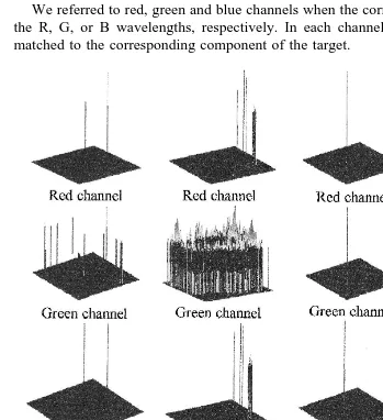
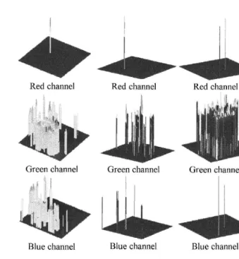
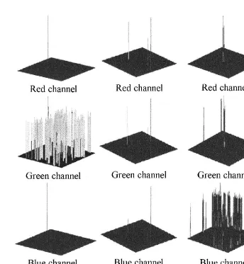
We referred to red, green and blue channels when the correlator was illuminated with the R, G, or B wavelengths, respectively. In each channel, the filter to be used was matched to the corresponding component of the target.
Ž . Ž . Ž .
Fig. 1. Photomicrographs of the typical intranuclear Cowdry type A inclusion bodies arrows in: a and b
Ž . Ž . Ž . Ž . Ž . Ž . Ž .
cuticular epithelium and connective tissue H&E 435.6X ; c in the gill H&E 435.6X ; d , e , f , g in
Ž . Ž .
mid-sagital sections of the ventral nerve cord H&E 732.6X ; h transversal section of a nerve cord embedded
Ž . Ž . Ž . Ž . Ž . Ž .
in reserve cells and H&E 435.6X ; i in connective tissue H&E 435.6X ; j , k , l in mid-sagital sections
Ž .
Ž .
Fig. 1 continued .
Ž .
Objects that have a determined component Ali x, y that is similar to the same Ž .
component of the target V x, y will give a maximum of correlation in this channell
i
Žli.. Only the target will simultaneously give a correlation maximum in each channel. So, an object is detected as the target if it simultaneously produces a correlation peak in the three channels.
Ž . Ž .
The filter used to detect the target V x, y was a phase-only filter. Let V x, y beli li Ž . < Ž .< w Ž .x
the inclusion body to be recognized and let V u, Õ sV u, Õ expyif u, Õ be its
Ž . Ž .
Fourier transform frequency information . The phase-only filter VPO F u, Õ is defined
Ž ) w Ž .x < Ž .<
as VPO F u, Õ sexpyif u, Õ , where V u, Õ is equal to 1, and u, Õ are the variables in the frequency domain.
Ž
Several authors Yu and Chao 1983; Yu 1985; Yu and Javidi 1986: Yu and Zhou .
Ž .
Fig. 1 continued .
shape and the spectral information of the object. This permits an improvement in Ž
discrimination sensitivity. In the last 10 years, several authors Millan et al., 1989, 1992;
´
.
Campos et al., 1991; Moreno et al., 1997 have obtained important results in this line. They have used some images like butterflies and letters of different colors.
3. Results and discussion
The selected CAIs were observed in the ventral cord of the nervous tissue, gills, the subcuticular epithelium and in connective tissue. Their digital results are presented for
Ž .
Ž . images. Phase-only filters were used in the three channels red, blue and green in the
Ž .
digital color correlation. Fig. 1 shows, first, the results RGB channels after reducing the noise or background to less than 60% of the highest value. The green channel showed more noise than the other channels in five cases and in three cases in the blue channel. Nevertheless, correct identification of the viral inclusions was obtained by simultaneous correlated peak in the three channels. In these results, invariant correla-tions, like scale and rotation, have not been considered. In all cases, the viral inclusions
were well identified. In Fig. 1b, a fourth peak was present which was not considered to constitute detection of a viral inclusion. If we obtained the correlation of the phase-only
Ž .
filter with the phase of the input image Fig. 1b in this case , we found that three peaks
Ž .
were well defined Fig. 2 .
As a first approximation, digital color correlation could be used to detect IHHN virus in shrimp tissue with a diagnosis time reduced to seconds.
Only a few transparencies with typical CAIs in some tissues were selected to demonstrate the possible use of digital processing. More information must be added to the system, including a wide range of variation in CAIs and to assure that the system will accurately diagnose this virus in less time than normal histological methods. This technique will also require adaptation to a microscope able to sweep the slides automatically and sent the images directly to the computer to read the results. This work demonstrates the potential of the technique in diagnosing not only IHHNV but other viruses and pathogens which could be included in a similar digital program.
Acknowledgements
The authors wish to thank Dr. Don Lightner for reviewing the manuscript. The authors also wish to thank Marıa Soledad Morales Covarrubias and Selene Abad Rosales
´
for skilled technical assistance during this study in histological techniques. Thanks also to Valerie for the English review.References
Bell, T.A., Lightner, D.V., 1988. A Handbook of Normal Shrimp Histology Special Publication No. 1. World Aquaculture Society, Baton Rouge, LA, USA, pp. 1–114.
Campos, J., Millan, M.S., Yzuel, M.J., Ferreira, C., 1991. Colour invariant character recognition and´
character-background colour identification by multichannel matched filter. SPIE 1564, Optical Information Processing Systems and Architectures III.
Gjedrem, T., Fimland, E., 1995. Potential benefits from high health and genetically improved shrimp stocks.
Ž .
In: Graig, L., Browdy, J. Eds. , Swimming Through Troubled Water: Proceedings of the Special Session on Shrimp Farming. The World Aquaculture Society, pp. 60–65.
Ž .
Lightner, D.V., 1983. Diseases of cultured penaeid shrimp. In: Mcvey, J.P. Ed. , CRC Handbook of Mariculture. Crustacean Aquaculture, vol. 1. CRC Press, Boca Raton, FL, USA, pp. 289–320.
Lightner, D.V., 1996. A Handbook on Shrimp Pathology and Diagnostic Procedures for Diseases of Cultured Penaeid Shrimp. World Aquaculture Society, Baton Rouge, LA, USA.
Ž .
Lightner, D.V., Redman, R.M., Bell, T.A., 1983. Infectious hypodermal and hematopoietic necrosis IHHN , a newly recognized virus disease of penaeid shrimp. J. Invertebr. Pathol. 42, 62–70.
Lightner, D.V., Bell, T.A., Redman, R.M., Mohney, L.L., Natividad, J.M., Rukyani, A., Poernomo, A., 1992. A review of some major diseases economic significance in penaeid prawnsrshrimps of the Americas and
Ž .
Indopacific. In: Shariff, I.M., Subasinghe, R.P., Arthur, J.R. Eds. , Diseases in Asian Aquaculture. Fish Health Section, Asian Fisheries Society, Manila, Philippines, pp. 57–80.
Millan, M.S., Campos, J., Ferreira, C., Yzuel, M.J., 1989. Matched filter and phase only filter performance in´
Ž .
colour image recognition. Opt. Commun. 73 4 , 277–284.
Millan, M.S., Yzuel, M.J., Campos, J., Ferreira, C., 1992. Different strategies in optical recognition of´
Ž .
Moreno, I., Ahouzi, E., Campos, J., Yzuel, M.J., 1997. Real-time binary-amplitude phase-only filters. Appl.
Ž .
Opt. 36 29 , 7428–7432.
Yu, F.T.S., 1985. White-light Optical Signal Processing. Wiley, New York, pp. 208–217.
Yu, F.T.S., Chao, T.H., 1983. Color signal correlation detection by matched spatial filtering. Appl. Phys. B:
Ž .
Photophys. Laser Chem. B 32 1 .
Yu, F.T.S., Javidi, B., 1986. Experiments on real-time polychromatic signal detection by matched-spatial
Ž .
filtering. Opt. Commun. 56 6 .
Ž .