In vitro inhibitive effect of antibiotic-loaded beads to chronic
osteomyelitis: Hand-made vs commercial beads
Hermawan N. Rasyid1,2,3, Kuspriyanto1, Tati R. Mengko1, Soegijardjo Soegijoko1, Jim R. van Horn4, Henny C. van der Mei3, Henk J. Busscher3, and Daniëlle Neut3,4
Biomedical Engineering Program1, School of Electrical Engineering and Informatics, Institut Teknologi Bandung; Department of Orthopaedic and Traumatology Padjadjaran University– Dr. Hasan Sadikin Hospital2, Bandung; Departments of Biomedical Engineering3 and Orthopaedic Surgery4, University Medical Center Groningen, and University of Groningen, The Netherlands.
Submission to Bandung Medical Journal
*Corresponding author; Hermawan N. Rasyid
Biomedical Engineering Program
School of Electrical Engineering and Informatics Institut Teknologi Bandung
Jalan Ganesha 10 Bandung 40132 Indonesia
Tel/Fax: (022) 253-4117
Local antibiotic-loaded beads have been approved for standard treatment of orthopaedic pathogens, especially chronic osteomyelitis. Septopal®, the only commercial local antibiotic bead available on the market, is expensive and contains only gentamicin. This study aimed to compare the in vitro inhibitive effect of hand-made antibiotic-loaded beads (fosfomycin-sodium) with Septopal® for ten bacterial strains of chronic osteomyelitis by diffussion technique at various indicated time points. The results showed that Septopal® could inhibit all bacterial strains except CNS 7334 and CNS 7391. Hand-made fosfomycin beads were only slightly effective in two of the ten bacterial strains is used (Staphylococcus aureus 7323 and CNS 7391). In conclusion, this study demonstrated that Septopal® can inhibit common orthopaedic pathogens better than hand-made antibiotic beads in vitro study, however, good clinical results achieved with hand-made fosfomycin beads must come from the combination of thorough debridement together with the systemic antibiotics.
Key words: Antibiotic-loaded beads ● Chronic osteomyelitis ● Septopal® ● Fosfomycin sodium ● Inhibitive effect
EFEKTIFITAS ANTIBIOTIC-LOADED BEADS TERHADAP OSTEOMIELITIS KRONIS (IN VITRO): PERBANDINGAN ANTARA HAND-MADE BEADS DENGAN KOMERSIAL
ABSTRAK
Di bidang orthopaedi, campuran antibiotik-semen tulang berbentuk beads (antibiotic-loaded beads) telah diterima sebagai pengobatan standar untuk mematikan kuman patogen pada osteomielitis kronis. Satu-satunya campuran antibiotik-semen tulang yang ada di pasaran adalah Septopal®, berharga mahal dan hanya berisi gentamicin. Tujuan dari penelitian ini adalah membandingkan secara in vitro daya mematikan bakteri antara hand-made antibiotik-semen tulang (fosfomycin-sodium) dengan yang ada di pasaran yaitu Septopal® terhadap sepuluh jenis bakteri patogen pada osteomielitis kronis, pada waktu yang ditetapkan. Hasil menunjukkan bahwa Septopal® efektif mematikan delapan dari sepuluh bakteri. Sedangkan
hand-made fosfomycin beads hanya efektif terhadap dua jenis bakteri yaitu Staphylococcus aureus 7323 dan CNS 7391. Dapat diambil kesimpulan bahwa Septopal® dapat mematikan sebagian besar jenis bakteri dibandingkan dengan hand-made fosfomycin beads. Walaupun efektifitas hand-made fosfomycin beads ini minimal, keberhasilan klinis sangat didukung oleh ketelitian pada saat tindakan operasi dan pemberian antibiotik secara sistemik.
Kata Kunci: Antibiotic-loaded beads ● Osteomielitis kronis ● Septopal® ● Fosfomycin sodium ● Efektifitas beads
The uses of polymethylmethacrylate (PMMA) for delivery of local antibiotics for the treatment of musculoskeletal infection has become increasingly popular for a variety reasons.1,2,3 In vitro and in vivo study have shown amounts of the antibiotics incorporated in bone cement are actually released.4 High local levels of antibiotics facilitate delivery of antibiotics by diffusion to avascular areas of the bone and hypovascular areas of surrounding soft tissue that are inaccessible by systemic antibiotics and in many circumstances of organisms that are resistant to drug concentrations achieved systemically.5,6
The concept of antibiotic delivery by incorporation of gentamicin into acrylic bone cement was introduced in 1970.5 This concept has been approved for their efficacy and effectiveness for treatment of orthopaedic infections, especially chronic osteomyelitis, which is caused mostly by Gram positive bacteria such as Staphylococcus aureus.7,8
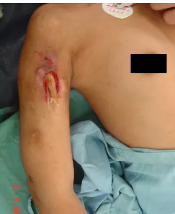
In developing countries, such as Indonesia there are many poor people that always make use of the service of traditional medicine like bonesetter when dealing with open factures of the bone. In treating these fractures, initially they reduce the fracture without cleaning the wound, then directly covered the wound by herbals, then, tighten up using wood as a slab on the affected side. After several months, the patient comes to the orthopedic clinic with persistent sinus tract and active pus, meaning that chronic osteomyelitis occurred (see Figure 1).
Figure 1. This photograph shows a large draining sinus tract with huge sequester exposed in a patient with chronic osteomyelitis of the right humerus.
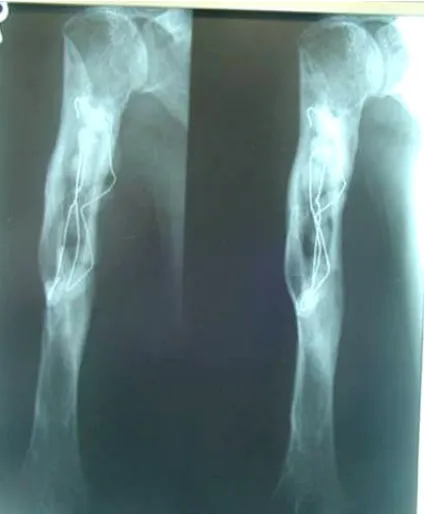
This is despite the fact that the release mechanisms of antibiotic from bone cements is poorly understood and controlled. These beads are left in situ, as shown in Figure 2, and are removed at a second procedure after fourteen days, when the cavity should be filled with bone graft, for instance in infected bone for bone consolidation.
Antibiotic-loaded PMMA beads are commercially available under the name Septopal®, however, the price of these beads is far too high for most patients. Fosfomycin is the commonly used antibiotic in these hand-made beads, because of its costs, its wide antibacterial spectrum, its small molecule (molecular weight 138), and its ability to remain stable up to the high temperatures reached during polymerization of the bone cement.9,10
Figure 2. A chain of beads has been inserted down the right humeral shaft, after removal of infected materials.
The usage of hand-made fosfomycin-loaded beads in Indonesia yields good clinical result, the infected bone is cleared from all infected materials, and eventually it has been returned to healthy bone. Successful treatment of an overcome osteomyeltis is still unknown whether the good clinical results come from these beads or from systemic antibiotics, or from the combination of both. The aim of this study is to evaluate several currently used hand-made fosfomycin-loaded beads by determination of antibiotic spectrum, kinetics of the fosfomycin release. All characteristics were compared with those of the commercially available gentamicin-loaded PMMA beads (Septopal®).
Six beads were made by different mixing techniques from different orthopaedic surgeons. Beads#1 were made of 40g of Zimmer bone cement (Zimmer, Inc, USA) and 4g of fosfomycin-sodium powder (Fosmicin, MEIJI, Japan). Bone cement was prepared by mixing 40g of Zimmer bone cement powdered polymethylmethacrylate (PMMA) with 4g of fosfomycin-sodium powder in a sterile bowl. After both materials are stirred up homogenously by spatula, then 2 ml of methylmethacrylate (MMA) liquid was poured into these mixed materials. When the mixture became doughy, then, they poured into three templates and wait until hardening phase of the PMMA cements within ten minutes. Each template consists of ten round holes to build spherical shapes. It shows that all beads are spherical shape and in same size. These beads were not connected on a small diameter of stainless steel wire.
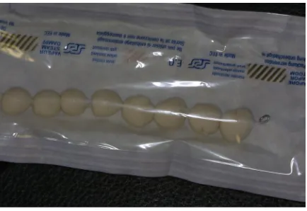
Beads#2 to #6 made of commercially available Simplex® P bone cement (Stryker Howmedica OSTEONICS, Howmedica International S, Limerick, Ireland). Bone cement was prepared by mixing the powdered PMMA with MMA liquid monomer in a bowl with a spatula. After both materials are mixed homogenously and doughy, then 2g of fosfomycin-sodium was added manually. When the mixture became doughy and non-sticky within four minutes, spherical shape beads were hand rolled in a various diameters between 5 to 10 mm’s. The beads were threaded onto a small diameter of stainless steel wire and the ends of wire were knotted (see Figure 3). All procedures were carried out under sterile conditions. By different orthopaedic surgeons handrolled fosfomycin-loaded bone cement bead sets were built in spherical shape. The size and weight of these beads are varied between 1.57 cm and 2.84 cm, with a weight range of 0.51g to 2.44 g.
Figure 3. Hand-made fosfomycin-loaded PMMA beads; ten handrolled beads attached on a wire.
Beads#1 to beads#6 and Septopal® beads were immersed in 10 ml of sterile Phosphate Buffer Saline, PBS (NaCl 8.76 g/l, K2HPO4 43.5 g/l, KH2PO4 34.5 g/l, pH 7.4) and incubated at
370C. After 24h of incubation 15 µl samples were taken and these were placed on seeded
Trypton Soya Brooth (TSB) agar plates. The zones of inhibition were established by measuring the clear areas around one drop of elution fluid. Absence of an inhibition zone was taken as a sign that the antibiotic concentration was to low to inhibit bacterial growth. For this study we used ten clinical strains isolated from patients with an implant-related infection of the University Medical Center Groningen, The Netherlands. Distribution of the strains are according to the occurrence of osteomyelitis.11,12 Each strain was diluted in 4 ml of 0.9% saline to concentration approximately of 108 bacteria/ml. After overnight incubation at 370C of
the seeded agar plates the inhibition zone was scored (size in mm). The zone of inhibition was considered as clear area around of a drop of elution medium in which bacteria were not able to grow.
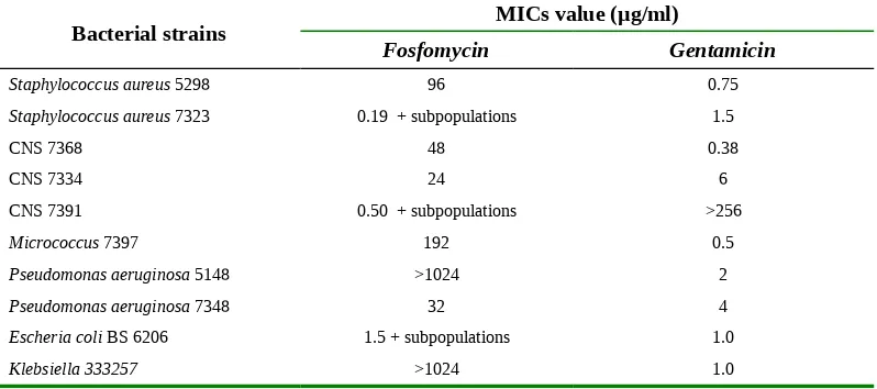
In addition, the minimal inhibitory concentration (MIC; µg/l) of the isolated bacteria against fosfomycin and gentamicin and possible subpopulations (see Table 1) were determined by an E-test (AB Biodisk, Dälvagen,Sweden).
Table 1.
MICs values of the ten bacterial strains used in this study.
2.3. Antibiotic release
Six different handmade beads and Septopal® beads were immersed in 10 ml sterile PBS (NaCl 8.76 g/l, K2HPO4 43.5 g/l, KH2PO4 34.5 g/l, pH 7.4) and incubated at 370C. At
indicated time points (24h, 48h, 72h, and 144h), the beads were transferred to fresh 10 ml PBS and again incubated at 370C. Duration of fosfomycin release was established by taking
15 µl samples at the indicated time points and placing this drop on TSB agar plates seeded with the ten clinical bacterial strains showing any antibacterial efficacy. The antibiotics release was determined by measuring the clear areas in four quadrants on one agar plate. Each quadrant contained one drop (15 µl) of the elution fluid taken at the indicated time points. The
Bacterial strains MICs value (µg/ml)
Fosfomycin Gentamicin
Staphylococcus aureus 5298 96 0.75
Staphylococcus aureus 7323 0.19 + subpopulations 1.5
CNS 7368 48 0.38
Escheria coli BS6206 1.5 + subpopulations 1.0
ten bacterial strains were diluted in 4 ml of 0.9% saline to concentration 108 bacteria/ml. After
24h incubation at 370C the inhibition zone was scored (size in mm). Again, absence of an
inhibition zone was taken as a sign that the antibiotic concentration was to low to inhibit bacterial growth.
3. RESULTS
3.1. Antibacterial efficacy
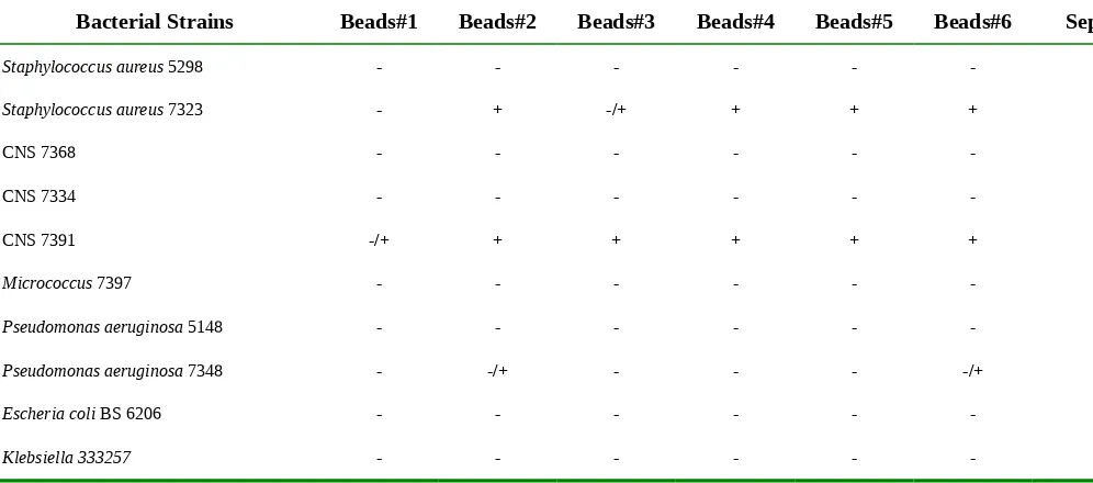
Table 2 shows that handmade fosfomycin-loaded beads were only slightly effective in two of the ten bacterial strains is used, namely Staphylococcus aureus 7323 and CNS 7391. In
Pseudomonas aeruginosa 7348, very partial efficacy of hand-made fosfomycin-loaded beads could be seen at 24h in only beads#2 and beads#6. Antibacterial efficacy of Septopal® beads could be seen in all bacterial strains used, except for CNS 7334 and CNS 7391, both gentamicin-resistent strains. In brief, Septopal® beads showed efficacy against eight of the ten bacterial strains, while the hand-made fosfomycin-loaded beads only showed efficacy against two of the ten strains.
Bacterial Strains Beads#1 Beads#2 Beads#3 Beads#4 Beads#5 Beads#6 Septopal®
- =no zone; -/+ = partial inhibition zone; + = clear inhibition zone
3.2. Antibiotic release
Table 3 presents the measured clear zones achieved with elution fluid from the hand-made fosfomycin-loaded beads. For both strains, beads#1 was not effective and beads#2-beads#6 showed only effectiveness at 24h. Subpopulations appeared at 24h with CNS 7391. With beads#2-beads#6, CNS 7391 showed partial inhibition after 48h, 72h, and 144h, meaning there was some release but not high enough the create a clear inhibition zone. Overall, the hand-made fosfomycin-loaded beads showed short-lived fosfomycin release.
Table 3. Clear zones of inhibition (mm) around elution fluid of the fosfomycin-loaded PMMA
beads for two strains showing any bacterial efficacy.
Staphylococcus aureus 7323 CNS 7391
24h 48h 72h 144h 24h 48h 72h 144h
Beads#1 - - - -* -* -*
Beads#2 16 - - - +subpopulations25 -* -* -*
Beads#3 - - - - +subpopulations15 -* - -*
Beads#4 11 - - - +subpopulations20 -* -* -*
Beads#5 7 - - -* +subpopulations25 +subpopulations20 - +subpopulations20
Beads#6 13 - - - +subpopulations25 -* -* -*
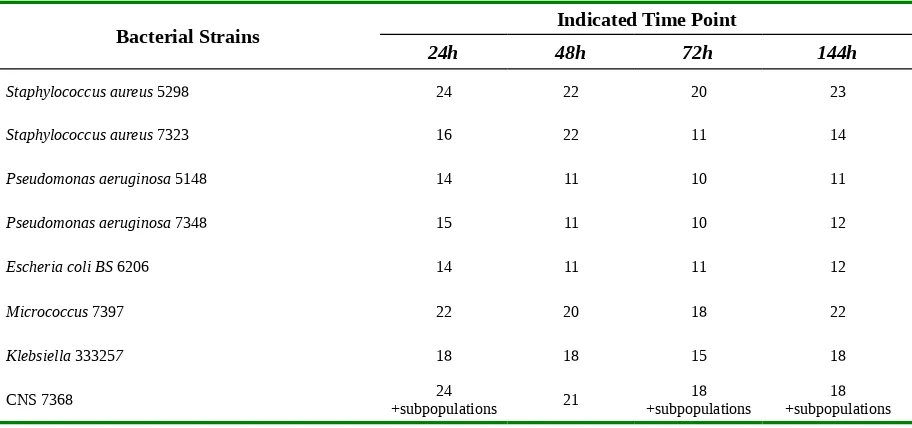
Table 4 presents the measured clear zones achieved with elution fluid from Septopal® beads. These beads showed the same release kinetics for all eight bacterial strains used, a high initial release and thereafter the antibiotic release gradually diminished for the next hours, with a small increase at 144h because of the antibiotic build-up of three days. Overall, Septopal® beads showed longlasting release of gentamicin. Note that subpopulations appeared in one strain, CNS7368.
Table 4.
Clear zones of inhibition (mm) around elution fluid of Septopal® beads for
eight strains showing any bacterial efficacy.
Bacterial Strains Indicated Time Point
24h 48h 72h 144h
Staphylococcus aureus 5298 24 22 20 23
Staphylococcus aureus 7323 16 22 11 14
Pseudomonas aeruginosa 5148 14 11 10 11
Pseudomonas aeruginosa 7348 15 11 10 12
Escheria coli BS 6206 14 11 11 12
Micrococcus 7397 22 20 18 22
Klebsiella 333257 18 18 15 18
CNS 7368 +subpopulations24 21 +subpopulations18 +subpopulations18
4. DISCUSSION
higher than after systemic application of the same antibiotic and far above the minimal inhibitory concentrations of most common pathogens.
The elutions of fosfomycin-loaded beads were only effective in two of the ten bacterial strains tested, while the elutions of Septopal® beads were effective against eight of the ten strains used. An E-test determined the MIC values of the clinical strains for both fosfomycin and gentamicin. Four of the ten clinical strains turned out to be fosfomycin resistant, meaning a MIC value above 64 µg/ml. On the other hand two of the ten strains were considered gentamicin resistant as their MICs value was above 4 µg/ml. Accordingly, the fosfomycin-loaded beads did not show efficacy against four of the fosfomycin sensitive bacteria. Most of hand-made fosfomycin beads revealed partial inhibition, meaning there is some sensitivity but the release of fosfomycin is not high enough to fully inhibit growth of the bacteria.
The large inhibition zone seen with beads#2 can be explained by the fact that these are the biggest beads in the study. Since these beads are huge when compared to the others in this study, they contain much more antibiotics. Together with a large surface, they were able to release more antibiotics as antibiotic release is mostly a surface phenomenon (van de Belt 2001). Moreover, prolonged release from bone cement is only possible when the cement is very porous.16 The release rate of antibiotic from PMMA is important clinically. Porosity/permeability of the PMMA is an important factor in determining the release rate.
The evaluation of fosfomycin-loaded beads are using clinical strains isolated from patients with an infected joint replacement in Dutch patients, these situations mostly due to deep infection. It must be different with clinical strains focused on chronic osteomyelitis hematogenously. Retrospective study was done in 56 osteomyelitis patients at the Dept. of Orthopedic and Traumatology Hasan Sadikin Hospital, Bandung, Indonesia. The microorganism pattern of osteomyelitis is S. aureus 26.9% of all cases, and P. aeruginosa
11.0% of all cases.17
We may conclude from the presented observations that a mixture of fosfomycin and PMMA bone cement is not an effective one. Efficacy could only be seen in two of the ten bacterial strains. We assume that hand-made beads were not porous enough as porosity of bone cement is the key to high antibiotic release. Therefore we have to try procedures to increase the porosity, like adding glycine as in currently done in the Septopal® beads.18 Also trying an other antibiotic is an option, together with changing the antibiotic carrier from PMMA bone cement to calcium phosphate.19 One big advantage of calcium phosphate above PMMA bone cement is that calcium phosphate is biodegradable, so no second operation is needed to remove the beads.
systemic antibiotics given and the locally very low fosfomycin concentrations released by the bone cement beads.
REFERENCES
1.Buchholz, H.W., and Engelbrecht H. Uber die Depotwirkung einiger Antibiotica bei Vermischung mit dem Kunstharz Palacos. Chirurg 1970;41:511-15.
2.Klemm, K. Gentamycin-PMMA-Kugeln in der Behandlung abszedierender Knochen und Weichteilinfektionen. Zentral bl. Chirurg 1979;104:934-42.
3.Brinker, M.R. Antibiotic Beads in General Principles of Trauma, in: Review of Orthopaedic Trauma 2001:31-43.
4.Patzakis, M.J., Wilkins, J., Kumar, J., Holtom, P., Greenbaum, B., and Ressler, R. Comparison of the results of bacterial cultures from multiple sites in chronic osteomyelitis of long bones: A prospective study. J Bone Joint Surg Am 1994;76:664-666.
5.Walenkamp, G.H.I.M., Coombs, R., and Fitzgerald, R.H. Gentamicin– PMMA beads: Pharmacokinetics and Toxicology in: Infection in the Orthopaedic Patient 1989:163-66. 6.Wahlig, H., Dingeldein, E. Antibiotics and bone cements. Experimental and clinical long term observations. Acta Orthop Scand 1980;51:495.
7.Brinker. M.R. Antibiotic Beads in General Principles of Trauma in: Review of Orthopaedic Trauma 2001:31-43.
8.Bronzino, A.D. Polimeric Biomaterials, The Biomedical Engineeering Handbook, IEEE Press USA 1995:581-88.
9.Buccholz, H.W., Elson, R.A., and Engelbrecht, E. Management of deep infection of total hip replacement. J Bone Joint Surg 1981;63:342-353.
10.Seligson, D., and Henry, S.L. Newest knowledge of treatment for bone infection: antibiotic-impregnated beads. Clin Orthop 1993;295:2-18.
11.Schmidt, A.T., and Swiontkowski, M.F. Pathophysiology of infections after internal fixation of fractures. J Am Acad Orthop Surg 2000;8:285-291.
12.Alonge, T.O., Ogunlade, S.O., Omololu, A.B., Fashina, A.N., and Oluwatosin, A. Management of chronic osteomyelitis in a developing country using ceftriaxone-PMMA beads: an initial study. Int J Clin Pract 2002;56:181–3.
13.Bowyer, G.W., and Cumberland, N. Antibiotic release from impregnated pellets and beads. J Trauma 1994;36:331-5.
15.Van de Belt, H., Neut, D., Van Horn, J.R., Van der Mei, H.C., Schenk, W., and Busscher, H.J. Staphylococcus aureus biofilm formation on different gentamicin-loaded polymethylmethacrylate bone cements. Biomaterials 2001;22:1607-1611.
16.Saragih, J.R. Microorganisms Pattern and Its Sensitivity in Osteomyelitis Case at the Dept. of Orthopedic Surgery Dr Hasan Sadikin Hospital, Bandunng, Thesis 1999:50-51.
17.McLaren, A.C., Nelson, C.L., McLaren, S.G., and DeClerk, G.R. The effect of glycine filler on the elution rate of gentamicin from acrylic bone cement. A pilot study. Clin Orthop
2004;427:25-27.