Apr.-Jun. 2014, Volume 11, No. 2 (Serial No. 94) pp. 68-73 Journal of US-China Medical Science, ISSN 1548-6648, USA
The Optimal Dose of Euphorbia milii Extracts in NKp46
Expression against Mice Infected with Mycobacterium
tuberculosis
Ni Made Linawati1, Ida Ayu Alit Widhiartini2, I Nyoman Wande3, Ida Bagus Nyoman Putra Dwija4, I Gusti Sri Wiryawan1, I Gusti Kamasan Arijana1, Wayan Sugiritama1, I Gusti Ayu Ratnayanti1, Ida Ayu Ika Wahyuniari1 and I Gusti Ngurah Mayun1
1. Department of Histology, Faculty of Medicine, University of Udayana, Denpasar 80232, Bali, Indonesia
2. Department of Pharmacy, Faculty of Medicine, University of Udayana, Denpasar 80232, Bali, Indonesia
3. Department of Clinical Patology, Faculty of Medicine, University of Udayana, Denpasar 80232, Bali, Indonesia
4. Department of Microbiology, Faculty of Medicine, University of Udayana, Denpasar 80232, Bali, Indonesia
Abstract: Objective: To compare the effect of EEM (Euphorbia milii) extract in various dose for NKp46 expression in mice infected with MTB (Mycobacterium tuberculosis). Methods: An experimental study with 24 mice Balbc were divided into 8 groups of treatment which is observed at weeks I and II. All groups were infected with Mycobacterium tuberculosis strain H37Rv then each successive group was given sterile distilled water (P0.1 and P0.2), extract of EEM 5 mg/20 grbw (gram body weight) (P1.1 and P1.2), 10 mg/20 grbw (P2.1 and P2.2), and 15 mg/20 grbw (P3.1 and P3.2). Then, termination in each group at weeks I (P0.1, P1.1, P2.1 and P3.1) and II (P0.2, P1.2, P2.2 and P3.2). Pulmonary organ harvesting conducted for immunohistochemistry of NKp46 expression. Result: Quantitative results of NKp46 expression from successive groups (1 to 8) are 48.33, 25.33, 58.67, 46.67, 35.67, 58.67, 61.33 and 40.67, respectively. One Way ANOVA showed significant difference between groups (P < 0.05). Conclusions: EEM with doses of 5 mg/20 grbw and 15 mg/20 grbw are able to increase the expression of NKp46 in the second week, whereas a dose of 10 mg/20 grbw is able to increase expression of NKp46 on the first and second week. It is make an opinion if EEM with 10 mg/20 grbw is the optimal dose to increase the expression of NKp46 in mice infected with Mycobacterium tuberculosis.
Key word: Lung tuberculosis, natural killer cells, Euphorbia milii, NKp46.
1. Introduction
According to WHO data in 2010, Indonesia ranked fifth in the country with the highest tuberculosis burden in the world. The mechanism of pulmonary parenchymal tissue damage which is manifested as pulmonary tuberculosis is based on immunopathogenesis. Characteristic lesions of pulmonary tuberculosis is granulomas, due process based on the reaction of cell interactions including macrophages, lymphocytes, epithelioid cells, NK cells (natural killer cells), and neutrophils against
Corresponding author: Ni Made Linawati, M.D., research fields: immunology and histology. Email: [email protected].
Mycobacterium tuberculosis [1, 2]. NK cells are
effector in the innate immune response to patrolling against pathogens and tumor invasion. The main biological function of NK cell activity include natural cytotoxic and cytokine secretion that are directly or indirectly control infections and tumors as well as regulate the immune system. Through the dual role of NK cells in the secretion of IL-22 which activate macrophages infected with Mycobacterium
tuberculosis and NKp46 expression which lyse
macrophages infected with Mycobacterium
tuberculosis. NKp46 is a natural cytotoxic receptors [3-5].
Active NK cells are expected to work optimally in eliminating MTB in macrophages infected either by
DAVID PUBLISHING
increasing the activation of macrophages and lyse infected macrophages so that tuberculosis can be controlled. Euphorbia milii is a species of “euphorbiaceae” contains flavonoids (phenols and phenolic glycosides), saponins (terpenoids and steroids), and tannins (carbohidrat). Several studies have shown antibacterial effects, inhibitors against aspergillus, mollusidal, as well as non-teratogenic [6-8]. This study aimed to investigate the effect of EEM (Euphorbia milii extract) increased expression of NKp46 of the lungs of mice infected with
Mycobacterium tuberculosis.
2. Material and Methods
2.1 Preparing Suspension of Mycobacterium tuberculosis Strain H37Rv
Mycobacterium tuberculosis strain H37Rv (ATCC27294) were grown in liquid medium Middlebrook 7H9 for 2 weeks, then stored at -70 °C and is ready to used for research. Before use bacterial suspension was thawing at 37 °C and sonification for 10 s to dissolve the clot of bacteria became homogeneous and separate one cell to another. To infected the mice we inoculated intranasally 60 μL suspension contain 105 bacteria per mL [9].
2.2 Preparing Ethanol Ekstrak of Euphorbia milii
(EEM)
Euphorbia milii flowers are taken from Ketewel
(region of Gianyar, Bali). These flowers are taken in a fresh state, colored pink with the whole look, subsequently collected and aerated to dry. Once dried, they are crushed with a blender, and then weighed 100 g.
E. milii flower powder was added 300 mL of methanol
and stirred with a magnetic stirrer for 1 h at room temperature. Then, filtered with Whatman paper No. 42 in order to obtain the filtrate 1. Obtained residue was re-extracted filtrate thus obtained 2. Filtrates 1 and 2 mixed filtrate was then evaporated with a rotary evaporator.
2.3 Mice Infection with Mycobacterium tuberculosis
Strain H37Rv
Mice were infected with 105 bacteria per mL intranasally, initially created as suspension equivalent to 107 McFarland 1, then diluted until a concentration of 105 bacteria, as many as 60 µL of suspension in the nostrils of mice inoculated with a micropipette. Then, the mice were placed in the cabinet for further observations [9].
2.4 Immunohistochemistry NKp46 Expression in Mice Lung Tissue
Right lobe of the lung tissues were fixed in 10% formaldehyde solution in PBS for 2 days and then soaked in paraffin and cutting is done along the widest area of each lobe. This is done for all the examined lung tissue so that all the lung tissue of mice examined in the same area. Lung tissue with 5 µm thickness
separated 100 μm each other then process with
immunohistochemical staining technique based on rabbit anti-mouse NKp46 (Bioss, Bs-2417R) then using secondary antibody from Daco (LSAB + systems-HRP). Then see below the light microscope CX3100 with 100× magnification. Numbers of positive NKp46 cells expression were counted in 5 fields then calculated the average per 100 cells.
2.5 Data Analysis
Data quantity of NKp46 expression were tabulated then analyzed with SPSS 14.0 programme. The homogeneity by Lavene test with a confidence level
3.
Results
3.1 The Effect of EEM against NKp46 Expression in
Lung Tissue of Mice Infected with Mycobacterium tuberculosis
The experiment was conducted in June 2013 at Department of Histology, Faculty of Medicine and Department of Pathology, Faculty of Veterinary, University of Udayana. Primary antibody kit of anti-mouse NKp46 from Bioss (Bs-2417R) and secondary antibody of anti-rabbit from Dako were used. Immunohistochemistry results of anti NKp46 in samples which were terminated on day 8 and 15 consist of 8 groups: P0.1 is the control 1, terminated at day 8; P1.1 is the treatment 1, with 5 mg/20 grbw EEM, terminated at day 8; P2.1 is the treatment 2, with 10/ 20 grbw EEM, terminated at day 8; P3.1 is the treatment 3, with 15 mg/20 grbw EEM, terminated at day 8; P0.2 is the control 2, terminated at day 15; Group P1.2 is treatment 4, with 5 mg/20 grbw EEM, terminated at day 15; P2.2 is the treatment 5, with 10 mg/20 grbw EEM, terminated at day 15; P3.2 is treatment 6, with 15 mg/20 grbw EEM, terminated at day 15. The example of NKp46 imunostaining in lung mice was showed in Fig. 1.
3.2 Histopathological Picture of the NKp46 Expression in the Study Group
The immunohistochemistry staining of NKp46 expression in the above 8 groups are presented.
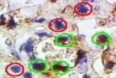
The fourth picture in Fig. 2 is the result of NKp46 imunostaining in mice lung tissue which were terminated on day 8. The fourth picture in Fig. 3 shows the result which terminated on day 15. Positive cells expressing NKp46 is the cell with the cell membrane brown with or without brown nuclei. Whereas, cells that do not express NKp46 are mononuclear cells with blue nuclei, cytoplasm and cell membrane. In Fig. 1, positive cells showed brown nuclei is marked with a green circle while negative cells showed blue nuclei which is marked with a red circle. The number of
positive cells in Figs. 2 and 3 were counted in five fields of view using Optilab camera cell count programme. Each group contained three replications. Then the mean of NKp46-positive cells per 100 cells of lung tissue were calculated.
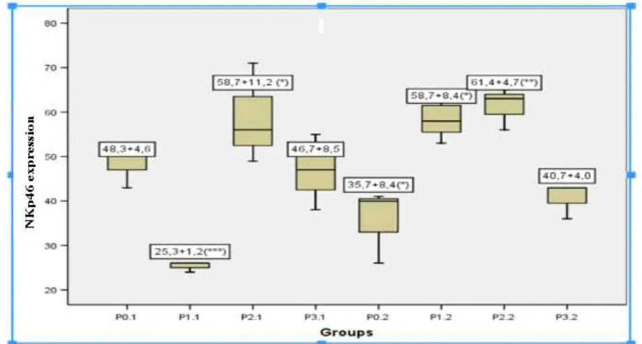
Fig. 4 showed the mean and standard deviation of NKp46-positive cells per 100 cells of lung tissue, with its significantly different.
4. Discussion
Based on the results, there are different expressions of NKp46 between eight groups. In the first week, NKp46 was expressed successively from the lowest group to the groups as high as P1.1, P3.1, P0 .1 and P2.1. When compared with the control group (P0.1), the expression of NKp46 on lower P1.1 group is probably due to EEM at a dose of 5 mg/20 grbw within 1 week which is not optimal for increasing the expression of NKp46. A dose of 15 mg/20grbw may cause resistance to the receptors so that the expression is not increased. However, at a dose of 10 mg/20 grbw (P2.1), the expression is increased compared to the control group although the Scheffe test showed no significant increase (P = 0.82), which suggested the possibility of an optimal dosage for increased activity NK cells in NKp46 expression .
Fig. 2 The NKp46 imunostaining (Bioss) and secondary antibody (Daco) of mice lung from 4 treated group in the first week. (CX3100 Olympus Microscope, magnification 100×, Optilab doc, Histology Department, Medical Faculty of Udayana University).
Fig. 3 The NKp46 imunostaining (Bioss) and secondary antibody (Daco) of mice lung from 4 treated group in the second week. (CX3100 Olympus Microscope, magnification 100×, Optilab doc, Histology Department, Medical Faculty of Udayana University).
Fig. 4 The mean (with standard deviation) of NKp46 expression in each groups of study. (*): Significant different with another group. P2.1 is significantly different with P1.1, and P1.2 is also significantly different with P1.1. (**): Significant different with the other two groups. P2.2 is significantly different with P0.2 and also P1.1. (***): Significant different with the other three groups. P1.1 is significanly different with the other three group such as P2.1, P1.2 and P2.2.
P0.2 P1.2 P2.2 P3.2
In the second week, the lowest expression of NKp46 is in the control group (P0.2), while the highest is in P2.2 (dose 10 mg/ 20 grbw). It shows EEM flower extract in different doses at 2-week administration was able to increase the expression of NKp46 compared to the control group. Overall, it shows the content of the active substance in the EEM on duration of 2 weeks were able to increase the expression of NKp46 at all doses (Fig. 4). Although when it is analyzed by Scheffe test, it showed a significant increase (P = 0.03) only at doses 10 mg/20 grbw (P2.2) when compared with the control group (P0.2). NKp46 expression was deteced in all groups, because the infection with Mycobacterium
tuberculosis is a potent inducer for NK cells that are
activated to express NKp46. It is in accordance with the experiments performed by Vankalayapati et al. [10] who found an increase in the expression of NKp46 in NK cells and macrophages co-cultures which are infected with Mycobacterium tuberculosis, then causing lysis of macrophages infected with
Mycobacterium tuberculosis. In addition to infection
with Mycobacterium tuberculosis, Culley [3] also found that NKp46 receptor plays an important role in infection control against influenza virus hemagglutinin which binds to NKp46 expressed in cells infected with influenza virus and causes lysis of the infected cells. Increased expression of NKp46 in the group with the dose of EEM 10 mg/20 grbw in both first and second week suggests the possibility of optimal dose is 10 mg/20 grbw. There was an increase in the second week of NKp46 expression at all doses when compared to the control group EEM. It is because of the active substance content in the EEM, containing a variety of active substances such as flavonoids, saponins, triterpenoids, phenols, and alkaloids.
Flavonoid is one of the compounds contained in
Euphorbia milii. Flavonoid compounds are said to have
immunomodulatory effects on several immune cells. In NK cells, it was found to have immunostimulatory effects such as IFN-γ synthesis and inhibitory action on the cytotoxic activity. These different effects can be
caused by the concentrations or conditions of different flavonoids from natural materials [11].
Increased expression of NKp46 is an inducer for NK cells to produce IFN-γ. Flavonoids increase the activity of NK cells through the high expression of NKp46, which can increase the secretion of IFN-γ. The content of flavonoids in Euphorbia milii flowers gives the possibility to act as an immunomodulator for NK cells. Increased expression of NKp46 of NK cell would increase IFN-γ levels many times higher after cytokine stimulated [12].
6. Conclusions
Ethanol extract of flowers of Euphorbia milii (EEM) at a dose of 5 mg/20 grbw; 10 mg/20 grbw, and 15 mg/20 grbw are able to increase the expression of NKp46 in lung tissue of mice infected with
Mycobacterium tuberculosis in the second week. EEM
at dose of 10 mg/20 grbw is an optimal dose because it increase the expression of NKp46 in lung tissue of mice infected with Mycobacterium tuberculosis in the first and second week.
Acknowledgment
Thank to the Higher Education Grant Programme through Decentralization Competitive Grant 2013 for funding this research, and the research centre in Udayana University.
References
[1] R.S. Wallis, M. Broder, J. Wong, A. Lee, L. Hoq, Reactivation of latent granulomatous infections by infliximab, Clin. Infect. Dis. 41 (Suppl. 3) (2005) 194-198. [2] N. Wiweko, Global Tuberculosis Control, WHO Report,
2010.
[3] F.J. Culley, Natural killer cells in infection and inflammation of the lung, Immunology 128 (2) (2009) 151-163.
[4] R. Dhiman, M. Indramohan, P.F. Barnes, R.C. Nayak, P. Paldipally, L.V. Rao, et al., IL-22 produced by human NK cells inhibits growth of Mycobacterium tuberculosis by enhancing phagolysosomal fusion, Journal Immunol. 183 (10) (2009) 6639-6646.
[5] R. Vankayalapati, The Role of NK cells in human M.
http://www.labome.org/grant/r01/ai/the/role/the-role-of-n k-cells-in-human-m--tuberculosis-infection-7538343.html (accessed Feb. 25, 2013).
[6] B. Sermsart, S. Sripochan, T. Suvajeejarun, R. Kiatfuengfoo, The molluscicidal activities of some Euphorbia milii hybrids against the snail indoplanorbis exustus, Southeast Asian J. Trop. Med. PublicHealth 36 (Suppl. 4) (2005) 192-195.
[7] S. Dalimartha, The atlas of medicinal plants in Indonesia, www.perpustakaan.depkes.go.id/.../opac-search.pl? (accessed Oct. 13, 2011). (in Indonesian)
[8] Euphorbia milii in Philippine medicinal plants, 2011, http://stuartxchange.com/CoronaDeEspina.html (accessed Nov. 26, 2011). (in Indonesian)
[9] B.M. Saunders, C. Cheers, Intranasal infection of beige mice with Mycobacterium avium complex: Role of
neutrophils and natural killer cells, Infecction and Immunity 64 (10) (1996) 4236-4241.
[10] R. Vankalayapati, B. Wizel, S.E. Weis, H. Safi, D.L. Lakey, O. Mandelboim, et al., The NKp46 receptor contributes to NK cell lysis of mononuclear phagocytes infected with an intracelular bacterium, The Journal of Immunology 168 (7) (2002) 3451-3457.
[11] E. Middleton, C. Kandaswami, T.C. Theoharides, The Effect of plant flavonoids on mammalian cells: Implications for inflammation, heart disease and cancer, Pharmacological Review 52 (4) (2000) 678-751. [12] K.H. Mair, S.E. Essler, M. Patzl, A.K. Storset, A.