Research Journal of Pharmaceutical, Biological and Chemical
Sciences
Comparison of Sol-Gel And Hydrothermal Synthesis Of Zinc Ferrite (ZnFe
2O
4)
Nanoparticles.
Syukri Arief*, Rahmayeni, and Zulhadjri.
Department of Chemistry, Faculty of Mathematic and Natural Sciences, Andalas University, Kampus Limau Manis – Padang, West Sumatera, Indonesia.
ABSTRACT
Two solution-based techniques (sol-gel and hydrothermal synthesis methods ) for the preparation of ZnFe2O4 nanoparticles have been investigated. In sol-gel method, ethanol is used as thesolvent, while citric
acid as thechelating agent. The hydrothermal process used NaOH solution as the precipitator and controlling alkaline conditions. The synthesized nanoparticles are characterized by X-ray diffraction analysis, Scanning Electron Microscopy and Transmission Electron Microscopy. Spherical of ZnFe2O4 nanoparticle with a size of 10
nm can be obtained using hydrothermal method, which is smaller than nanoparticle resulted from sol gel method (35 nm). The results depict that formation methods play an important role on the particle size and quality of the ZnFe2O4nanoparticles.
INTRODUCTION
Ferrite compounds (MFe2O4) where M = transition metal or alkaline earth is a magnetic material with
spinel structure. Recently, ferrite compounds has attracted the attention of researchers for potential applications such as data storage, biosensors, drug delivery and diagnosis of the disease, gas sensors, electronic devices, catalysts and others [1-2]. In the photocatalytic process MFe2O4 particles have been used to
degrade some of the volatile organic compounds in the water[3-4]. MFe2O4 with a narrow band gap can be
used to enhance the ability of ZnO in the photocatalytic process, because this material can narrow band gap of ZnO and expanding absorption area in the visible light region. In addition, ferrite with magnetic properties can be separated easily from the liquid by using the influence of the external magnetic field [5-6].
On the other hand, some spinel material such as ZnFe2O4known as semiconductorcompound
withrelatively smallenergy gap (about 2 eV). The semiconductor properties show a very good response to the visible light, good photochemical stability, as well as magnetic profitable. Until now, ZnFe2O4nanoparticles has
been synthesized using various methods, such as co-precipitation [7], sol-gel [8], the reaction of solid-state [9], the combustion reaction using glycine [10] and urea as a reducing agent [11], hydrothermal synthesis [12], solvothermal and microwave solvothermal synthesis [13], a high-energy ball-milling [14], microwave combustion method [15]. Most of the research was done independently. The current study made and
compared between solgel and hydrothermal methodson the preparation of ZnFe2O4nanoparticles. The use of
sol-gel and hydrothermal methods on the structural, morphological and magnetic properties of the prepared ZnFe2O4 nanoparticle is presented.
MATERIALS AND METHODS
The materials used in this study was zinc nitrate tetra hydrate Zn(NO3)2. 4H2O (Merck),
Fe(NO3)3.9H2O(Merck), citric acid C6H8O7 (Merck), NaOH (Merck), ethanol p.a (Merck), ethanol, and distilled
water. Equipment used in the form of glassware, Teflon autoclave, furnace and spectrophotometers. Characterization of the samples was done by means of X-ray diffraction (XRD), Scanning Electron Microscopy-Energy Dispersive X-ray (SEM-EDX), and Transmission Electron Microscopy (TEM).
Synthesis by sol-gel method:
A total of 10 mmol of Zn(NO3)2.4H2O and 20 mmol Fe(NO3)3.9H2O mixed in 40 mL ethanol p.a, then 30
mmol of citric acid was dissolved in 40 mL ethanol P.A. All of the solution is mixed with a mole ratio of Zn + 2: Fe + 3: citric acid is 1: 2: 3. The mixture was stirred at 500 rpm for 1 hour at 70°C to form a wet gel. The gel formed is dried in an oven at a temperature of 120°C for 24 hours to form a dry gel. Then, dry gel is crushed with a mortar to form more fine powders. The resulting powder heated in a furnace at a temperature of 600°C for 2 hours, and then characterized by XRD equipment, SEM-EDX, and TEM.
Synthesis by hydrothermal method:
Some of 10 mmolZn(NO3)2.4H2O and 20 mmol Fe(NO3)3.9H2O dissolved in 100 mL of distilled water
and stirred until homogeneous. 2 M NaOH was then added to the solution gradually and keep stirring until pH 12. The result was a mixture of suspension. The mixture was poured into tube autoclave and heated in an oven at a temperature of 180°C for 3 hours. The nanoparticles were filtered then rinsed with distilled water until pH 7-8 and dried. The resulting powders were characterized by means of XRD equipment, SEM-EDX, andTEM.
RESULTS AND DISCUSSION
Fig 1: The photograph of ZnFe2O4 powders which were synthesized by a) sol-gel and b) hydrothermal
methods
XRD samples were prepared by the method ZnFe2O4 solgel (a) and hydrothermal (b) as can be seen in
Figure 2. The diffractograms for both samples showed that every sample has a cubic spinel structure according
to the standard JCPDS No. 89-1012. Diffraction peaks at θ of 0.0 °, . °, . °, . °, . ° and . 0°
can be ascribed to the reflection spinel ZnFe2O4field at (220), (311), (400), (422), (511) and (440). On the other
hand, the difference in this preparation method gives different peak sharpness and heights. Peaks obtained formsolgel method weresharper and higher than hidrotemal method. Based on the equation Scherer, a sharp and heightpeak give crystal size was larger than the width and low peak. This can be proved later by SEM and TEM.
Fig 2: XRD pattern of ZnFe2O4 which are synthesized by a) sol-gel process and b) Hydrothermal
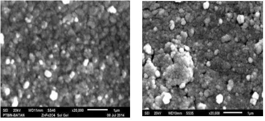
The results of the SEM image (see Fig. 3) showed that the sample ZnFe2O4 with solgel method (a) and
hydrothermal (b) formed by the agglomeration of the particles. Smaller particle size was obtained from ZnFe2O4prepared by solgel method. This data is matched with the visualization of the powder before and
approved also agreed by the XRD data. The fine particles obtained by using hydrothermal, since this methodsused lower temperature during processing. The use ofsolgel method to get a good ofcrystallinitymust be done at temperatures above 500oC. When the temperature is raised, in general, the growth of crystallinity
will also increase.
b)
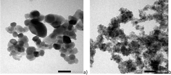
Representative of TEM pictures of solgel and hydrothermal samples are shown in Fig. 4. Most of the ZnFe2O4 nanoparticles are spherical in these pictures. It can be seen that differences in the shape and size of
ZnFe2O4nanoparticles were synthesized via sol-gel method (Fig.4a) and hydrothermal (Fig. 4b). Particles
prepared by sol gel method have a larger size with an average size of about 35 nm. This may be caused by high temperatures in the formation of ZnFe2O4 crystals (calcination at 600oC). This condition has caused the
incorporation of particles or small crystals to form a larger particles.While particles prepared by hydrothermal method has a smaller size that is below 10 nm. Moreover, particles obtained by hydrothermal method has a size that is much more refined and homogeneous
a) b)
Fig 4: TEM images of ZnFe2O4 which are synthesized by a) sol-gel process and b) Hydrothermal
CONCLUSION
Two solution-based techniques for the preparation of ZnFe2O4 nanoparticles have been presented:
sol-gel and hydrothermal synthesis methods. The physical and chemical properties of the powders obtained from both methods have been compared by using a range of techniques. Comparison of these two methods showed that the powder obtained by hydrothermal process is much smaller than solgel method.The sol-gel synthesis method produced ZnFe2O4 nano-particles which were spherical particles with average of 35 nm in
size. Whereas by using hydrothermal synthesis produced a very fine powder with an average of 10 nm.
ACKNOWLEDGMENTS
Authors would like to acknowledge the financial support fromthe KemenristekdiktiGovt. of Indonesia
and Andalas University, under the BOPTN grant number. 45/UN.16/HKRGB/LPPM/2016.
REFERECES
[1] Sharifi I., H. Shokrollahi, S. Amiri; Journal of Magnetism and Magnetic Materials, 2012324, 903–915. [2] Sivakumar P. R. Ramesh , A. Ramanand , S. Ponnusamy C. Muthamizhchelvan; Journal of Alloys and
[6] Ogasawara, T.; Nóbrega, M.C.S.;Mater. Res. 2006, 9, 257–262
[7] Li, Q.; Bo, C.; Wang, W; Mater. Chem. Phys. 2010, 124, 891–893.
[8] Liu, H.; Guo, Y.; Zhang, Y.; Wu, F.; Liu, Y.; Zhang, D; Mater. Sci. Eng. B 2013, 178, 1057–1061.
[9] Jesus, C.B.R.; Mendonça, E.C.; Silva, L.S.; Folly, W.S.D.; Meneses, C.T.; Duque, J.G.S ; 2014, 350, 47–49. [10] Patil, J.Y.; Nadargi, D.Y.; Gurav, J.L.; Mulla, I.S.; Ceram. Int. 2014, 40, 10607–10613.
[11] Costa, A.C.F.M.; Tortella, E.; Neto, E.F.; Morelli, M.R.; Kiminami, R.H.G.A.; Mater. Res. 2004, 7, 523–528. [12] Han, L.; Zhou, X.; Wan, L.; Deng, Y.; Zhan, S.; J. Environ. Chem. Eng. 2014, 2, 123–130.
[13] Blanco-Gutierrez, V.; Climent-Pascual, E.; Torralvo-Fernandez, M.J.; Saez-Puche, R. Fernandez-Diaz, M.T,
[14] Banerjee, A.; Bid, S.; Dutta, H.; Chaudhuri, S.; Das, D.; Pradhan, S.K.; Mater. Res. 2012, 15, 1022–1028. [15] Lemine O. M., ; International Journal of Physical Sciences, 2013, 8(10) : 380-387