World Health House Indraprastha Estate, Mahatma Gandhi Marg, New Delhi-110002, India
Website: www.searo.who.int SEA-HLM-401
per se does not kill the infected individuals. Instead, it weakens the body's ability to fight disease. Infections which are rarely seen in those with normal immune systems can be deadly to those with HIV. People with HIV can get many infections (known as opportunistic infections, or OIs). Many of these illnesses are very serious and require treatment. Some can be prevented. Of the several OIs that cause morbidity and mortality in HIV infected individuals those belonging to various genera of fungi have assumed great importance in recent past. Since these OIs were earlier considered as nonpathogenic, the diagnostic services for confirmation of their causative role need to be strengthened. This document is an endeavour in this direction and hopefully shall be useful in
establishing early diagnosis of fungal OIs in HIV infected people thus assuring rapid institution of specific treatment.
Laboratory manual for diagnosis of
fungal opportunistic infections in
HIV/AIDS patients
All rights reserved.
Requests for publications, or for permission to reproduce or translate W H O publications – whether for sale or for noncommercial distribution – can be obtained from Publishing and Sales, W orld H ealth O rganization, Regional O ffice for South-East Asia, Indraprastha Estate, M ahatma Gandhi M arg, New Delhi 110 002, India (fax: + 91 11 23370197; e-mail: [email protected]).
The designations employed and the presentation of the material in this publication do not imply the expression of any opinion whatsoever on the part of the W orld H ealth Organization concerning the legal status of any country, territory, city or area or of its authorities, or concerning the delimitation of its frontiers or boundaries. Dotted lines on maps represent approximate border lines for which there may not yet be full agreement.
The mention of specific companies or of certain manufacturers’ products does not imply that they are endorsed or recommended by the W orld H ealth O rganization in preference to others of a similar nature that are not mentioned. Errors and omissions excepted, the names of proprietary products are distinguished by initial capital letters.
All reasonable precautions have been taken by the W orld H ealth O rganization to verify the information contained in this publication. H owever, the published material is being distributed without warranty of any kind, either expressed or implied. The responsibility for the interpretation and use of the material lies with the reader. In no event shall the W orld H ealth O rganization be liable for damages arising from its use.
Contents
Page
Abbreviations... iv
Chapter 1: HIV/AIDS and fungal infections...1
Chapter 2: Common opportunistic fungal infections in HIV/AIDS...8
Chapter 3: Overview of diagnostic mycology laboratory ...31
Chapter 4: M icroscopy...39
Chapter 5: Culture ...60
Chapter 6: Special confirmatory tests ...70
Chapter 7: Serology ...76
Chapter 8: Antifungal susceptibility tests...78
Further reading ...85
Annexes 1. Yeast Identification Scheme...86
2. List of contributors...87
Abbreviations
AIDS acquired immunodeficiency syndrome
ARDS acute respiratory distress syndrome
ART antiretroviral therapy
BAL bronchoalveolar lavage
BHI brain heart infusion
BHIA brain heart infusion agar
CFT complement fixation test
CGB canavanine glycine bromothymol
CM A cornmeal agar
CSF cerebrospinal fluid
CNS central nervous system
CT computed tomography
DM SO dimethylsulfoxide
EIA enzyme immunoassay
FNAC fine needle aspiration cytology
GM S Gomori’s methenamine silver
GT germ tube
H&E hematoxylin eosin
HCI hydrochloric acid
HIV human immunodeficiency virus
IA invasive aspergillosis
ID immunodiffusion test
KOH potassium hydroxide
LA latex agglutination
LCB lactophenol cotton blue
NSA Niger seed agar
OI opportunistic infection
PAS periodic acid Schiff
PCP pneumocystis carinii pneumonia
PCR polymerase chain reaction
PLHA people living with HIV and AIDS
PLHIV people living with HIV
QC quality control
SDA Sabouraud dextrose agar
SEA South-East Asia
YCB yeast carbon base
Chapter 1: H IV/AID S and fungal infections
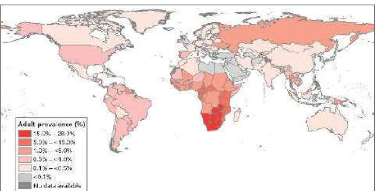
Ever since its discovery, the human immunodeficiency virus (HIV) has emerged as a global disaster. Around the world acquired immunodeficiency syndrome (AIDS) has led to more than 20 million deaths. O ver 33 million people are living with HIV today (Fig 1) and by 2010 it is estimated that more than 40 million children will have one or both parents dead from AIDS. People in productive age groups are predominantly affected by AIDS and hence in some countries the impact of AIDS has led to a major decrease in gross national product. To date the only tool available to ascertain the presence of HIV in an otherwise healthy-looking individual is a laboratory assay.
A
Fig. 1: A global view of H IV infection
33 million people [30–36 million] living with HIV, 2007
H IV/AID S in the South-East Asia Region
The South-East Asia (SEA) Region bears the second highest burden of HIV/AIDS, with 3.6 million people estimated to be living with the virus. There is wide variation in the number of people living with HIV in countries in this Region (Table 1).
Table 1: People living with H IV (PLH A) in countries of South-East Asia Region
There is a wide variation in the number of PLHA from less than 100 in M aldives to 2.4 million in India
Country
Estimated number of people living with H IV
(PLH A)
% of adult population infected with H IV
Bangladesh 12 000 < 0.1
Bhutan < 500 < 0.1
DPR Korea NA NA
India 2 400 000 0.3
Indonesia 270 000 0.2
M aldives < 100 < 0.1
M yanmar 240 000 0.7
Nepal 70 000 0.5
Sri Lanka 3800 < 0.1
Thailand 610 000 1.4
Timor-Leste < 100 < 0.1
Source: Report on Global AIDS Epidemic, UNAIDS, 2008
H IV and opportunistic infections
HIV does not kill those who are infected directly. Instead, it weakens the body's ability to fight disease. Infections which are rarely seen in those with normal immune systems can be deadly to those with HIV.
O Is are caused either by organisms of low or no virulence which are nonpathogenic in individuals with an intact immune system, or by known pathogens which present in a different way than usual in immunodeficient individuals, e.g. in the form of increased virulence, recurrence, multidrug resistance or atypical presentation.
The spectrum of OIs has been found to vary from continent to continent and region to region. W ith the unprecedented increase in the number of AIDS cases, O Is are also increasing. Several of these O Is are recognized as case-defining entities in HIV/AIDS patients (Table 2).
Table 2: WH O clinical staging of H IV/AID S for adults and adolescents with confirmed H IV infection (fungal O Is highlighted)
Clinical Stage 1
Asymptomatic
Persistent generalized lymphadenopathy
Clinical Stage 2
M oderate unexplained weight loss (< 10% of presumed or measured body weight)
Recurrent respiratory tract infections (sinusitis, tonsillitis, bronchitis, otitis media, pharyngitis) H erpes zoster
Angular cheilitis
Recurrent oral ulceration Papular pruritic eruptions Seborrhoeic dermatitis Fungal nail infections
Clinical Stage 3
Unexplained severe weight loss (> 10% of presumed or measured body weight) Unexplained chronic diarrhoea for longer than one month
Unexplained persistent fever (intermittent or constant for longer than one month) Persistent oral candidiasis
O ral hairy leukoplakia Pulmonary tuberculosis Lymph node TB
Severe bacterial infections (e.g. pneumonia, empyema, pyomyositis, bone or joint infection, meningitis, bacteraemia)
Acute necrotizing ulcerative stomatitis, gingivitis or periodontitis Unexplained anaemia (< 8 g/dl), neutropenia (< 0.5 x 10⁹
Clinical Stage 4
H IV wasting syndrome
Pneumocystis pneumonia (caused by Pneumocystis jiroveci) Recurrent severe bacterial pneumonia
Chronic herpes simplex infection (orolabial, genital or anorectal of more than one month’s duration or visceral at any site)
O esophageal candidiasis (or candidiasis of trachea, bronchi or lungs) Extrapulmonary tuberculosis
Kaposi’s sarcoma
Cytomegalovirus infection (retinitis or infection of other organs) Central nervous system toxoplasmosis
HIV encephalopathy
Extrapulmonary cryptococcosis (including meningitis) Disseminated non-tuberculous mycobacteria infection Progressive multifocal leukoencephalopathy
Chronic cryptosporidiosis Chronic isosporiasis
Disseminated mycosis (extrapulmonary histoplasmosis or coccidiomycosis) Recurrent septicaemia (including non-typhoidal Salmonella)
Lymphoma (cerebral or B cell non-Hodgkin) Invasive cervical carcinoma
Atypical disseminated leishmaniasis
Symptomatic H IV-associated nephropathy or HIV-associated cardiomyopathy
The early diagnosis of these infections is vital for better management and preventive measures. OIs can be bacterial, viral, parasitic or fungal, and it is in this context that the establishment and strengthening of existing mycology laboratories gains paramount importance. The ability to easily, cheaply and quickly diagnose these and other potential OIs is crucial.
O pportunistic fungal infections
Even though all fungal infections are “ opportunistic” , i.e. they cause disease in a host whose resistance is lowered, the true “ opportunistic” fungal infections only infect those who are already sick or immunocompromised.
seen in geographically restricted areas, the latter being reported frequently from northeast India. Blastomycosis and paracoccidioidomycosis have been reported to cause severe and disseminated disease in AIDS patients, though there has been no significant increase in the number of infections occurring in such patients. Unlike the above diseases, which occur primarily due to the defect in cell mediated immunity, aspergillosis and zygomycosis are now being increasingly encountered in advanced AIDS cases with neutropenia. Neutropenia in these patients also increases the risk of disseminated candidiasis, and invasive infections due to miscellaneous hyaline and dematiaceous (melanized) fungi. In Asia, paracoccidioidomycosis has only been reported from Japan, and only two authentic cases of coccidioidomycosis were reported from India (both were imported cases from an endemic area).
Antiretroviral therapy (ART) can lower the incidence of OIs in patients with AIDS. However, this therapy has not been available to most HIV-positive individuals due to its cost. M oreover, patients are not diagnosed in the early stages in a substantial number of cases. Hence there has been no let-up in incidence of the often fatal, invasive mycotic infections in the SEA Region, unlike the change in prevalence of these infections in developed countries.
CD
4T lymphocytes and fungal infections
CD4 counts, a useful prognostic marker in HIV/AIDS patients, also have critical levels below which certain invasive fungal infections start appearing frequently (Table 3).
Table 3: Relationship between CD 4 T lymphocytes and fungal O Is
CD4 count O pportunistic fungal infection
< 350 cells/µl M ucocutaneous candidosis
< 200 cells/µl Pneumocystosis
< 150 cells/µl H istoplasmosis
< 100 cells/µl Cryptococcosis and Penicilliosis marneffei
Clinical criteria provide only a presumptive diagnosis of fungal infection. Usually there are few specific signs and symptoms indicating fungal infections. The lesions and radiological findings are sometimes characteristic enough (crescent sign and halo sign in invasive aspergillosis) to strongly suggest a fungal etiology but the accompanying immunosuppresion frequently results in atypical presentations. In most circumstances, early diagnosis considerably increases the chance of successful treatment. Thus, it is important that the possibility of fungal infection be considered from the outset so that appropriate mycological investigations can be carried out.
Diagnostic mycology is also rapidly moving into the mainstream of clinical medicine as a result of the convergence of several independent developments. First, dramatic progress in antifungal therapeutics has increased the need for specific viral diagnosis. Second, the number of patients at risk of accruing opportunistic viral infections has expanded greatly as a result of the HIV/AIDS epidemic. Finally, modern management of HIV infection is providing a new paradigm for the integration of molecular techniques into management of chronic fungal infections. All these developments are not only increasing the use of diagnostic mycology techniques but have enabled the detection of a variety of fungal OIs. These guidelines are therefore intended to provide the laboratory techniques in diagnosis of fungal O Is at different levels of health laboratories.
Scope
This manual covers key aspects of diagnosing fungal infections. The manual describes the technique for processing the specimens and also can be of use to each laboratory in developing its own standard operating procedures. Although a variety of standard textbooks is available for these techniques, an effort has been made to consolidate relevant information in this manual.
M anual development process
developing countries in establishing practical diagnostic facilities for fungal infections based upon a pragmatic approach and available scientific evidence. The draft manual was first reviewed by W HO staff and subsequently peer-reviewed by two mycologists. The manual was revised by experts in W HO CC in accordance with the reviewers’ observations. The manual also provides information on the procurement of products that can be used in laboratories. However, this information is only suggestive, and W HO does not specifically endorse these products.
M anual development team
Chapter 2: Common opportunistic fungal infections
in H IV/AID S
Candidiasis
Candidiasis is commonly an endogenous opportunistic infection. Occasionally exogenous acquisition has also been proven. Of the causative agents, the most common species is Candida albicans. O ther non-albicans Candida species, especially C. tropicalis are increasingly being reported.
Candidiasis is the most common fungal infection found in HIV/AIDS patients. Extensive esophageal candidiasis is an AIDS-defining infection. But oral candidiasis, unless very extensive and causing symptoms unequivocally, is not diagnostic of AIDS. It is of prognostic value only as its presence indicates progression of immunodeficiency. Vulvovaginal candidiasis, though not unequivocally shown to occur more frequently in AIDS patients, nevertheless affects a considerable proportion of HIV-positive women with extensive disease. In advanced AIDS cases, with neutropenia and very low CD4 counts, disseminated candidiasis is certainly a possibility.
Clinical considerations
Clinical manifestations include:
Ø oropharyngeal candidiasis – up to 90% of untreated, advanced HIV cases develop this disease, with more than 60% getting > 1 episode per year.
Ø vaginal candidiasis – 27%-60% women of childbearing age develop this disease, and the rates are similar for HIV-infected and non-infected patients
Ø non-healing extensive skin ulcers (seen in Indian patients)
Ø intertriginous candidiasis*
Ø hospital-acquired UTI*
Ø superinfection of bacterial abscesses/infections in those treated for prolonged period with broad-spectrum antibiotics*
Ø candida lung infection (very rare)
Ø disseminated candidiasis (rare in HIV patients)
D iagnosis
Specimens depend on the site of lesion. Usual specimens for investigation of candidiasis are:
Ø esophageal brushing
Ø blood for culture, central venous line tip
Ø urine (suprapubic, specially in children; or aseptic aspiration from Foley’s) and other body fluids (CSF only in very rare cases)
Ø fine-needle aspiration samples
Ø biopsy samples
Yeast cells Candida albicans
*
Since candidais commensal, demonstration of a few cells or isolation from non-sterile specimens such as sputum, or swabs from mucosal surfaces, has no significance. However, the presence of a large number of organisms in a fresh specimen may have some diagnostic significance, especially in a symptomatic patient when other diseases have been ruled out.
In blood, urine (suprapubic or collected with aseptic precautions), cerebrospinal fluid (CSF) and sample from closed inflammatory foci, the presence of candida, with any number and any species is of pathogenic significance. It is wise, however, to request repeat specimens, wherever possible, to rule out possible contamination from the skin when the specimen was obtained. A central venous catheter may get colonized and serve as a portal for candidemia in hospitalized patients. The catheter should be removed in such patients and sent to the laboratory for culture isolation by rolling and flushing methods. Urinary catheter colonization may not however represent infection; these catheter tips are not recommended for microbiology processing, and instead properly and freshly aspirated urine through the catheter is recommended.
Candida albicans in culture
Candida species are readily isolated on most laboratory media (including blood agar), of which the most commonly used medium is Sabouraud Dextrose Agar (SDA). Since antibacterials do not inhibit fungi, including candida, the use of media containing antibacterials is helpful in the isolation of candida, especially from specimens which are not sterile (e.g. skin, sputum and urine). M ost strains grow well at 37°C or at room temperature (22-25°C).
does not break off from the mother cell. O n the other hand, true mycelium is formed by the elongation and branching of a germ-tube produced by the mother cell and formation of true septa. Identifying features in pseudohyphae are constrictions between cells, invariable presence of septa at points of branching and the smaller and rounded terminal cell. True hyphae on the other hand have no constrictions between two cells, may or may not have septa at points of branching and typically the subterminal cell is smaller than the terminal cell, which is usually cylindrical; true septa are normally refractive in nature.
Yeasts can also be cultivated from clinical specimens on the selective and differential medium called CHROM agar Candida, which uses a chromogenic mixture that allows selective isolation of yeasts and simultaneously identifies colonies of C. albicans, C. tropicalis, C. glabrata and Candida krusei.
This medium has proven to be useful for detection of mixed cultures of Candida sp. within a single specimen.
The rapid detection of C. tropicalis and C. krusei, which tend to be more resistant to antifungal agents, is important to optimize antifungal treatment especially for immunocompromised patients. Simple and easy detection of mixed yeast cultures is particularly useful to detect antifungal resistant strains that may be present in the sample as a minor population.
Serology (Antigen or antibody detection tests)
Ø Antibody detection test – no special use except in candida endocarditis.
Ø Antigen detection test – M annan and Enolase antigens have been evaluated, but require further validation.
M etabolites and D NA detection
Ø D -arabinitol (metabolite) detection – /L-arabinitol ratio or D-arabinitol/creatinine ratio helps in indirect diagnosis, but requires sophisticated equipment. The test is yet to be validated for routine use.
Cryptococcosis
The etiologic agent is Cryptococcus neoformans, the only pathogenic species of the genus Cryptococcus. There are five serotypes (A, B, C, D and AD), and it exists in three varieties, var. grubii (A), var. gattii (B & C) and var.
neoformans (D). The current thinking on varieties, serotypes and
pathogenesis of Cryptococcus neoformans is depicted in the box below:
Cryptococcus neoformans - varieties, serotypes and pathogenesis
• The encapsulated basidiomycete Cryptococcus neoformans was recently divided into 2 species
- C. neoformans (Cn) and - C. gattii (Cg).
• Cn consists of 2 varieties - grubii (CnVG) and neoformans (CnVN ), which are opportunistic pathogens and predominantly infect immunocompromised persons
• CnVG is the major causative agent of cryptococcosis worldwide, except in central Europe, where CnVN infection is most prominent
• In contrast, Cg is a primary pathogen, which predominantly infects immunocompetent persons.
- Cg was previously thought to be restricted to tropical and subtropical climates with a special ecologic niche on Eucalyptus trees.
- H owever, the recent outbreak of Cg infection in healthy humans and animals in the temperate climate of Vancouver in Canada, and its isolation from several species of trees other than Eucalyptus have raised the strong possibility that this fungus might have broader geographic distribution
• The mechanisms underlying pathogenic and environmental differences between Cn and Cg are not known
• W ithin Cn species, CnVN infections are more likely to display skin involvement and to afflict older patients
• CnVG infections are reported to have a higher mortality rate
• In contrast, infections caused by Cg result in a lower mortality rate but are frequently complicated by neurologic sequelae and require surgery and prolonged therapy
disseminated. C. neoformans has a predilection for the central nervous system (CNS), and patients mostly present with symptoms of chronic meningitis. In the present era of AIDS pandemic, WHO has considered cryptococcal meningitis as one of the AIDS-defining infections. Incidence varies from 4%-7% in African patients to < 1% in the developed world. Introduction of fluconazole and ART at reduced cost in developing countries like Thailand and Brazil has helped to reduce incidence in those countries. The effects are yet to be observed in other countries of the SEA Region.
Clinical considerations
CNS cryptococcosis/D isseminated cryptococcosis
Clinical manifestations include:
Ø onset is insidious; symptoms are present for weeks
Ø headache with minimal or no neck rigidity
Ø fever of unknown origin
Ø drowsiness and alteration in sensorium, with advancement of disease process
Ø chronic meningitis
Ø may result occasionally in cerebral cryptococcal granuloma
Ø M ay be associated with visual loss (rapid or slow), osteolytic bony lesions, secondary cutaneous cryptococcosis, cryptococcemia and cryptococciuria – called disseminated cryptococcosis. In recent years a few cases of cryptococcal peritonitis have also been reported.
Pulmonary cryptococcosis
Ø Often asymptomatic.
D iagnosis
Specimens should be collected according to the patient’s symptoms. M ost common specimen is CSF; other body fluids, sputum, bronchoalveolar lavage (BAL), bronchial washings, biopsy tissues, prostatic fluid (in case of relapse) and blood for culture are used where indicated. If the initial demonstration/isolation was from an extra-pulmonary site, attempts should be made to examine CSF irrespective of clinical presentation, to rule out asymptomatic meningeal involvement.
Diagnosis is by conventional methods as described above: direct demonstration and culture. Direct demonstration by India ink/Nigrosin wet mount is possible when the yeast cell count is more than 105 cells/ml of CSF. Since in HIV-positive patients the usual cell count is 107-109 cells/ml, direct microscopy is positive in 90% of cases. In tissue sections, mucicarmine stain and alcian blue stain are useful besides periodic acid Schiff (PAS) or methenamine silver-nitrate stain. Blood culture is helpful especially in the disseminated condition.
C. neoformans in CSF
On SDA, both at 25°C and 37°C, colonies may appear within 48 hours, but may take longer depending on the fungal load. If chloramphenicol is incorporated in media, delayed growth is observed. The colonies are soft and creamy in texture, or mucilaginous if considerable capsular material is present. In India ink mounts, C. neoformans appears as round budding yeasts 5-20µm in diameter surrounded by a prominent capsule (seen as a halo in India ink mounts). Rarely, acapsular, larger, smaller or short pseudohyphae-forming variants may be seen in samples. These yeasts are urease positive (Christensen’s urea agar or broth containing 2.9% urea), nitrate negative, do not ferment any sugars and produce a chocolate-brown pigment on bird seed agar.
Serology
capsular polysaccharide antigens of C. neoformans in CSF and serum. It is the test of choice, with very high sensitivity and specificity, and is considered one of the best serological tests for antigen detection in the field of infectious diseases.
Ø Antibody detection – not useful in immunocompromised patients like patients with AIDS.
Pneumocystis jiroveci
infections
Pneumocystis carinii was officially recognized as a fungus in 2001, and renamed as Pneumocystis jiroveci, though the disease continues to be referred as PCP (Pneumocystis carinii pneumonia). PCP is the most common AIDS-defining condition, though compared to the developed world it is less commonly reported from South-East Asia. It is nonetheless a major OI in HIV-infected individuals in the Region. The likelihood of developing PCP in AIDS patients increases as the CD4 count falls below 200/µl. Thrush, persistent fever and presence of other AIDS-defining OIs are other independent risk factors for PCP in these patients.
M ost healthy adults have been exposed to Pneumocystis jiroveci at some point of time in life, though asymptomatic carriage is quite common. The lifecycle of pneumocystis, though incompletely understood, contains at least two stages – the cyst and the trophozoite. The walls of the cyst forms take up the GM S and Toluidine Blue O stain, round-, cup- or typically “ deflated-ball” -shaped. In Giemsa stain the trophozoites are also seen and the cysts show eight sporozoites within them.
Clinical considerations
Ø Clinical presentation is often insidious with slow but steady progression of fatigue, fever, chills, sweats and exertional dyspnoea. The features are characteristic of atypical pneumonia. Circumoral, peripheral and mucus membrane cyanosis may be apparent in extensive infection.
D iagnosis
Ø Among the biochemical tests, serum LDH is high.
Ø Pulmonary function tests and arterial blood gas analysis are very important adjuncts in the management of these cases. In fact, these are the tests used first to screen patients for PCP.
Ø Suggestive chest X-ray, high resolution computed tomography (CT) scan, diffusion capacity of the lung for carbon monoxide, and nuclear imaging studies using Ga67 scan also aid in the diagnosis of these patients.
Ø Specimens include BAL, transbronchial biopsy, sputum and open lung biopsy. BAL and sputum should always be stained for acid-fast bacillus and other fungi also. Staining methods for Pneumocystis jiroveci include:
(1) Gomori’s M ethenamine Silver (GM S) stain
(2) Rapid GM S
(3) Toludine Blue O stain
(4) Giemsa stain
(5) Diff Q uick (Rapid Giemsa) stain
(6) Fluorescent antibody staining with monoclonal antibody: this is more specific and sometimes more sensitive when a low number of organisms is present in the specimen.
Ø Although polymerase chain reaction (PCR) technology has been studied as a diagnostic aid, further evaluation and validation are required before such tests can be applied widely.
H istoplasmosis
Two varieties of H . capsulatum are recognized in human diseases, depending on the clinical disease: var. capsulatum and var. duboisii (the African type). H . capsulatum var. duboisii causes disease restricted to Africa and M adagascar, and no case has been described from South-East Asia. Histoplasmosis due to var. capsulatum has a worldwide distribution.
Clinical considerations
Ø All stages of this disease may mimic tuberculosis. In some clinical situations, it can be confused with sarcoidosis.
Ø Since this intracellular pathogen resides in macrophages, the ideal specimen for direct demonstration and culture is bone marrow. The organism is best grown when inoculated in the medium at the bedside immediately after collection from the patient.
Ø In patients with AIDS, histoplasmosis presents as a progressive disseminated infection in 95% of cases. The majority of such cases have CD4 counts < 150 cells/µl and a median cell count of 50 cells/µl.
Ø Patients with disseminated disease usually present with fever, weight loss and malaise over a period of several weeks. In about half, respiratory symptoms are reported.
Ø Hepatosplenomegaly and generalized lymphadenopathy are encountered in 15%-30% of cases.
Ø A syndrome representing septicemia has been well documented. The patients usually present with hypotension, acute respiratory distress syndrome (ARDS), hepatic failure, renal failure and disseminated intravascular coagulation. It represents a later manifestation of disseminated histoplasmosis and can occur in 10%-15% cases of disseminated histoplasmosis in AIDS.
Ø M ucocutaneous involvement is very common in patients from South-East Asia, particularly India.
Ø Gastrointestinal and osteo-articular lesions are also found. Adrenal insufficiency may be a presentation of disseminated histoplasmosis.
Ø Patients who experience immunologic improvement following anti-retroviral therapy may demonstrate atypical pathology. This has been referred to as immune reconstitution inflammatory syndrome and is characterized by focal inflammatory histology, elevation of hepatic enzymes, hepatic abscesses, lymphadenopathy, arthritis, uveitis and intestinal obstruction.
D iagnosis
Ø Specimens – Bone marrow, tissue or body fluid are usually sent to the laboratory. In disseminated infection, samples can be taken from blood, bone marrow, liver, skin lesion, or any other site of infection.
Ø Lysis centrifugation (Isolator Tube) system has been shown to be more sensitive than automated or conventional culture for growing H . capsulatum from blood.
Ø W hen sputum or BAL fluid is sent for culture, a selective medium containing ammonium hydroxide on the surface of agar to increase pH is helpful to inhibit the growth of commensals and allow the slow growing H . capsulatum to grow.
Ø For direct microscopy of smear from samples or tissue sections, routine 10% potassium hydroxide (KOH) wet mount or hematoxylin and eosin stains generally will not allow visualization of tiny yeasts. Smear or section should be stained with methenamine silver or PAS stain to best visualize H. capsulatum. Distinctive 2-4 µm, oval, narrow-based budding yeasts within macrophages, but also seen free in tissue sections, allow tentative diagnosis of histoplasmosis.
Ø Two types of conidia are produced on hyphae: tuberculate macroconidia (8-15 µm in diameter) having characteristic projections on their surface; and smooth-walled microconidia (2-4 µm in diameter). Identification of macroconidia allows only presumptive diagnosis of histoplasmosis as Sepedonium spp. produce similar macroconidia. The following tests verify the identification of H . capsulatum:
- Yeast conversion by repeated subcultures at 37°C on pine agar medium. This is a laborious and time-consuming process.
Yeast stage of H . capsulatum H . capsulatum in culture
- Exo-Antigen test can help in identification even in a contaminated culture, but it is also time-consuming and complicated.
- Commercially available chemiluminescent DNA probe (AccuProbe; Gen Probe, San Diego, USA) is used to identify H . capsulatum isolates in three hours. The test is highly specific though very rare false positives are reported.
H . capsulatum exhibits a thermal dimorphism by growing in living tissue or in culture at 37°C as budding yeast-like fungus, and in soil or cultures at temperatures below 30°C as a mould. On SDA at 25°C colonies are mostly slow-growing (2-3 weeks in many cases), white or buff brown, cottony with pale brown transverse folds. Sometimes colony types may be glabrous or verrucose in appearance.
conjugated to biotin, and horseradish peroxidase. Both assays are more sensitive in urine rather than serum. In AIDS patients, the antigen is detected in urine in 95% of patients and in serum in 85% of patients. The test can also help monitor response to therapy. There are few data on the usefulness of antigen assays for BAL fluid (detected in 70% of patients). False positive reactions are seen in patients who have other endemic mycoses (blastomycosis and paracoccidioidomycosis, but not with coccidioidomycosis). False positive reactions are also reported in patients with penicilliosis, which is prevalent in the South-East Asia region. The test is yet to be commercialized.
Antibody detection – It is not useful in AIDS patients, as they mount a poor antibody response.
Penicilliosis marneffei
The dimorphic fungus Penicillium marneffei causes the disease, which is an emerging systemic mycosis in AIDS patients. Penicillium marneffei is endemic in Thailand, Northeastern India (M anipur and Nagaland states), southern China, Hong Kong, Vietnam and Taiwan. Bamboo rats and soil are considered the reservoir of the disease though the agent has never been isolated from soil except near bamboo rat burrows.
Penicilliosis marneffei, after tuberculosis and cryptococcosis, is the third most common opportunistic infection in patients with AIDS in the South-East Asia Region, and is therefore considered an AIDS-defining illness. Persons affected by penicilliosis usually have AIDS with low CD4 counts, typically < 100/µl. The average CD4 count at presentation is 63.5/µl.
Clinical considerations
Various types of manifestations include:
Ø fever of unknown origin, loss of weight, generalized lymphadenopathy, anemia
Ø hepatomegaly with or without splenomegaly
Ø skin lesions - characteristic generalized papular eruptions, central umbilicated papules resembling those of molluscum contagiosum, or acne like lesions and folliculitis over face, trunk, and extremities
Ø pharyngeal and palatal lesions also can be seen
Ø subcutaneous nodules may be seen
Ø chest radiographic abnormalities typically manifest as diffuse reticulonodular infiltrates, and cavitations
D iagnosis
Specimen: Bone marrow aspirate, blood, lymph node biopsy, skin biopsy, sputum, BAL, pleural fluid, liver biopsies, CSF, pharyngeal or palatal ulcer, scrapings, urine, stool, kidney biopsy, pericardium, stomach or intestinal specimens.
Ø Giemsa, W right, GM S or PAS stain shows characteristic intracellular (within neutrophils or tissue histiocytes) round to oval yeast-like cells, which may divide by crosswall formation. The crosswall formation can differentiate yeast cell of P. marneffei from those of H. capsulatum. Elongated sausage-shaped extracellular forms are also seen.
Ø Direct immuno-fluorescence test is the test of choice for specific diagnosis; this would be done at reference laboratories, since it requires a fluorescent microscope. Several types of antibodies have been used for detection of P. marneffei in paraffin embedded, formalin fixed tissue. M onoclonal antibody directed to Aspergillus galactomannan can be used to stain P. marneffei due to identical epitope between the two fungi (the difference in morphology helps to distinguish them). Rabbit antiglobulin against P. marneffei mycelial culture filtrate antigens is also proven to be specific for detection of P. marneffei in tissue section.
Ø P. marneffei exhibits thermal dimorphism by growing in living tissue or in culture at 37°C as a yeast-like fungus, and in culture at below 30°C as a mould. On SDA (without cycloheximide) at 25°C colonies are fast-growing, suedelike to downy, white with yellowish-green conidial heads. Colonies become grayish-pink to brown with age and produce a characteristic diffusible brownish-red to wine-brownish-red pigment. Conidiophores are hyaline, smooth-walled and bear terminal verticils or whorls of 3-5 metulae, each bearing 3-5 phialides. The conidiophores are described as biverticillate or irregularly monoverticillate. Conidia are globose to subglobose, 2-3 µm in diameter, smooth-walled and are produced in basipetal succession from the phialides.
Penicillium sp
Ø O ther biverticillate, red-pigment-producing Penicillium species include P. purpurogeum and P. rugulosa, from which these can be definitely differentiated by demonstration of dimorphism (mould to yeast conversion is achieved by subculturing on brain heart infusion agar [BHIA] at 37°C).
Ø Several serological methods for detection of antibodies or antigens are attempted and produce conflicting results especially in AIDS patients. No standard kit is available to date. Commercially available Pastorex Aspergillus (Latex Agglutination test kit) or Platelia Aspergillus (EIA kit) for detection of Aspergillus antigen in serum or body fluid cross-react with P. marneffei and are positive in patients with penicilliosis marneffei at a low titre.
Ø A specific PCR assay is under evaluation and might be useful as an alternative test for rapid diagnosis of P. marneffei infection.
Invasive aspergillosis (IA)
Exposure to Aspergillus is universal, but aspergillosis is uncommon unless the host immune defense is compromised. The aspergilli producing infection are many but more than 95% of disease is caused by A. fumigatus and A. flavus; the latter is especially more common in the tropics. The disease usually occurs when phagocytic host defenses by granulocytes and macrophages are quantitatively or functionally suppressed. Despite the severe immunosuppression that results from advanced HIV infection, there are relatively few cases of aspergillosis in patients with HIV disease. The overall incidence varies from 1.1 to 3.5 episodes per 1,000 person-years. The incidence of aspergillosis in AIDS patients is significantly high in the following settings: age = 35 years old, men who have sex with men, white blood count < 2,500 cells/µl, CD4 count < 100/µl, prior history of an OI, and prescribed medications associated with neutropenia.
In patients with AIDS, aspergillosis is usually restricted to the lungs with a variety of distinct manifestations, including thick-walled cavitary disease of the upper lobes, diffuse unilateral or bilateral infiltrates, ulcerative tracheobronchial disease, and obstructive bronchitis. The diagnosis is made often upon postmortem and relies on the histologic or microbiologic identification of the fungus in infected tissue.
Clinical considerations
Ø Pulmonary – most common site of involvement.
diffuse or nodular; pseudo-membranous tracheo-bronchitis, extensive bronchial hemorrhage.
(2) Noninvasive, obstructing bronchial aspergillosis – fungal plugs block the airways, leading to breathlessness, cough and chest pain. Chest X-ray shows hazy infiltrates, representing segmental or lobar atelectasis.
Ø Extrapulmonary aspergillosis – CNS aspergillosis is very uncommon in AIDS patients and may present as focal abscesses or hemorrhagic or mycotic aneurysms. In rare cases it may present as a focal neurologic deficit or intracerebral space-occupying lesion. The lesion usually extends from the sinuses; in fact it is merely an extension of the mycotic disease into the contiguous cranial cavity.
Ø Very rarely, unusual manifestations in the AIDS patients include endocarditis and myocarditis, esophagitis, lymphadenitis, musculoskeletal and skin abscess, liver and spleen involvement, and renal and pancreatic abscesses.
Aspergillus
D iagnosis
Specimens: Biopsy, fine needle aspiration cytology (FNAC), BAL, sputum, sinus mucosa.
Ø Because aspergilli are ubiquitous, growth on culture alone may not represent actual disease. M uch controversy surrounds the specimens useful for diagnosis of IA in AIDS patients. A positive sputum or nasal secretion in an AIDS patient with consistent respiratory symptoms should merely invite suspicion. Probable diagnosis is established when BAL sample is positive for aspergilli, but definitive diagnosis is only established on observing acute angled, septate, 3-6 µm wide true hyphae in tissue samples followed by the growth of aspergilli in culture. M icroscopy alone cannot distinguish between Aspergillus and other hyalohyphomycetes, most notably Fusarium and Pseudallescheria species.
Ø The fungus is rarely cultured from blood, CSF, bone marrow or other organs (brain, kidney and liver). In such a situation, the diagnosis is entertained only if the patient has a clinical history and presentation compatible with IA or if the fungus is isolated from another site.
Ø Aspergilli are identified on the basis of colony morphology (on M alt Extract Agar or Czapek Dox Agar at 37°C and 25°C) and microscopic examination (foot cells and vesicle are characteristic of the genus; colony morphology, size, colour on both media at two temperatures; and the microscopic arrangement of conidia; presence of sexual fruiting body; hülle cells, etc.).
Ø M onoclonal and polyclonal antibody staining of tissue sections has been used as an adjunct, but there are problems of poor specificity with these techniques.
Ø Enzyme-linked immunosorbent assay (ELISA) to detect galactomannan polysaccharides in serum of IA patients is a useful procedure for diagnosis of aspergillosis. Cross-reaction and lack of consistent results are major drawbacks of this assay. However the EIA technique is far better than the previous latex agglutination (LA) test available.
zygomycosis. However, it cannot identify the etiological agent of the disease. The test also requires further validation.
Ø PCR from BAL and blood samples is being standardized and validated in reference laboratories for the early detection of IA.
Ø Chest X-ray and CT scan are important adjuncts to diagnosis.
Zygomycosis
Though the disease is rare in AIDS patients, there has been an increase in the number of cases of zygomycosis in recent years, especially in tropical countries. Advanced AIDS with neutropenia is now recognized as a risk factor for acquiring zygomycosis, a disease caused by Zygomycetes belonging to the order M ucorales. Rhizopus oryzae, Rhizopus rhizopodiformis, Rhizomucor pusillus, Absidia corymbifera, Apophysomyces elegans, M ucor circinelloides, Cokeromyces recurvatus and Saksenaea vasiformis are the commonly recognized human pathogens causing this fatal disease. Fungi invade vascular tissues and cause extensive necrosis in these cases, and disseminated disease is common in the immunosuppressed. These fungi are characterized by wide (5-20 µm diameter), aseptate or sparsely septate, coenocytic hyphae with rightangled branching along with a predominantly necrotic and acute neutrophilic tissue reaction. In culture, these fungi show unrestricted cottony mycelia mostly white to grey and black in colour.
Clinical considerations
Zygomycosis may affect any or more than one organ complex and the disease is categorized as:
(1) Rhino-orbito-cerebral (most common and carries a poor prognosis) – nasal congestion, dark-blood-tinged rhinorhea, epistaxis, sinus tenderness, retro-orbital headache, fever, malaise, facial swelling, blurred vision, lacrimation, chemosis, proptosis, loss of vision. Examination of nasal cavities may reveal a black eschar on septum or turbinates.
(3) Pulmonary – hemoptysis, fever and cough – fatal and frequently diagnosed post mortem.
(4) Gastrointestinal – necrotizing lesions.
(5) Renal – though isolated renal disease is yet to be seen in patients with AIDS, the disease is prevalent in South-East Asia.
(6) Cerebral – as a part of rhino-cerebral disease, or disseminated disease or isolated cerebral zygomycosis. Isolated disease presents as space-occupying lesion, encephalopathy or strokelike symptoms. Unlike in the west, where isolated disease mainly occurs among injecting drug users, in South-East Asia it occurs mainly in immunocompetent individuals or diabetics.
(7) Disseminated – where more than one organ system is involved.
D iagnosis
Specimen – Biopsy, scrapings, aspirates
Ø Direct examination in KO H mount of sample from tissue, and histopathological examination of the same samples are the gold standard for diagnosis.
Ø Culture in SDA at 37°C and 25°C is sufficient to isolate the fungus. W hile processing, the tissue should not be crushed or ground, but rather cut into small pieces and inoculated. (The viability of the fungi is lost by crushing it.)
Ø M ost fungi can be identified on routine lactophenol cotton blue (LCB) mounts; however, water agar may be required to induce sporulation, especially in Apophysomyces elegans and Saksenaea vasiformis.
Tissue demonstration of aseptate hyphae by a variety of staining techniques (H&E, GM S, KOH mount, PAS is considered adequate for the diagnosis of zygomycosis. Culture is a useful adjunct to diagnosis. However, being ubiquitous fungi, growth in culture only does not conclusively prove the disease.
Radiographic findings of infection are often suggestive and also help in delineating the extent of disease. The agents are angioinvasive and often produce infarction in tissues. There are as yet no validated immunological or PCR-related diagnostic techniques available for zygomycosis.
Blastomycosis
Blastomycosis, caused by a dimorphic fungus, Blastomyces dermatitidis, is a systemic fungal infection endemic in North America, but cases have been reported worldwide in Africa and the M iddle East as well as South-East Asia. Frequently associated with point-source exposure, it occurs more commonly in men than in women, which may reflect a larger number of male occupational exposures. However, in more recent reports, occupational exposure accounts for a smaller proportion of cases, presumably as recreational exposures increase. Immunosuppressed patients typically develop infection following exposure to the organism, but reactivation may also occur.
Blastomycosis is uncommon among people infected with HIV and is not recognized as an AIDS-defining illness. In the setting of AIDS or other marked immune suppression, the disease is usually more severe with multiple-system involvement, including the CNS, and progresses rapidly to a fatal course.
Clinical considerations
Ø Acute disease typically presents as flu-like illness characterized by fever, malaise, fatigue, weight loss, and pulmonary involvement.
Ø Rarely, pneumonia with fever, chills, purulent sputum and hemoptysis may occur. ARDS may occur very rarely.
Ø Individuals with underlying lung disease may develop chronic pneumonia with symptoms lasting 2-6 months: weight loss, night sweats, fever, chest pain, and productive cough mimicking tuberculosis.
Ø Cutaneous blastomycosis is very common – lesions vary from nodules to verrucous lesions and may become ulcerative, mimicking squamous cell cancer and keratoacanthoma.
Ø Prostatitis, female genital tract infections, osteolytic lesions involving vertebrae, skull, ribs and long bone may also be seen.
Ø Disseminated disease has been found in the brain, skeletal system, prostate, myocardium, pericardium, sinuses, pituitary, and adrenal glands and also invades the reticuloendothelial system. Epidural or cranial abscesses as well as meningitis occur in up to 40% of patients with AIDS and chronic blastomycosis.
D iagnosis
Diagnosis depends on direct examination of tissue or the isolation of Blastomyces dermatitidis on culture.
Specimens - Sputum, BAL, transbronchial biopsy, pus, aspirates and biopsy from skin lesions, urine for antigen detection.
Ø Sputum as a specimen has diagnostic sensitivity of over 80% while samples obtained via bronchoscopy have > 90% positivity rates. Samples from cutaneous lesions yield the organism in most cases.
Ø Conversion from mould to yeast form and exoantigen testing are essential for confirming the identity as B. dermatitidis.
Ø The yeast phase is characteristic, with yeast cells exhibiting unipolar, broad-based budding. The budding yeast cells, in both tissue and in culture, are described as “ dumbbell-shaped” . Initially the colony appears yeast-like at room temperature and then develops hyphal projections, eventually becoming a fluffy white mould. The colonies are moderately slow growing, requiring up to three weeks’ incubation.
Ø Antigen detection in urine has been recently developed and is commercially available, being positive in 70%-80% of cases of disseminated disease and 100% of pulmonary cases. Crossreactivity with Histoplasma capsulatum is a problem. Serum antigen detection is positive in only 50% of cases.
Ø Antibody detection in AIDS cases is of limited utility because of the accompanying immunosuppression.
Chapter 3: O verview of diagnostic mycology laboratory
Laboratory procedures
Laboratory procedures in diagnostic mycology are directed mainly towards:
(1) Direct demonstration of the pathogenic fungi in clinical specimens (microscopy)
(2) Successful isolation of pathogenic fungi (culture)
(3) Supportive evidence of specific fungal infection (antigen, antibody, cell wall markers such as ß 1-3 Glucan and metabolite detection). However, PCR for detection of fungal DNA is yet to be validated for routine diagnosis of any fungal infection.
(4) Prediction of possible therapeutic outcome (antifungal susceptibility)
(5) Tracing the source of infection (epidemiology)
Accurate diagnosis relies heavily on a combination of microbiological, histopathological and serological evidence. Since in recent years newer methods have been continuously added, introduction of sophisticated instruments in day-to-day working of the laboratory invariably increases the cost of laboratory investigations. Therefore, at the primary level, the mycologist has to fall back upon simple established standard procedures. No single procedure may help in definitive diagnosis, since each has its own limitations. All conventional procedures should therefore be incorporated in a standard mycological laboratory as far as practicable.
Prerequisites for laboratory diagnosis
For a proper diagnosis it is essential to have brief details of:
(a) clinical history
(c) the patient’s residence (Penicilliosis marneffei is endemic in South-East Asia, but only in M anipur and Nagaland states in India) and occupation (agricultural labourers engaged in soil-disturbing activities are predisposed to histoplasmosis and coccidioidomycosis, gardeners for sporotrichosis, etc.)
(d) any history of travel or residence abroad (coccidioidomycosis in south-west USA, i.e. Arizona and California)
(e) history of activities which result in exposure to soil, dust and organic debris
(f) the source of the specimen
(g) how and when the specimen was collected and transported
It is necessary that adequate amounts of appropriate and properly collected specimens are received in the laboratory. The procedures of mycological investigation that can be undertaken at the primary level are simple, and no elaborate equipment is required. For advanced techniques, specimens should be referred to better-equipped reference laboratories designated for this purpose.
Safety and training
W ith regard to protective measures against blood exposure, the laboratory safety officer should ensure that all staff members, particularly new staff, receive adequate instructions. These instructions should ensure that the safety aspects of new practices and the equipment used are understood and that standard practices are being followed.
A laboratory safety manual should be maintained and updated at least annually. Supervisory staff must ensure that their staff adhere to the documented safe working practices. Staff should formally report needle stick injuries or other episodes of exposure to blood or body fluids.
Safety considerations
Specimen collection Universal precautions should be followed
Specimen transport/storage Sealed plastic bag (preferably double-layered) with proper legible labelling
Specimen processing Containment level II or III lab
Specimen collection
Optimum time of specimen collection
• Ideally as close to the onset of symptoms as feasible
• Before initiation of antifungal therapy, where possible
• M orning sample for inpatients Correct specimen
type
• Appropriate specimen according to the site of lesion, in adequate amount (as mentioned below), collected with sterile implements and precautions
Recommended quantity*
• CSF > 2 ml
• Sterile body fluids (pleural, pericardial, synovial, ascitic fluid) > 10 ml
• Lower respiratory (BAL, bronchoscopic washings/aspirate) > 1 ml (ensure it contains lower respiratory tract specimen)
• Upper respiratory – oral swab/saline wash
• Nasal sinuses – surgical removal of sinus contents
• Urine (midstream) 10-20 ml (for catheter urine, clamp hub of Foley’s catheter distally, clean hub sequentially with 70% alcohol, iodine and 70% alcohol, then aspirate collected urine from the hub with sterile needle and syringe)
• Sputum 2-5 ml
- Coughed-out sputum (not saliva) in a wide-mouthed sterile container; Q score is calculated from a gram stained smear and only
representative samples are accepted
- In case of dry cough, induced sputum in sterile container • Stool 1-5 g (rarely used, only to know colonization by Candida)
• Vaginal Swab
• Prostatic Fluid – Bladder emptied followed by prostatic massage
• Blood 20 ml-10 ml in each bottle; for pediatric patients 4-10 ml divided equally between two bottles
- Candidemia is known to be intermittent, so it is essential to obtain
three samples to rule out yeast sepsis
- Inoculated directly to biphasic blood culture bottle maintaining a ratio
of 1:10 of blood to broth
- Serum - additional 2-5 ml of blood to be put in clean, dry vials,
preferably leak-proof screw-capped for serum separation
• Subcutaneous sites
- Abscess – Aspirate with sterile needle and syringe, if needed sample
base of lesion and abscess wall (scrape, punch biopsy)
- Open wound – Aspirate or swab deeply, especially base and margins
- Tissue biopsy specimen – surgical collection, punch biopsies may be
used for skin lesions
• Repeated sampling from the same site (for authentic diagnosis of OI repeated demonstration/isolation of same organism from same site is essential)
Specimen transport and storage
Time between specimen collection and transport
• Specimens should be transported to the specific laboratory as soon as possible
• M aximum time allowed for transport is 24 hours at room temperature from sterile site; specimen from non-sterile site may be transported at 4°C, if time for transport is > 1 hour
Storage • Specimen should be processed in the laboratory as soon as possible
• Delay of more than four hours in processing of
unrefrigerated specimens is undesirable. W here there is a delay in processing, specimens should be refrigerated except CSF and specimens for isolation of Cryptococcus
• As a general principle, sterile samples should never be refrigerated, while samples expected to contain commensal should be refrigerated if there is delay
General techniques used in medical mycology
The techniques used in clinical mycology are in general similar to those used in clinical bacteriology. However, certain differences in the techniques should be noted.
Basic equipment required
(1) A straight nichrome wire with a long handle – used for stabbing inoculation.
(2) A bent (right angle) nichrome wire with long handle – used for handling the mycelial types of fungal cultures.
(3) Pair of short stiff teasing needles – helpful in pulling apart dense masses of mycelium on the slide for better microscopic examination.
(5) A pair of scissors, for cutting biopsy tissues into small pieces.
(6) Pair of forceps.
Test tubes should be used in preference to petri dishes for primary culture of organism from clinical specimen, or transferring fungal cultures. Test tubes afford the following advantages:
(a) easy handling
(b) easy storage
(c) less frequent breakage
(d) greater protection to worker
(e) less drying of medium
Cotton plugs in the test tubes for cultures are preferred over screw caps as they:
(1) allow the surface of the slants to remain dry;
(2) help better development of aerial mycelium and sporulation due to proper aerobic condition;
(3) help in better pigment production;
Test tubes of fairly large diameter (18-25 x 150 mm) are preferable to narrow tubes. These allow a thick butt of agar, which will withstand drying during the several weeks of incubation often necessary for the growth of fungal cultures. At all times, for the protection of personnel and for securing and maintaining pure cultures, precise sterile techniques are to be employed.
Specimen processing
Proper documentation upon specimen receipt
• M aintenance of laboratory records with lab number
Initial processing • Depends upon the specimen and suspected etiologicorganism
• Blood culture - two bottles incubated at 37°C and 25°C (one each)
• CSF and other body fluids should be centrifuged in a separate sterile test tube or centrifuge tube at 2000-2500 rpm for 10 minutes, and supernatant poured back to the container of collection and stored for antigen detection. Pellet is used for smear and wet mount and for subsequent culture.
• Tissue – M incing, grinding, or use of stomacher (do not grind if zygomycosis is suspected)
• Exudates, pus, drainage – washing, centrifugation and crushing of granules, if any
• Sputum:
- Sterile glass beads are added and vortexed briefly; for detection of
Actinomyces and Nocardia, samples are plated onto Blood Agar and M odified Lowenstein Jensen M edia at this point
- Equal volume of freshly prepared sodium citrate (2.94%) and 0.5
g% N-Acetyl L- Cysteine (or Sputolysin) added to specimen and vortexed again for 10-30 seconds depending upon the consistency of the specimen
- Dilute mixture in PBS by adding double the volume and centrifuge
at 1 000 g for 15 minutes
- Use the sediment to prepare smears for direct microscopy and
inoculate culture media
Direct microscopy Procedure employed depends upon specimen
Wet mount:
• KOH preparation
• India ink/Nigrosin wet mountsfor screening Cryptococcus
• LCB wherever applicable Stain/smears
• Gram staining (for yeasts)
• Giemsa/W right staining (for histoplasma, blastomyces and Penicillium marneffei); however PAS/GM S staining preferable
• M ucicarmine, Alcian Blue for Cryptococcus Additional microscopic procedures
• Direct fluorescence (Calcofluor W hite) where applicable
• Direct and indirect immunofluorescence where applicable
Histopathology • H &E stain
• PAS stain
• GM S stain
• M ucicarmine stain/Alcian Blue Culture Given in a separate chapter
Levels of laboratory for diagnosis and reporting
Level Specimen Process
Reporting of positives
(stating if appropriate further report will be issued)
Comments
KO H mount
Tube KO H for extended clearing of tissue samples
Same-day reporting presence of fungal filaments with characteristic branching (septate or aseptate, acute-angled or wide-acute-angled branching and thin or wide hyphae) and yeasts
positive yeast cells with hyphae and
pseudohyphae
Same-day reporting: Yeast cells seen, suggestive of fungal infection (except samples from respiratory tract contaminated with upper respiratory tract flora)
M ycelial fungi may not stain well, show only Intracellular yeast cells seen suggestive of H istoplasma capsulatum Intracellular yeast cells seen suggestive of P. marneffei
Take care to
GM S/Toluidine Blue and Rapid Giemsa:
characteristic cysts and Trophozoites of P. jiroveci
Same day reporting: Positive for P. jiroveci
Giemsa does not stain cysts of pneumocystis
Antigen detection for presence of C. neoformans
titre ≥ 8 confirmatory repeat test after 1 week index criterion 0.5 or 1.5)
Repeat samples
ß-D Glucan Assay Same-day reporting: Sample positive for Beta-D-Glucan
Level Specimen Process
Reporting of positives
(stating if appropriate further report will be issued)
Comments
Immunofluorescent Staining for P. jiroveci
Same-day reporting: Positive report to the doctor concerned as soon as possible
Antigen detection for presence of H. capsulatum*
Same-day reporting: Sample positive for H istoplasma
antigen
Antigen detection for presence of Blastomyces dermatitidis precipitin bands;result in three days.
CFT: Same day result, CFT titre > 32 significant
Antibody
testing Antigen detection for presence of Penicillium marneffei
Same-day reporting: Sample positive for P. marneffei
antigen
Not commercially available
* At present available only in USA
Chapter 4: M icroscopy
Direct microscopic examination of the clinical material provides valuable information about the fungal aetiology. Various methods and staining techniques are available and in wide use. Some of the commonly used techniques are:
KO H mount preparation
(1) Potassium hydroxide is a strong alkali used as a clearing agent to observe fungi in the specimen in a wet mount preparation.
(2) 10%-20% KOH is usually used depending on the specimen; occasionally 40% is also used when not cleared by 10%-20% KOH.
(3) Used for specimen such as sputum, pus, urine sediment, homogenate from biopsy tissue, nail, hair, etc., to clear cell debris.
Preparation of the mount
(1) Take a clean grease-free glass slide.
(2) Place a large drop of KO H solution with a Pasteur pipette.
(3) Transfer small quantity of the specimen with a loop or the tip of a scalpel into the KOH drop.
(4) Put a clean cover slip over the drop gently so that no air bubble is trapped.
(5) Place the slide in a moist chamber, and keep at room temperature.
(6) Tissue usually takes 20-30 minutes; sometimes overnight contact with KO H is useful for getting a positive result.
O bservation
(1) Examine the clear specimen under low power (10X or 20X objective). Scan the entire cover slip from end to end in a zigzag fashion.
(2) If any fungal elements are suspected, examine under high power (40X objective).
(3) Reduce the light coming into the condenser while examining at high power.
(4) Look for budding yeast cells, branching hyphae, type of branching, the colour, septation and thickness of hyphae.
M odification
For more distinction, stains like methylene blue or Parker blue-black fountain ink may be used along with KOH. This will impart a colored background and fungal elements, if present, will show as prominent refractile objects.
Advantages
Simple, economical and rapid.
D isadvantages
Ø Pus and sputum may contain artifacts, which may superficially resemble hyphal and budding forms of fungi. These artifacts may be produced by cotton or wool fibres, starch grains (in pleuritis) or cholesterol crystals.
Ø It gives an idea about the presence of hyphal element, but cannot distinguish different fungi.
Precautions
(1) The drop of KO H should not be so large that the cover slip floats.
(2) If kept outside a moist chamber, the KOH dries and crystals form that restrict the visibility of the fungus.
(3) After clearing, pressure is to be gently applied on the top of the cover slip with a fold of filter paper or the handle of a teasing needle. This ensures even spreading of the material onto the slide.
(4) KO H should be kept in a closed container in small aliquots ready to use on the workbench.
Q uality assurance
Fungal spores or hyphae may contaminate the KOH solution kept in the laboratory and may give false positive results. A negative control should therefore be put up every day.
India ink or nigrosin preparation for
C. neoformans
(1) The preparation should be made in the centre of a clean, grease-free glass slide.
(2) Put 1 drop of India ink or Nigrosin on the centre of the slide. Too much stain makes the background too dark. (Upon examination, if the staining appears too dark, a small amount of water may be applied on the edge of the cover slip and the cover slip gently tapped: this will dilute the stain to some extent.)
(3) Put 1 loopful of the specimen or preferably centrifuge sediment from the fluid specimen to be tested (e.g. CSF, spinal fluid, urine, and other body fluids) close to the drop of the stain.
(5) Hold the cover slip vertically such that one edge just touches the fluid on the slide. The fluid will spread along the edge by surface tension.
(6) Keeping that edge in contact with the fluid surface, drop the cover slip gently on the fluid, so that no air bubble is trapped inside. If there are air bubbles, the surface of the cover slip may be gently tapped by the needlepoint, so as to move the bubbles towards the edge. But this should be avoided as far as possible.
(7) Examine slide immediately under the microscope. Since the stain tends to dry fast in air, if immediate examination is not feasible, the slide should be kept in a moist chamber.
(8) Scan the entire cover slip from end to end in a zigzag fashion. Encapsulated yeast cells (Cryptococcus neoformans) are seen under low power as luminous dots in an otherwise dark background. Under high power, the cells can also be seen, containing refractile bodies, and surrounded by the unstained thickness of the capsule. Characteristic pinched-off budding, when observed, is confirmatory for diagnosis.
Please note
(a) Besides the classical budding-yeast form, various unusual forms can also be seen, including elongated forms that look like pseudohyphae; this is mainly due to a very high multiplication rate of the organism in HIV/AIDS patients.
(b) In the very late stage in progressive AIDS, it may be difficult to differentiate the capsules of individual cells; the organism may remain enmeshed in a matting of the capsular material.
(c) M icro- or non-capsulated strains of the organism are also reported on rare occasions. In such cases, gram stain of the sample is helpful in identification.