Choledocholithiasis during Pregnancy:
Multimodal Approach Treatment
Hendra Koncoro*, Cosmas Rinaldi Lesmana**, Benny Philipi***
*Department of Internal Medicine, Sint Carolus Hospital, Jakarta**Division of Hepatobiliary, Department of Internal Medicine, Faculty of Medicine, Universitas Indonesia/Dr. Cipto Mangunkusumo General Nasional Hospital, Jakarta
***Department of Digestive Surgery, Sint Carolus Hospital, Jakarta
Corresponding author:
Cosmas Rinaldi Lesmana. Division of Hepatobiliary, Department of Internal Medicine, Dr. Cipto Mangunkusumo General National Hospital. Jl. Diponegoro No.71 Jakarta Indonesia. Phone: +62-21-31900924; Facsimile: +62-21-3918842. E-mail: [email protected]
ABSTRACT
Pregnancy is an important risk factor for growth of choledochal stones. Since choledocholithiasis encountered during pregnancy, which is also a possible cause of pancreatitis and cholangitis, may be the reason for serious morbidity and mortality both for the mother and the fetus, it should be treated. In this article, the results and reliability of endoscopic retrograde cholangiopancreatography (ERCP) application on a pregnant woman accompanied with percutaneous biliary procedures are presented.
We report a case of 33-year-old woman at 19th week of gestation with cholestatic jaundice due to a common
bile duct (CBD) stone managed by endoscopic retrograde cholangiopancreatography (ERCP). The patient had post ERCP pancreatitis which resolved with medical management. Percutaneous cholecystostomy was also performed to control source of infection in the gallbladder.
(5&3LVWKH¿UVWSURFHGXUHWKDWZLOOEHSUHIHUUHGLQWKHWUHDWPHQWRIFKROHGRFKROLWKLDVLVLQSUHJQDQF\ZLWKWKH
right indications provided that proper precautions have been taken. Possible harmful effects of ionized radiation
RQIHWXVGXULQJÀXRURVFRS\VKRXOGEHPLQLPDOL]HGE\JLYLQJLQVKRUWSHULRGVDQGORZGRVHV
Keywords: pregnancy, choledocholithiasis, endoscopic retrograde cholangiopancreatography (ERCP)
ABSTRAK
Kehamilan merupakan faktor risiko penting untuk pembentukan batu di saluran empedu. Koledokolithiasis yang dijumpai pada saat kehamilan, yang merupakan penyebab terjadinya pankreatitis dan kolangitis, dapat menjadi penyebab morbiditas dan mortalitas yang serius baik pada ibu maupun janin, sehingga harus mendapat terapi. Dalam artikel ini akan ditampilkan hasil dan penggunaan endoscopic retrograde cholangiopancreatography (ERCP) pada wanita hamil disertai dengan prosedur perkutaneus bilier.
Dilaporkan kasus seorang wanita usia 33 tahun dengan usia kehamilan 19 minggu dengan icterus obstruktif karena batu saluran empedu yang ditangani dengan ERCP. Pasien mengalami pankreatitis post ERCP yang membaik dengan terapi medis. Kolesistostomi perkutaneus dilakukan untuk mengendalikan infeksi di dalam kandung empedu.
ERCP merupakan prosedur pertama yang dipilih dalam terapi koledokolitiasis pada kehamilan. ERCP memiliki banyak keuntungan dibandingkan prosedur bedah maupun laparaskopik. Efek merugikan yang dapat
WHUMDGLDNLEDWUDGLDVLLRQLVDVLSDGDIHWXVVHODPDÀXRURVNRSLKDUXVGLPLQLPDONDQGHQJDQPHPEHULNDQSDSDUDQ
dengan durasi singkat dan dosis rendah.
INTRODUCTION
Biliary tract disease can occur in 4-10% of pregnant women and increases with gestational age.1 Despite the
rarity of the condition, complication of gallstones represent the second most common non-gynecologic condition requiring surgery in pregnancy.2 Choledocholithiasis can
cause cholangitis and gallstone pancreatitis or both and may lead to serious fatal results for mother and fetus, it should be treated.3+RZHYHUWKHUHDUHYDULRXVGLI¿FXOWLHV
in choledocholithiasis management during pregnancy.
7KH ¿UVW RSWLRQ IRU JDOOVWRQH WUHDWPHQW LQ QRQ
pregnant women is surgical treatment. On the contrary, with additional problems such as general anesthesia being applied to patients during surgery at pregnancy and applying T-tube after choledochal exploration,
VXUJHU\ GXULQJ SUHJQDQF\ LV QRW WKH ¿UVW WUHDWPHQW
option anymore. High fetal loss rates have been reported due to open cholecystectomy and choledochal examination during pregnancy.
Endoscopic retrograde cholangiopancreatography (ERCP) can be carried out safely during pregnancy providing that proper precautions are taken with right indications.4 However, there is a limited number of
data in the literature and most of them are case reports. In this study, we presented a case of pregnant patient diagnosed as choledocholithiasis and its multimodal approach treatment.
CASE ILLUSTRATION
A 33-year-old, gravida 5, para 2, abortus 2, female at 19 weeks gestation presented to our digestive surgery policlinic with a referral from other hospital with complaint of yellowish eyes and daily intermittent epigastric pain that radiated to the right shoulder for the
previous 5 days. The patient’s initial pain onset began 3 months previously. Initially, the pain was less severe, intermittent and most often occurring after meals. The morning of admission the patient experienced severe, right upper quadrant pain radiating to the scapula, and thoracolumbal region, the pain was associated with nausea and heartburn, but no vomiting. There were symptoms of dark urine and itchy in all over of her body. Fever or chills were denied at admission. Past medical history was abortion on her second and fourth child. The patient had no previous surgeries. She was a non-smoker, non-drinker, and had no allergies to medicines. Her only medications were prenatal vitamins and acetaminophen for abdominal pain. Review of systems
UHYHDOHGIUHTXHQWV\PSWRPVRIJDVWURHVRSKDJHDOUHÀX[
Physical examination revealed the patient to be 160 cm with a weight of 60 kg. She had-low grade fever
DQGWDFK\FDUGLD3HUWLQHQWSK\VLFDO¿QGLQJVVKRZHG
the patient was uncomfortable with pain, icteric sclera,
JUDYLGXWHUXV¿QJHUEUHDWKVEHORZWKHXPELOLFXV8SRQ
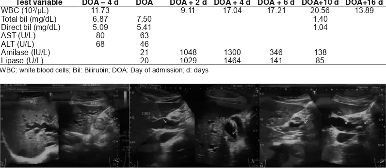
presentation to the hospital, her total bilirubin was 7.50 mg/dL with dominance of direct bilirubin (5.41 mg/dL) and slight increase of aspartate transaminase (63 IU/L) and alanine transaminase (46 IU/L). Laboratory tests of the patients while hospitalization were documented in Table 1.
Transabdominal ultrasound of the right upper quadrant revealed mild hepatomegaly with dilatation of intrahepatic bile duct, common hepatic duct, until distal common bile duct caused by choledocholithiasis sized 14.4 mm with sludge (Figure 1A, 1B, and 1C). The gallbladder wall thickness was increased with sludge inside the gallbladder. The sonographic murphy’s sign was positive. Sonographic fetal examination showed normal fetal heart rate, gestational age, and activity.
Table 1. Laboratory values of patient
Test variable DOA – 4 d DOA DOA + 2 d DOA + 4 d DOA + 6 d DOA+10 d DOA+16 d 3/μL)
Total bil (mg/dL) 6.87
Direct bil (mg/dL)
AST (U/L) 63
ALT (U/L) 68
Amilase (IU/L)
Lipase (U/L) 85
WBC: white blood cells; Bil: Bilirubin; DOA: Day of admission; d: days
The following day coagulation tests for preparation of ERCP were done showing normal results. Endoscopic ultrasound was performed to examine the bile duct. Echoendoscope showed enlargement of common bile duct with hyperechoic well-rounded stone throughout the common bile duct and sludge on distal part of the duct. Gallbladder wall also thickened with sludge inside the gallbladder (Figure 2A and 2B).
)LJXUH (QGRVFRSLF XOWUDVRXQG VKRZHG D WKLFNHQHG
gallbladder with sludge; (b) common bile duct stone
The treatment options for this patient included laparoscopic cholecystectomy or therapeutic ERCP. Laparascopic cholecystectomy was postponed to the postpartum period to avoid intrapartum surgical risks pertaining to the fetus and gravid uterus. Ultimately, a multidiscipline decision favored therapeutic ERCP option. The patient and spouse gave informed consent for ERCP understanding the risks to the mother, fetus and pregnancy.
The second hospital day she was taken to be done ERCP and placed under general anesthesia. Lead shielding was draped below and above the gravid uterus. A therapeutic lateral viewing video duodenoscope was passed per os and positioned in a favorable biliary position, looking upward toward the
DPSXOODU\RUL¿FH)LJXUH$&RQWUDVWZDVJHQWO\ PDQLSXODWHGZLWKLQWKHSDSLOODU\RUL¿FHXQWLOELOLDU\ FDQQXODWLRQZDVDFKLHYHGDQGFRQ¿UPHGE\DVSLUDWLRQ
of bile (Figure 3B).
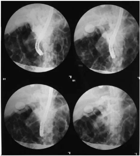
&RQWUDVWZDVLQMHFWHGDQGDVLQJOHVSRWÀXRURVFRSLF LPDJHWDNHQFRQ¿UPLQJELOLDU\GXFWDODQDWRP\)LJXUH
4).
The cannula was then gently advanced and biliary cannulation was performed. At first there
)LJXUH )OXRURVFRSLF LPDJH ZDV WDNHQ WR FRQ¿UP ELOLDU\
duct anatomy
Figure 3. Endoscopic retrograde cholangiopancreatography
VKRZHG D DPSXOODU\ RUL¿FH E %LOLDU\ FDQQXODWLRQ ZDV
performed
ZHUHGLI¿FXOWLHVGXHWRVXVSLFLRQRILPSDFWHGVWRQH
After guidewire was back loaded and advanced into
WKH ELOLDU\ WUHH DVSLUDWLRQ RI ELOH ZDV FRQ¿UPHG
Papillotomy was performed and pus was drained out
WKURXJKWKHRUL¿FH3ODVWLFELOLDU\VWHQWZDVSODFHGLQ WKHRUL¿FHZLWKVPRRWKELOHÀRZDFFRPSDQLHGE\SXV
(Figure 5A, 5B, and 5C). Stent was placed to ensure drainage and prevent recurrent cholangitis during the remainder of her pregnancy. Post ERCP, fetal monitoring demonstrated normal fetal movement and heart tones.
The day after ERCP, the mother had fever and
DEGRPLQDOSDLQ)DVWLQJZDVLPSOLHGZLWKÀXLGDQG
broad spectrum antibiotic was given. Post ERCP pancreatitis was diagnosed with increased of amylase and lipase. Leukocyte count and CRP showed high probability of biliary infection suggestive of cholecystitis. Due to the infection which still occurred, source control was performed. Besides infection in the common bile duct, gallbladder also can be the source of infection, especially when there is sludge inside the gallbladder. Daily observation showed improvement of pancreatitis. Meanwhile, ultrasound-guided percutaneous cholecystostomy was also performed at day 6 after admission due to cholecystitis which have not resolved. Bile culture showed Burkholderia cepacia as the causative agent of infection. After the procedure, patient showed improvement of her temperature and heart rate with low level of procalcitonin indicated resolution of infection. Patient then discharged about two weeks after admission with improvement of her condition.
DISCUSSION
Gallstone or common bile duct stone during pregnancy is not uncommon condition occurred. There are several reasons for this condition. Physiologic changes in pregnancy caused gallbladder volume doubles in the 2nd and 3rd trimesters. Gallbladder
emptying rate are slower compared to woman without pregnancy. Impairment of gallbladder motility, increase of residual volume, and increase bile stone formation, which becomes saturated with cholesterol, create a good environment for gallstone formation. US studies demonstrated up to 10% of pregnant patients have gallstones on routine obstetric ultrasound.1,5 Biliary
tract disease should, therefore, be considered in any pregnant woman with abdominal pain. Pain can be located in the epigastrium or right upper abdominal quadrant with associated nausea and vomiting. Fever suggests more serious complications, such as acute cholecystitis, cholangitis or pancreatitis. The development of biliary-tract complications during pregnancy has implications for maternal and fetal morbidity. This patient’s present with epigastric pain and cholestatic jaundice and consistent with the diagnosis of choledocholithiasis, which occurs in approximately up to 12% in pregnancy.
Abdominal ultrasound is the initial diagnostic test of choice for biliary tract disease, with a sensitivity of over 90% for gallstones. Deutchman et al found that
gallstones were found in 5.3% of the patients during routine obstetric ultrasound.6 Ultrasonography is also
useful to look for other intraabdominal causes of pain or complicated biliary disease. Measurement of serum transaminases, alkaline phosphatase, bilirubin, amylase, and lipase levels, and complete blood counts aids in the differential diagnosis. Early sonography of this patient revealed dilatation of intra- and extrahepatic bile duct accompanied by common bile duct stone consistent with cholestatic jaundice due to choledocholithiasis. Increase of total bilirubin with dominance of direct component strengthened the diagnosis. Endoscopic ultrasound also had been reported to be valuable in diagnosis of choledocholithiasis in pregnancy.7 In
this case, endoscopic ultrasound was also performed with image revealed stone in common bile duct and dilatation of common bile duct.
Clinical management varies, depending on gestational age and severity of symptoms. When facing a gravid patient with biliary tract disease, the outcomes of medical and surgical (open and laparoscopic cholecystectomy) management must be considered. Non-operative management of symptomatic cholelithiasis in pregnant patients led to a suboptimal clinical outcome in 38% of patients and higher rate of fetal death.2,8 There is
consensus that surgical intervention is indicated for obstructive jaundice, acute cholecystitis failing medical management, gallstone pancreatitis, or suspected peritonitis. When the common bile duct is
FOHDURIVWRQHVDQGWKHSUHJQDQF\LVZLWKLQWKH¿UVW
or second trimester, laparoscopic cholecystectomy is the usual recommended course of action.2 However
cholecystectomy during pregnancy was considered as a relative contraindication, mainly because of the lack of knowledge of the effects of CO2 to the fetus.9 Potential risks of laparoscopic surgery during
pregnancy are uterine lesion during trocar placement, induction of preterm labor and fetal acidosis. In the third trimester the gravid uterus can make laparoscopic
FKROHF\VWHFWRP\PRUHGLI¿FXOWLQWKLVFLUFXPVWDQFH
the option for therapeutic ERCP is dependent upon the availability of endoscopic expertise. In this setting of patient, aggressive management was chosen due to high complication rate in non-surgical management. Biliary drainage was preferred by using ERCP in this case.
ERCP is to relieve distal obstruction by sphincterotomy, stone extraction and/ or biliary stent placement. During ERCP the gravid uterus should be shielded with lead
DQGÀXRURVFRS\PLQLPL]HG,QWKHFDVHSUHVHQWHGLQ
this report, even though the patient was in her second trimester, therapeutic ERCP was preferred due to its safety issue to the mother and fetus and also because of stone which were located in common bile duct that
FDXVHGGLI¿FXOWLHVLQFRPPRQELOHGXFWH[SORUDWLRQ
by using laparascopic cholecystectomy. Simple “wait and watch” observation seemed to be a poor option due to symptomatic condition. Placement of biliary stent offered avoidance of biliary stasis as the focus of infection.
ERCP is one of the main non-surgical procedures applied in the treatment for choledochal stones and gallstones pancreatitis. Since it has low rate of mortality and morbidity theoretically, ERCP is the
¿UVWPHWKRGWRSUHIHULQWKHWUHDWPHQWRIFKRODQJLWLV
and pancreatitis due to choledocholithiasis during pregnancy. On the other hand, there are some limitations for applying ERCP. Endoscopic procedure
DQGULVNVVKRXOGEHDFFHSWHGE\SUHJQDQWZRPDQ¿UVW
Also, ERCP indication should be established precisely,
DQGLWLVVLJQL¿FDQWWRNQRZDWZKDWWULPHVWHULWZLOO
be done and at which position will she be during the
SURFHGXUH$QRWKHUVLJQL¿FDQWIDFWRUOLPLWLQJWRFDUU\
out ERCP is that the ERCP is carried out in company
ZLWKÀXRURVFRS\DQGWKHUHIRUHIHWXVLVH[SRVHGWR
ionized radiation during the procedure.
Caution must be taken for the complications of ERCP. In this case, pancreatitis occurred after ERCP procedure. However, this is not uncommon condition. Tiwari, et al, conducted a retrospective review of 19 studies including 214 ERCPs in pregnant women and the procedure related complications included spontaneous abortion (0.9%), fetal distress (0.6%), post procedure pancreatitis (4.6%) and preterm labor occurred in 4.6% of cases.11 Tang, et al, in 2009,
reported retrospective results of 68 ERCP performed in 65 pregnant women. Their study showed that term delivery of the baby was achieved in 53 patients (89.8%), but 11 patients (16%) suffered post-ERCP pancreatitis. The rates of pancreatitis were statistically higher than expected in the pregnancy group vs. the matched control group (12% vs. 5%; p < 0.001; OR = 2.8).12 Jamidar et al also found that post ERCP
pancreatitis occurred in one out of twenty-three pregnant patients in his study.13 Latest study by Lee
et al, found 1 post-ERCP hyperamylasemia out of 13 pregnant patients.14
Treatment options for cholecystitis were limited to laparoscopic cholecystectomy and observational management. However, in our case medical management didn’t show improvement of the clinical signs. After ERCP, patient had fever, abdominal pain, increased leukocytes, and positive biliary culture despites of pancreatitis resolution. Therefore, cholecystitis was suspected and ultrasound guided percutaneous cholecystostomy was performed. Described in 1980, a percutaneous drainage catheter can be placed with local anesthesia utilizing 2-dimensional B-mode ultrasound, without ionizing radiation, with excellent results. Furthermore, some case report has documented successfulness of percutaneous cholecystostomy in pregnancy that applied to a 33-year-old pregnant woman who failed ERCP and papillotomy and had multiple return visits for recurrent biliary infection.15
In conclusion, our case suggested that aggressive management of choledocholithiasis using ERCP and accompanied by percutaneous cholecystostomy can be carried out safely. Patient should be treated with less invasive procedures compared to laparascopic cholecystectomy. This result might allow us to change the original protocol and treat patient with endoscopic or percutaneous technique for biliary stone disease in pregnancy.
REFERENCES
1. Ko CW, Beresford SA. Incidence, natural history, and risk factors for biliary sludge and stones during pregnancy. Hepatology 2005;41:359-65.
2. Lu EJ, Curet MJ, El-Sayed YY, Kirkwood KS. Medical versus surgical management of biliary tract disease in pregnancy. Am J Surg 2004;188:755-9.
3. Scott LD. Gallstone disease and pancreatitis in pregnancy. Gastroenterol Clin North Am 1992;21:803-15.
4. Samara ET, Stratakis J, Enelo MJ, Mouzas IA, Perisinakis K, Damilakis J. Therapeutic ERCP and pregnancy: is the radiation risk for the conceptus trivial? Gastrointest Endosc 2009;69:824-31.
5. Maringhini A, Ciambra M, Baccelliere P, Raimondo M, Orlando A, Tine F, et al. Biliary sludge and gallstones in pregnancy: incidence, risk factors, and natural history. Ann Intern Med 1993;119:116-20.
6. Deutchman ME, Connor P, Hahn RG, Rodney WM. Maternal gallbladder assessment during obstetric ultrasound: results,
VLJQL¿FDQFHDQGWHFKQLTXH-)DP3UDFW
7. Girotra M, Jani N. Role of endoscopic ultrasound/ SpyScope in diagnosis and treatment of choledocholithiasis in pregnancy. World J Gastroenterol 2010;16:3601-2.
8. Jelin EB, Smink DS, Vernon AH, Brooks DC. Management of biliary tract disease during pregnancy: a decision analysis. Surgical Endoscopy 2008;22:54-60.
10. Al-Hashem H, Muralidharan V, Cohen H, Jamidar PA. Biliary disease in pregnancy with an emphasis on the role of ERCP. J Clin Gastroenterol 2009;43:58-62.
11. Tiwari P, Khan AS, Nass JP, Rivera RE, Romero RV, Antillon MR, et al. ERCP in pregnancy: a systematic review. Gastrointest Endosc 2011;73:AB392.
12. Tang SJ, Mayo MJ, Rodriguez-Frias E, Armstrong L, Tang L, Sreenarasimhaiah J, et al. Safety and utility of ERCP during pregnancy. Gastrointest Endosc 2009;69:453-61.
13. Jamidar PA, Beck GJ, Hoffman BJ, Lehman GA, Hawes RH, Agrawal RM, et al. Endoscopic retrograde cholangiopancreatography in pregnancy. Am J Gastroenterol 1995;90:1263-7.
14. Lee JJ, Lee SK, Kim SH, Kim GH, Park DH, Lee S, et al.
(I¿FDF\DQGVDIHW\RISDQFUHDWRELOLDU\HQGRVFRSLFSURFHGXUHV
during pregnancy. Gut Liver 2015;9:672-8.