Partial characterization and localization of a novel type of
antifungal protein (IWF6) isolated from sugar beet leaves
Anne Kroll Kristensen
b, Janne Brunstedt
a, John Erik Nielsen
a, Jette Dina Kreiberg
a,
Jørn Dalgaard Mikkelsen
a,*, Peter Roepstorff
c, Klaus Kristian Nielsen
daDanisco Biotechnology,Langebrogade1,P.O.Box17,DK-1001Copenhagen K,Denmark bProFound Pharma A/S,Agern Alle1,DK-2970Horsholm Denmark
cInstitute of Molecular Biology,Odense Uni6ersity,Campus6ej55,DK-5230Odense M,Denmark dDLF-Trifolium,Hoejerup6ej31,P.O.Box19,DK-4660Store Heddinge,Denmark
Received 28 February 2000; received in revised form 24 May 2000; accepted 2 June 2000
Abstract
An antifungal protein was isolated from the intercellular washing fluid (IWF) of leaves of sugar beet (Beta 6ulgarisL., cv.
Monova) and purified to homogeneity. The protein, IWF6, comprising 37 amino acids with six cysteines, was able to inhibit the growth of the pathogenCercospora beticola(Sacc.) in vitro, by 75% after 120 h of growth at a concentration of 20mg ml−1. The
amino acid sequence data were used to generate a polymerase chain reaction (PCR) clone, employed for the isolation of a corresponding cDNA clone. The cDNA encodes a precursor protein with an N-terminal putative signal sequence of 45 amino acids, followed by the mature protein of 37 amino acids. Antibodies raised against a synthetic peptide covering the complete sequence of IWF6 were used in immunolocalization studies. The protein was recognized by the antibody in nearly all leaf cell types except epidermal cells. In necrotic tissue, the protein was mainly recognized onC. beticolahyphae growing in a ‘pellet’ (ball-like) structure. The hyphal ‘pellets’ are primarily located beneath the stomata. IWF6 shows less than 26% identity to any previously described protein. © 2000 Elsevier Science Ireland Ltd. All rights reserved.
Keywords:Antifungal protein;Beta6ulgaris;Cercospora beticola
www.elsevier.com/locate/plantsci
1. Introduction
Plants are constantly exposed to pathogenic at-tack by different microorganisms and in order to protect themselves from these attacks, they posses several defense mechanisms. One of these defense mechanisms is the production of proteins with antifungal activity. Several groups of antifungal proteins have been characterized so far: one group of antifungal proteins consists of hydrolytic en-zymes like the chitinases [1 – 7] and the glucanases [8,9], while another group consists of small, cys-teine-rich proteins [10 – 12].
We have investigated the production of small, cysteine-rich proteins in sugar beet which show good activity against the causal agent of the leaf spot disease, Cercospora beticola. Previously, we have identified a number of cysteine-rich proteins isolated from the intercellular washing fluid (IWF) of leaves from sugar beet. The proteins, belonging to the group of plant defensin-like proteins [13], non-specific lipid transfer proteins [14] and chitin-binding proteins [15], all showed strong antifungal activity in vitro againstC. beticola. These proteins fall into three groups of antimicrobial proteins that vary with respect to size and cysteine-content. The plant defensins are 5 kDa proteins with eight cysteines, the chitin-binding proteins are 3.5 kDa proteins with six cysteines, while the non-specific lipid transfer proteins are 9 – 10 kDa proteins with Abbre6iations: FPLC, fast protein liquid chromatography; IWF,
intercellular washing fluid; PAGE, polyacrylamide gel electrophore-sis..
* Corresponding author. Tel.:+45-32662249; fax:+45-32662167.
E-mail address:[email protected] (J.D. Mikkelsen).
eight cysteines [12]. They are all basic, with isoelectric points of 9 to 10, and inhibit the growth of various pathogens at low concentra-tions [10].
Here we report the isolation and characteriza-tion of a new type of antifungal protein. It is a 37 amino acid protein with six cysteines present and has an isoelectric point of 7. The protein shows no homology to any of the hitherto characterized antifungal proteins.
2. Materials and methods
2.1. Biological materials and bioassay
Plants of sugar beet (Beta 6ulgaris L., cv.
Monova, Danisco Seed) were grown in growth chambers as described previously [1]. The in vitro inhibitory effects of protein fractions on the growth of C. beticola (Sacc.) (isolate ‘FC573’, Dr Earl G. Ruppel, USDA, Fort Collins, USA) were tested using the microtiter plate bioassay de-scribed previously [1]. In this assay, measuring the increase in absorbance at 620 nm follows the growth of submerged spore cultures. Cultures for microscopical analysis were grown likewise. The data presented are from one experiment, but the same growth curves and thus level of growth inhibition are observed in other experiments.
2.2. Purification of antifungal protein IWF6 from the intercellular washing fluid of sugar beet lea6es
The intercellular washing fluid (IWF) from 1 kg sugar beet leaves was isolated and fractionated as described previously [13]. In short, the IWF was applied to an ion exchange column containing 10 ml CM-Sepharose (Pharmacia LKB) equilibrated in 20 mM acetic acid, pH 4.5. Bound proteins were eluted by stepwise increasing the salt concen-tration in the starting buffer: 0.1, 0.3 and 0.5 M NaCl. The 0.3 M NaCl eluate comprised antifun-gal activity and was further purified by cation exchange fast protein liquid chromatography (FPLC) on a Mono S HR 5/5 column (Pharmacia LKB). The individual protein peaks from the Mono S column were further purified by reverse-phased HPLC on a Vydac C4 silica column (The
Separations Group, CA, USA). The solvent sys-tem was (A) 0.1% TFA in water and (B) 0.1%
TFA in acetonitrile. Proteins were eluted with a linear gradient from 5 to 45% of the B-buffer from 0 to 18 min after sample loading followed by 60% B-buffer for 2 min. Flow rate was 0.7 ml min−1. Protein was detected at 214 and 280 nm.
Discrete protein peaks were collected and freeze dried. After two washes with water followed by freeze drying, the protein peaks were resolved in 10 mM Tris – HCl, pH 8.0, for the analysis of purity (SDS – PAGE) and antifungal activity.
Prior to N-terminal sequencing, proteins were reduced and carboxymethylated and subjected to reversed-phase HPLC on a Vydac C4 column.
Amino acid sequencing was performed as de-scribed previously [1].
2.3. Polyacrylamide gel electrophoresis, antibodies and immunoblotting
Proteins were separated by SDS – PAGE on 16.6% Tricine gels as described [1,16]. Isoelectric focusing was performed using the Phast system (Pharmacia LKB) as described [2].
A full-length peptide covering the entire se-quence of IWF6 was synthesized by Research Genetics (Alabama, USA) using the Fmoc tech-nology [17]. The full length IWF6 peptide (37 amino acids) was conjugated to difteria toxoid before immunization as described [18].
Immunoblotting of proteins separated by Tricine SDS – PAGE was performed as outlined previously [1], using IWF6 antibodies diluted 1:100. Protein concentrations were determined us-ing the Bio-Rad protein assay kit with g-globulin as standard as described [13].
2.4. Immunolocalization of IWF6
For the immunolocalization studies of IWF6, the following sugar beet varieties were used: Monova, Marathon and +Tol (Danisco Seed). Infection with C. beticola was performed as de-scribed [1]. Samples were extracted from leaves of sugar beet (approx. 1 cm2) with necroses 12, 15
deviation was the use of xylen instead of petroleum ether. Prior to the immune procedure, samples were reduced by treatment with 50 mM DTT in TBS (0.5 M Tris – HCl, 0.15 M NaCl, 0.1% Triton X-100, pH 7.6) for 20 min in order to obtain optimal detection by the antibodies (anti-bodies were raised against a synthetic form of IWF6, i.e. randomly coiled). Variations in expo-sure time to DTT (10, 20 and 30 min) gave no significant difference. The IWF6 antibodies and preimmune control antibodies, were used in a 1:50 dilution. The immune procedure previously de-scribed [3] was used.
2.5. Mass spectrometry
Matrix-assisted laser desorption mass spec-trometry (MALDI MS) [17,18] was performed using a Voyager DE instrument (PerSeptive Biosystems, Framingham, MA) equipped with delayed extraction [20] in the linear configuration. All samples were analyzed in the positive mode using an acceleration voltage of 20 kV and ex-ternally calibrated. The matrix used was a-cyano 4-hydroxy cinnamic acid (Sigma) at a concen-tration of 15 mg ml−1in 70% CH
3CN, 0.1% TFA
[19]. Sample preparation was the dried droplet method [21] and an average of at least 100 shots were collected and used for analysis.
2.6. Cloning of IWF6 cDNA
The cDNA sequence of IWF6 was obtained by 3%and 5%RACE essentially as described previously
[14] using the following primers: 3% RACE Primers
QT: 5%
-CCAGTGAGCAGAGTGACGAGGA-CTCGAGCTCAAGC(T)17-3%
Q0: 5%-CCAGTGAGCAGAGTGACG-3%
Q1: GAGGACTCGAGCTCAAGC-3%
5% RACE primers
5%-Anchor: 5%
-GGCCACGCGTCGACTAGT-ACGGGGGGGGGG-3%
5%-UNI: 5%-GGCCACGCGTCGACTAGTACG
2.7. 3% RACE
The amino acid sequence of the IWF6 protein was used to construct two degenerated oligonucle-otide primers for the isolation of a partial cDNA clone by 3%RACE. Total RNA was purified from
sugar beet (cv. Monova) leaves 6 days after inocu-lation with C. beticola according to [22]. Reverse transcription followed by PCR was done with the RT-PCR kit from Perkin Elmer and according to their protocol. Briefly, 1 mg of total RNA and 2.5 pmol QT-primer was incubated at 42°C for
45 min with reverse transcriptase followed by in-cubations at 99°C for 5 min and 5°C for 5 min. In the first PCR 40 pmol of the primer Q0was used
as downstream primer and the upstream primer was 150 pmol of the degenerated primer P1 (5% - GG(ACGT)TA(CT)TG(CT)AA(CT)AT(ACT)-(CT)T: position 297 – 313 in the IWF6 cDNA se-quence). In the second nested PCR 50 pmol of the primer Q1 was used as downstream primer and
the upstream primer was 50 pmol of the degener-ated primer P2 (5%
-AA(CT)GT(ACGT)TG(CT)-TG(CT)GC(ACGT)GG: position 314 – 332 in the IWF6 cDNA sequence). The PCR conditions were: 1 min at 94°C, 2 min at 42°C, 1 min at 50°C and 5 min at 72°C for 1 cycle followed by 1 min at 94°C and 2 min at 42°C and 3 min at 72°C for 35 cycles followed by 10 min at 72°C. After the second PCR a single DNA product of 320 bp was obtained. The DNA product was cloned into the pT7Blue vector (Novagen) and sequenced us-ing a Termo Sequenase fluorescent cycle sequenc-ing kit (Amersham) and an ALF DNA sequencer (Pharmacia).
2.8. 5% RACE
The sequence of the 5%end of IWF6 cDNA was obtained by 5%RACE using the 5%RACE system
from Gibco BRL with 3 gene specific primers constructed from the partial cDNA sequence ob-tained by 3%RACE. Briefly, 1 mg of the same total RNA as used for 3%RACE and 2.5 pmol of a gene specific primer GSP6-1 (5%
-CATCAAGAAGTC-CATAATTGTCTAG: position 508 – 532 in the IWF6 cDNA sequence) was incubated at 70°C for 10 min followed by the addition of reverse tran-scriptase and incubating at 42°C for 30 min, 70°C for 15 min and the addition of RNaseH and incubating further 10 min at 55°C. The cDNA was dC-tailed according to the protocol of Gibco BRL. The tailed cDNA was subjected to two rounds of PCR. In the first PCR 20 pmol of the 5%-Anchor primer was used as upstream primer and the downstream primer was 20 pmol of the gene specific primer GSP6-2 (5%
473-498 in the IWF6 cDNA sequence). In the second PCR 50 pmol of the 5%-UNI primer was used as upstream primer and the downstream primer was 50 pmol of the gene specific primer GSP6-3 (5% - ACAGACACGCTAGTTAGATGACTAAGC:-position 456 – 482 in the IWF6 cDNA sequence). The condition for the first PCR was: 1 min at 94°C and 1 min at 51°C and 2 min at 72°C for 35 cycles followed by 10 min at 72°C. The condition for the second PCR was: 1 min at 94°C and 1 min at 55°C and 2 min at 72°C for 35 cycles followed by 10 min at 72°C. The single 510 bp DNA product was cloned into the pT7Blue vector (No-vagen) and sequenced using a Termo Sequenase fluorescent cycle sequencing kit (Amersham) and an ALF DNA sequencer (Pharmacia).
2.9. Southern blotting
Genomic DNA was isolated from sugar beet leaves according to [23], digested with appropriate restriction enzymes and separated on a 0.8% agarose gel. A DNA probe of IWF5 cDNA was
32P-labelled by random priming using the
Ready-To-Go Labelling Kit from Pharmacia. Southern transfer and hybridization was performed accord-ing to [24] usaccord-ing Hybond N+ membranes (Amer-sham) following the recommendations of the manufacturer.
3. Results
3.1. Purification and biochemical characterization of IWF6
The 0.3 M NaCl eluate from the CM-Sepharose column possessed strong antifungal activity. When this fraction was subjected to cation exchange chromatography, six major and a number of smaller protein peaks were eluted. All peaks dis-play antifungal activity, and have been shown to contain a number of chitinases [2 – 4], plant de-fensins [13] and non-specific lipid transfer proteins [14]. Strong antifungal activity was also found in peak 4 and SDS-polyacrylamide gel electrophore-sis revealed that this protein peak contained a number of different proteins. When peak 4 was further separated by RP-HPLC on a Vydac C4
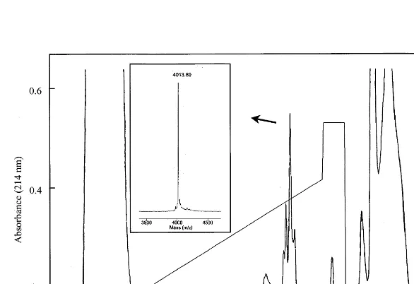
column, it separated into 9 – 10 distinct protein peaks on the RP-HPLC column (data not shown). One of these exhibited strong antifungal activity and was subsequently chromatographed by re-versed-phase HPLC using a Vydac C4 column
(Fig. 1). Only the major peak was collected and used in the subsequent work. The quality of the purification was evaluated by MALDI MS (Fig. 1, inset) and N-terminal amino acid sequencing, and only one protein was present. The complete N-ter-minal amino acid sequence of the protein was
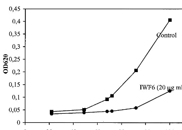
Fig. 2. Biological assay showing the growth of spore cultures ofC.beticola. The fungal growth was measured as increase in A620in the absence (rectangles) or presence (circles) of 20mg
ml−1IWF6.
A620) started 40 – 50 h after incubation start (Fig. 2).
When 20 mg ml−1 IWF6 was added to the spore
cultures, culture start was strongly delayed, and detectable growth not observed until 70 – 80 h after growth start. Furthermore, the growth rate of the culture was substantially slower (Fig. 2). Micro-scopical examination revealed that also the mor-phology of the IWF6 treated fungal hyphae was affected: the restricted hyphae were shorter and somewhat more branched (not shown), an effect similar to that exerted by another antifungal sugar beet peptide, IWF4 [15]. The difficulties in obtain-ing reproducible concentration values make the actual concentration uncertain and probably much lower than stated (the indicated concentrations are the maximium values obtained by protein determi-nation). The antifungal activity of IWF6 was confi-rmed in repeated experiments.
3.3. Localization of IWF6
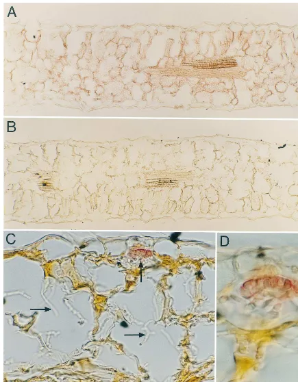
IWF6 was detected in all leaf cell types except epidermal cells [Fig. 3(A)], in various paraffin sections of non-infected sugar beet leaves by pre-treatment with DTT before employing the normal immune procedure. Without treatment with DTT, the same plants show only a faint positive reaction in xylem and stomata. In sugar beet leaves infected with C. beticola, IWF6 was only found in signifi-cant amounts in tissue probably about to disinte-grate due to infection, as well as in the established necrosis (Fig. 3(C) and (D)). Almost no IWF6 was found in the anatomical normal tissue surrounding the necroses. In the established necrosis (Fig. 3(C) and (D)), the IWF6 was detected to Cercospora
hyphae present in a ball-like structure, ‘pellet’ [25] and not to single hyphae. The hyphal ‘pellets’ are mainly placed under stomata. These reactions onC.
beticolacould also be seen without DTT treatment, but not as intensively stained. No significant differ-ence was observed in the distribution of IWF6 in the necroses from day 12 – 19. Preimmune controls were all negative.
3.4. Sequence homology
A database search was performed (BLAST vers. 2.0.10) in order to identify potential homology to other published protein sequences.
IWF6 shows no significant similarity to any protein sequence in the database. Only, the C-ter-obtained by automated Edman degradation. The
protein consisted of 37 amino acids, six of which were cysteines. The SDS – PAGE analysis indicated an apparent molecular mass of 3.5 kDa which is in agreement with the observed molecular weight (Mr) of 4012.89 as determined by MALDI MS.
An isoelectric point of 7.0 was deduced when IWF6 was subjected to isoelectric focusing using IEF Phast gels with a pH range of 3 – 9 (not shown). This result was in agreement with a predicted isoelectric point (pI) of 7.5 (when all cysteines take part of disulfide bonds) as calculated from the amino acid sequence (PC/Gene, version 6.85).
The IWF6 protein was present in the intercellular washing fluid (IWF) at extremely low concentra-tions (typically 100 mg in 2 kg leaves), and several IWF isolations and purifications were required in order to obtain sufficient amounts of protein (mg quantities) for amino acid sequencing and (limited) assaying for biological activity. Furthermore, it proved very difficult to determine the exact protein concentration as different methods gave different values. The stated concentrations are thus maxi-mum values and the actual concentration is proba-bly lower than stated.
Antibodies were raised against a synthetic 37-mer comprising the entire sequence of IWF6. In order to obtain maximum detection by the anti-body it proved necessary to reduce the protein with DTT prior to Western blotting.
3.2. Antifungal acti6ity
Fig. 3. IWF6 immuno-visualized (red) on cross sections of sugar beet leaves after pretreatment with DTT (20 min) prior to immunolocalization with alkaline phosphatase-coupled secondary antibodies and Fast Red development. (A), (C) and (D) IWF6 antiserum (1:50). (B) control (pre-immune serum 1:50). Fig. 3(A) show a broad distribution of IWF6 in the leaf except in the epidermal cells. Fig. 3(C) and (D) Necrose infected with Cercospora, IWF6 is detected on the hyphal ‘pellets’ typically found beneath stomata (vertical arrow). No IWF6 was detected on separate hyphae (horizontal arrows). (A) and (B) 108×, (C) 470×
minal part of IWF6 shows a low level of homol-ogy to agelenin, a neurotoxin of 35 amino acid residues isolated from the venom of the spider
Agelena opulenta [26]. The optimal identity is ob-tained by introducing a gap of two residues in IWF6, by which the six cysteines present in both proteins can be aligned. Hence, an identity of 35% (or 26% without gap) is found between the two proteins on the amino acid level (Fig. 4).
The RPRPRP-motif present in the N-terminal of IWF6 has not been observed in any of the hitherto characterized antifungal proteins. A simi-lar motif is present in the C-terminal part of a presumed Mycobacterium leprae antigen [27]. In addition, the hinge region of some chitinases is rich in proline, thus giving this region its flexible structure [4].
3.5. Characterization of IWF6 cDNA
The nucleotide sequence of the full-length IWF6 cDNA clone, obtained by a combination of 3%and 5%RACE, and the deduced amino acid sequence is
shown in Fig. 5. The IWF6 cDNA contains 593 nucleotides including a polyadenylation site (AATAAA) located 82 nucleotides upstream of a
poly(A) tail of 24 nucleotides. The sequence con-tains an open reading frame encoding 82 amino acid residues with the first in frame ATG at posi-tion 129 and a stop codon at posiposi-tion 375. In comparison with IWF6, the cDNA encode a pre-cursor protein with an N-terminal putative signal sequence of 45 amino acid residues followed by the mature protein of 37 amino acid residues. In accordance herewith, the preprotein contains a putative cleavage site between the alanine at posi-tion 45 and the arginine at posiposi-tion 46 [28]. The cDNA derived sequence is identical to the amino acid sequence of the isolated IWF6 except for the amino acid Ala to Val change at position 35 of the mature protein. This amino acid change was ob-served in both the 3% as well as the 5% RACE
derived clones, thus making it unlikely that it is due to a PCR introduced error.
3.6. Southern blotting
Genomic DNA from sugar beet was digested by the restriction enzymes BamHI or XbaI and sub-jected to Southern hybridization analysis using IWF6 cDNA as probe. Each restriction enzyme digest resulted in only one hybridizing band, indi-cating that only one gene (or a small number of genes) for IWF6 is present in the genome of sugar beet (data not shown).
4. Discussion
A novel type of antifungal protein was isolated from the IWF of sugar beet leaves using cation exchange chromatography and reverse-phased HPLC. The protein, IWF6, was present in very low amounts and several purifications were neces-sary to obtain sufficient material for sequence determination and growth inhibition assays. The complete amino acid sequence of 37 residues was obtained by N-terminal sequencing. The calcu-lated molecular weight (Mr) of IWF6 (based on the amino acid sequence) is 4012.59 assuming the six cysteines present form disulfide bridges. This correlates well with the MALDI MS data (ob-served molecular weight: 4012.8990.4) and indi-cates that all six cysteines take part in disulfide bonds.
The isoelectric point of 7.0, as determined by isoelectric focusing of IWF6 and in agreement
Fig. 4. Comparison of the amino acid sequence of IWF6 with agelenin, a neurotoxin from the venom of the spider A. opulenta[26]. In order to optimize the alignment, one gap has been introduced in IWF6. The character to show that two aligned residues are identical is’’. Identity: 35.1% (with gap) or 25.7% (without gap).
with the predicted pI of 7.5, is very different from that of all other antifungal proteins isolated from sugar beet so far which are all basic of nature and have isoelectric points of 9 or above [13 – 15].
When spore cultures of C. beticola were grown in the presence of 20 mg ml−1 (highest estimated
concentration, see results) of IWF6, the growth of the fungus was strongly inhibited. After 90 h of growth, a 90% growth inhibition is observed and after 120 h the fungus is still more than 70% inhibited. Although assay conditions not being directly comparable, the antifungal potency of IWF6 is judged to be at the level of other classes of antifungal peptides, e.g. the plant defensins and the chitin-binding proteins, for which the level of protein needed for 50% growth inhibition is in the range of 5 – 10 mg ml−1, depending on the fungal
species investigated [12]. These levels are found when assaying in low ionic strength media, whereas when adding divalent cations the activity of the proteins is severely decreased [10]. Due to the limited amount of material, it was not possible to test the effect of divalent cations on the antifun-gal potency of IWF6.
Antibodies raised against a synthetic 37-mer comprising the entire sequence of IWF6 were used to localize the native protein in paraffin embedded sections of sugar beet leaves. To obtain maximum labeling, the sugar beet leaves had to be reduced by DTT before recognition by the antibody. This is most likely because the antibody was raised against a randomly folded, synthetic peptide. The recognition of IWF6 in all leaf cell types (except epidermal cells in non-infected leaves) with no obvious induction during fungal infection, may suggest that IWF6 does not play a major role in the plant defense response. However, it cannot be excluded that the present plants were stressed by other factors (i.e. handling procedure) and thus reacted by expressing the protein in many cell types.
In the established necrosis, the IWF6 seemed to be localized only on the surface of C. beticola
when the hyphae grow in ‘pellet’ but not to single hyphae. This is an interesting observation since the antifungal proteins IWF4 and especially AX2 in previous investigations have been shown to reduce the growth of C. beticola in vitro and induce morphological changes resulting in hyphal struc-tures resembling the ‘pellet’ of hyphae observed in the necroses [13,15]. Although various sugar beet
antifungal proteins have been localized to the hy-phae present in necroses [3,14], the IWF6 localiza-tion is different because it was found only in the ‘‘pellet’’ of hyphae. Intensive staining for IWF6 was observed in some uninfected leaves (possibly stressed) and in infected leaves with possible initia-tion of infecinitia-tion, whereas almost no IWF6 was observed proximal to the necroses. These findings in combination with the intensive IWF6 detection on hyphal pellets beneath the stomata, could sug-gest that IWF6 participates in an early defense response asC.beticolais known to invade through stomata.
IWF6 is a new type of antifungal protein and shows no homology to other known antifungal proteins. A low level of identity (26%) is observed to agelenin, a neurotoxin isolated from the venom of the spider A. opulenta [26]. Agelenin belongs to the group of cysteine-rich neuropeptides and is an irreversible presynaptic toxin [2]. This resembles the actions of v-toxins [29] which act by blocking P-type (high-threshold) calcium channels [29 – 31]. Members of the plant defensin family have been shown to induce rapid membrane responses in fungi possibly via a receptor mediated response or by direct insertion of the protein in the membrane thus forming an ion channel [32].
Although the biological role of the different antifungal proteins isolated so far is still unknown, growing evidence suggest a role in plant defense mechanism against invading pathogens [12]. This is supported by the fact that they are mostly predominant in the peripheral cell layers or cells exposed to infection (stomata and stigma) or cells exposed to spreading infection (xylem) [10,14,33]. This ‘peripheral’ localization could be important in forming a primary antifungal defense against pathogens. The broad distribution of IWF6 in sugar beet leaf cells except epidermis suggest that it takes part of a second line of defense mechanism against invading pathogens, although it seems to disappear from the anatomical normal tissue quite early in the infection and is only found in the necroses in combination with C. beticola hyphae displaying reduced growth.
Acknowledgements
We thank Susanne Dreboldt, Lene D. Hansen and Jette Skovsgaard for their excellent technical assistance. Clive P. Walthers is greatly acknowl-edged for sequencing the protein. The work is supported by Novartis Seeds (Stanton, USA) and by a grant EF590 from the Danish Academy of Technical Sciences (ATV).
References
[1] K.K. Nielsen, J.D. Mikkelsen, K.M. Kragh, K. Bojsen, An acidic class III chitinase in sugar beet: induction by Cercospora beticola, characterization and expression in transgenic tobacco plants, Mol. Plant-Microbe Interact. 6 (1993) 495 – 506.
[2] K.K. Nielsen, P. Jørgensen, J.D. Mikkelsen, Antifungal activity of sugar beet chitinase against Cercospora beti-cola: an autoradiographic study on cell wall degradation, Plant Path. 43 (1994) 979 – 986.
[3] J.E. Nielsen, K.K Nielsen, J.D. Mikkelsen, Immunohisto-logical localization of a basic class IV chitinase inBeta
6ulgaris leaves after infection with Cercospora beticola,
Plant Sci. 119 (1996) 191 – 202.
[4] J.D. Mikkelsen, L. Berglund, K.K. Nielsen, H. Chris-tiansen, K. Bojsen, Structure of endochitinase genes from sugar beets, in: C.J. Brine, P.A. Sandford, J.P. Zikakis (Eds.), Advances in Chitin and Chitosan, Elsevier Applied Science, Oxford, 1992, pp. 344 – 353.
[5] D.B. Collinge, K.M. Kragh, J.D. Mikkelsen, K.K. Nielsen, U. Rasmussen, K. Vad, Plant chitinases, Plant J. 3 (1993) 31 – 40.
[6] J.J. Beintema, Structural features of plant chitinases and chitin-binding proteins, FEBS Lett. 350 (1994) 159 – 163. [7] A. Stintzi, T. Heitz, V. Prasad, S. Wiedermann-Merdi-noglu, S. Kauffman, P. Geoffroy, M. Legrand, B. Fritig, Plant ‘pathogenesis-related’ proteins and their role in defense against pathogens, Biochemie 75 (1993) 687 – 706. [8] T.E. Gottschalk, J.D. Mikkelsen, J.E. Nielsen, K.K. Nielsen, J. Brunstedt, Immunolocalization and character-ization of ab-1,3-glucanase from sugar beet, deduction of its primary structure and nucleotide sequence by cDNA and genomic cloning, Plant Sci. 132 (1998) 153 – 167. [9] K.K. Nielsen, K. Bojsen, D.B. Collinge, J.D. Mikkelsen,
Induced resistance in sugar beet againstCercospora beti-cola: induction by dichloroisonicotinic acid is independent of chitinase andb-1,3-glucanase transcript accumulation, Physiol. Mol. Plant Pathol. 45 (1994) 89 – 99.
[10] F.R.G. Terras, K. Eggermont, V. Kovalena, N.V. Raikhel, R.W. Osborn, A. Kester, S.B. Rees, S. Tor-rekens, F. Van Leuven, J. Vanderleyden, B.P.A. Cammue, W.F. Broekaert, Small, cysteine-rich antifungal proteins from radish: their role in host defense, Plant Cell 7 (1995) 573 – 588.
[11] F. Garcı´a-Olmedo, A. Molina, A. Segura, M. Moreno, The defensive role of non specific lipid-transfer proteins in plants, Trends Microbiol. 3 (1995) 72 – 74.
[12] W.F. Broekaert, B.P.A. Cammue, M. De Bolle, K. Thevissen, G.W. De Samblanx, R.W. Osborn, Antimicro-bial peptides from plants, Crit. Rev. Plant Sci. 16 (1997) 297 – 323.
[13] K.M. Kragh, J.E. Nielsen, K.K. Nielsen, S. Dreboldt, J.D. Mikkelsen, Characterization and localization of new anti-fungal cysteine-rich proteins fromBeta6ulgarisL, Mol. Plant – Microbe Interact. 8 (1995) 424 – 434.
[14] K.K. Nielsen, J.E. Nielsen, S.M. Madrid, J.D. Mikkelsen, New antifungal proteins from sugar beet (Beta6ulgarisL.) showing homology to non-specific lipid transfer proteins, Plant Mol. Biol. 31 (1996) 539 – 552.
[15] K.K. Nielsen, J.E. Nielsen, S.M. Madrid, J.D. Mikkelsen, Characterization of a new antifungal chitin-binding pep-tide from sugar beet leaves, Plant Physiol. 113 (1997) 83 – 91.
[16] H. Schagger, G. Von Jagow, Tricine-sodium dodecyl sulfate-polyacrylamide gel electrophoresis for the separa-tion of proteins in the range from 1 to 100 kDa, Anal. Biochem. 166 (1987) 368 – 379.
[17] J.P. Tam, Synthetic peptides vaccine design: synthesis and properties of a high-density multiple antigenic peptide system, Proc. Natl. Acad. Sci. USA 85 (1988) 5409. [18] J. Marcussen, C. Poulsen, A nondestructive method for
peptide bond conjugation of antigen haptens to a diphthe-ria toxoid carrier, exemplified by two antisera specific to acetolactase synthase, Anal. Biochem. 198 (1991) 318 – 323.
[19] M. Mann, G. Talbo, Developments in matrix-assisted laser desorption/ionization peptide mass spectrometry, Curr. Opin. Biotechnol. 7 (1996) 11 – 19.
[20] M. Vestal, P. Juhasz, S.A. Martin, Delayed extraction matrix-assisted laser desorption time-of-flight mass spec-trometry, Rapid Commun. Mass Spectrom. 9 (1995) 1044 – 1050.
[21] M. Kussmann, E. Nordhoff, H.R. Nielsen, S. Haebel, M.R. Larsen, L. Jakobsen, J. Gobom, E. Mirgorodskaya, A.K. Kristensen, L. Palm, P. Roepstorff, Matrix-assisted laser desorption/ionization mass spectrometry sample preparation techniques designed for various peptide and protein analytes, J. Mass Spectrom. 32 (1997) 593 – 601. [22] D.B. Collinge, D.E. Milligan, J.M. Dow, G. Scofield, M.J. Daniels, Gene expression inBrassica campestrisshowing a hypersensitive response to the incompatible pathogen Xanthomonas campestris pv. Vitians, Plant Mol. Biol. 8 (1987) 405 – 414.
[23] S.L. Dellaporta, J. Wood, J.B. Hincks, A plant DNA minipreparation: Version II, Plant Mol. Biol. Rep. 1 (1983) 19 – 21.
[24] T. Maniatis, E.F. Fritsch, J. Sambrook, Molecular Cloning: A Laboratory Manual, Cold Spring Harbor Laboratory, New York, 1982.
[25] J.L. Prosser, A.J. Tough, Growth mechanisms and growth kinetics of filamentous microorganisms, Biotechnology 10 (1991) 253 – 274.
[27] R.A. Hartskeerl, R.M. van Rens, L.F. Stabel, M.Y. de Wit, P.R. Klatser, Selection and characterization of recombinant clones that produceMycobacterium leprae antigens recognizes by antibodies in sera from household contacts of leprosy patients, Infect. Immun. 58 (1990) 2821 – 2827.
[28] G. Von Heijne, A new method for predicting signal sequence cleavage sites, Nucl. Acids Res. 14 (1986) 4683 – 4690.
[29] M.E. Adams, I.M. Mintz, M.D. Reily, V. Thanabal, B.P. Bean, Structure and properties ofv-Agatoxin IVB, a new antagonist of P-type calcium channels, Mol. Pharm. 44 (1993) 681 – 688.
[30] M.E. Adams, E.E. Herold, V.J. Venema, Two classes of channel-specific toxins from funnel web spider venom, J. Comp. Physiol. A 164 (1989) 333 – 342.
[31] I.M. Mintz, V.J. Venema, K.M. Swiderek, T.D. Lee, B.P. Bean, M.E. Adams, P-type calcium channels blocked by the spider toxinv-Aga-IVA, Nature 355 (1992) 827 – 829. [32] K. Thevissen, A. Ghazi, G.W. De Samblanx, C. Brownlee, R.W. Osborn, W.F. Broekaert, Fungal membrane re-sponses induced by plant defensins and thionins, J. Biol. Chem. 271 (1996) 15018 – 15025.
[33] S. Thoma, Y. Kaneko, C. Somerville, A non-specific lipid transfer protein from Arabidopsis is a cell wall protein, Plant J. 3 (1993) 427 – 436.