Development of screening systems for drugs against human
papillomavirus-associated cervical cancer: based on
E6-E6AP binding
Young-Sik Cho, Cheong-Weon Cho, Ok Joung, Kyung-Ae Lee, Sue-Nie Park,
Do-Young Yoon*
Cellular Biology Lab,Korea Res. Institute of Bioscience and Biotechnology,Taejon 305-600, South Korea
Received 7 March 2000; accepted 25 May 2000
Abstract
Human papillomavirus (HPV) E6 protein forms a ternary complex with the cell-cycle regulator p53 and the E6-associated protein (E6AP) known as an E3 ubiquitin protein ligase, leading to the degradation of p53 via the ubiquitination pathway. As an attempt to employ interaction between HPV viral oncogene E6 and a cellular protein E6AP for in vitro screening system of drugs against HPV infection, we primarily investigated the E6AP-E6 binding through pull down assay and enzyme-linked immunosorbent assay (ELISA). E6AP immobilized on the resin produced specifically complexes with bacterially expressed E6 in a dose-dependent manner, as determined by immunoblot analysis. This result was collinear with that shown in ELISA, which is a useful system for mass-screening potential drugs with rapidity and cheapness. Screening system based on the interaction between E6AP and E6 may be a promising system in the development of drugs against cervical cancer caused by HPV infection. © 2000 Elsevier Science B.V. All rights reserved.
Keywords:HPV; E6AP; ELISA
www.elsevier.com/locate/antiviral
1. Introduction
Human papillomaviruses (HPVs) have been recognized as the primary cause of cervical cancer
because of detection of HPV genomes in about 90% of all cervical cancers and transforming properties of their encoding proteins in cell cul-ture and in situ (zur Hausen and de Villiers, 1994; Howley, 1996). HPV 16 is the most common HPV types in malignant neoplasia and is found in about 60% of all cervical carcinoma, while other HPV types account for another 30% of these malignancies and are often associated with benign neoplasia, such as genital warts (zur Hausen and de Villiers, 1994; Howley, 1996). More than ten
Abbre6iations: E6AP, E6-associated protein; ELISA, en-zyme-linked immunosorbent assay; GST, glutathioneS -trans-ferase; His, histidine; HPV, human papillomavirus.
* Corresponding author. Tel: +82-42-8604218; fax: + 82-42-8604593.
E-mail address:[email protected] (D.-Y. Yoon).
million American women are infected with high-risk HPVs, such as HPV-16 and HPV-18, and an estimated 15 000 American women are diagnosed with cervical cancer each year (zur Hausen and de Villiers, 1994; zur Hausen and Schneider, 1987). While routine Papanicolaou (Pap) screening has significantly reduced the rate of cervical cancer in developed countries, this cancer remains a leading cause of death in many other parts of the world. There are approximately 500 000 cases of cervical cancer worldwide each year; about one-third of them are fatal (de Villiers, 1989). HPVs have circular, double-stranded DNA genomes that are approximately 8 kb in size and encode eight genes, of which E5, E6 and E7 have transforming properties. These proteins have pleiotropic func-tion, such as transmembrane signaling, regulation of the cell cycle, transformation of established cell lines, immortalization of primary cell lines, and chromosomal stability (Filatov et al., 1998; Cru-sius et al., 1999; Syrjanen and Syrjanen, 1999; Zwerschke and Jansen-Durr, 2000). Specifically, E6 has been reported to activate or repress tran-scription, to stimulate telomerase, to immortalize primary cell cultures, and to interfere with the differentiation of human keratinocytes (Sherman et al., 1997). Although limited success is achieved with immune modulator like interferon against HPV 11- and HPV 6-associated lesions, current treatment for HPV 16-associated lesions is surgery (Stellato et al., 1997; Trobs et al., 1998). Preven-tion of HPV infecPreven-tion by vaccinaPreven-tion and immune therapy is under investigation but not yet estab-lished. Therefore, prevention through early detec-tion and abladetec-tion of dysplastic tissues is the best management of cervical cancer.
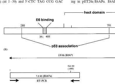
E6 oncoprotein can form a ternary complex with the cell-cycle regulator p53 and the E6-asso-ciated protein (E6AP), leading to the degradation of p53 via the ubiquitination pathway (zur Hausen and de Villiers, 1994; Howley, 1996). E6AP is a member of a family of proteins, known as E3 ubiquitin-protein ligases, which have been proposed to play a role in defining the substrate specificity of the ubiquitin-proteasome degrada-tion system. The schematic funcdegrada-tional domains of E6AP are depicted in Fig. 1 as reported previ-ously (Huibregtse et al., 1993). The 100-kDa
E6AP protein contains an 18-amino acid region (amino acids 391 – 408) that is sufficient for bind-ing to E6. The E6AP-dependent bindbind-ing of p53 involves amino acids 280 – 781, a domain encom-passing the E6-binding region. Finally, an intact COOH terminus hect (homologous to E6AP C-terminal) domain is necessary for E6-mediated p53 ubiquitination. E6 can also bind E6-binding protein (E6-BP, also known as ERC-55), a cal-cium-binding protein localized in the endoplasmic reticulum, with possible consequences for intracel-lular signaling (Chen et al., 1995). E6 can change cellular morphology by interacting with paxillin and disrupting the actin cytoskeleton (Tong and Howley, 1997).
Recently, with extensive knowledge of molecu-lar mechanism by HPV oncoproteins, a new drug has been designed to suppress infections by the HPV so as to prevent the development of cervical cancer (Beerheide et al., 1999). Therefore, an in vitro screening system for development of drugs to treat HPV infection is primarily required to search compounds with desirable properties. In our report, we established in vitro screening sys-tem, using interaction between E6 and E6AP, and its application for the ELISA system could be available for the development of potential drugs against HPV infection.
2. Materials and methods
2.1. Construction and o6erexpression of E6 and E6AP
digestion of pBluescript/E6 withBamHI/SalI and subcloned into corresponding expression vectors, pET28a (Novagen) and pGEX4T-1 which were prepared by digesting with BamHI/SalI, respec-tively. Transformation of pGEX/E6 and pET28a/
E6 into DH5a or BL21 (DE3 pLys) was performed to express GST-tagged and 6× His-Tagged E6, respectively. To prepare E6AP cDNA, two halves of cDNAs with overlapping region in the middle of full ORF were amplified using two different primer sets by RT-PCR from total RNA isolated from CaSki, cervical car-cinoma cell line. The primers of the former half in E6AP (E6APa) are 5%-AGA TCT ATG AAG CGA GCA GCT GCA AAG CAT CTA ATA-3% (sense) (nt 1 – 30) and 5%-CTC TAG CCG GAC
AAG TGC ATC ATC TAT GAT-3% (antisense) (nt 1524 – 1553), and the latter half primers (E6APb) are 5%-AGC GAG CTG ACA CTT CAG GAA CTT TTG GGA-3%(sense) (nt 1143 – 1172) and 5%-TTA CAG CAT GCC AAA TCC TTT GGC ATA CGT-3% (antisense) (nt 2529 – 2558). The respective PCR products, E6APa and E6APb were ligated to PCR®
2.1-TOPO cloning vector (Invitrogen, Carlsbad, CA) and then desig-nated as PCR®2.1-TOPO
/E6APa and PCR® 2.1-TOPO/E6APb, respectively. To construct bacterial expression vector for wild type E6AP, E6APa was excised from PCR®2.1-TOPO
/E6APa by digestion with BglII/HindIII, and ligated into pET28a predigested with BamHI/HindIII, result-ing in pET28a/E6APa. E6AP was finally
structed by ligation of E6APb insert, excised by digestion of PCR®2.1-TOPO
/E6APb with EcoRI, into pET28/E6APa which was predigested with
EcoRI. For glutathione S-transferase (GST)-fused E6AP, E6APa prepared from PCR® 2.1-TOPO/E6APa by predigestion with BglII/EcoRI was subcloned into pGEX4T-1 vector digested with BamHI/EcoRI to construct pGEX/E6APa. The resulting product pGEX/E6APa was digested with EcoRI and subsequently combined with E6APb insert obtained from digestion of PCR®
2.1-TOPO/E6APb with same restriction en-zymeEcoRI. In addition, to examine the specific-ity of E6AP-E6 binding, lysates derived from
Escherichia coliexpressing E7 or vector were pre-pared with pET28 system (Novagen, Madison, WI). E7 insert was amplified from total RNA extracted from CaSki cell lines by RT-PCR using primer set comprising 5% -CACCATGGCATG-GCATGGAGATACACCT-3% (sense) and 5% -TTATGGTTTCTGAGAACA-3% (antisense) and inserted into T-vector prepared from pBluscript KS. E7 fragment was cloned into pET28 vector to construct 6×His-fused E7 as an insert BamHI/ SalI. GST-fused proteins were expressed inE.coli
DH5a by induction with 1 mM isoprophyl-b -D-thiogalactopyranoside (IPTG), Cells were har-vested and lysed in phosphate-buffered saline (PBS) containing 0.5% Triton X-100, 0.5 mM phenylmethylsulfonyl fluride (PMSF) and 10 mg/
ml of aprotinin, and followed by sonication. Proteins expressed in pET28 vector system were prepared in BL21 (DE3 pLys) through same pro-cedures as described above, except for using lysis buffer, pH 8.0, containing 50 mM NaH2PO4 and 150 mM NaCl. The resulting mixture was cen-trifuged for 30 min at 12 000 rpm to remove pellet containing cell debris. Supernatants were used as cell lysate for binding assay. For immobi-lization of GST-E6AP on a resin, the superna-tants were incubated with glutathione (GSH)-Sepharose (Glutathion Sepharose™4B, Phamacia Biotech, Sweden) for 1 h at 4°C. His-tagged E6AP proteins were purified by incubating supernatant with Ni-NTA Agarose (Peptron Co., Taejon, South Korea) and following extensive washing with buffer (20 mM imidazole in lysis buffer). The beads were collected and washed
three times with PBST buffer for utilizing as a bait of binding assay. Also, E7 lysates as histidine tagging protein were prepared in the same proce-dure as described above and their concentrations of lysates, GST-E6, His-E6 and His-E7, were approximately 9.7, 8.2 and 7.5 mg/ml, respec-tively, as determined by Bradford reagents (Bio-Rad Lab., Hercules, CA).
2.2. E6AP and E6 binding assay
E6 binding assays were performed by combining 6×His-E6AP or GST-E6AP fusion protein immobilized on nickel- or GSH-Sepharose beads, respectively, with various amounts of bac-terially expressed GST-E6 or 6×His-E6 superna-tants (50, 100 and 200 ml). The mixtures were rotated in microcentrifuge tubes at 4°C for 1 h. The Sepharose beads were then collected by cen-trifugation, washed three times with 1 ml of lysis buffer, and then boiled for 5 min in sodium dodecyl sulfate (SDS) gel loading buffer. The amounts of E6 protein that bound to the beads were determined by SDS-polyacrylamide gel elec-trophoresis (PAGE; 12% acrylamide) and West-ern blot analysis. Gels were transferred to Immobilon-P membranes (Millipore, Bedford, MA) at 50 V for 1.5 h at room temperature and were blocked by soaking into methanol for 5 min and drying at room temperature. The membrane was probed with goat E6 monoclonal anti-body (Santa Cruz Biotechnology, Santa Cruz, CA) diluted 1:1000 in 3% skimmed milk, followed by an alkaline phosphatase-conjugated anti-goat antibody (Sigma), and visualization was achieved with NBT/BCIP substrate kit (Bio-Rad Lab.).
2.3. Optimization of ELISA
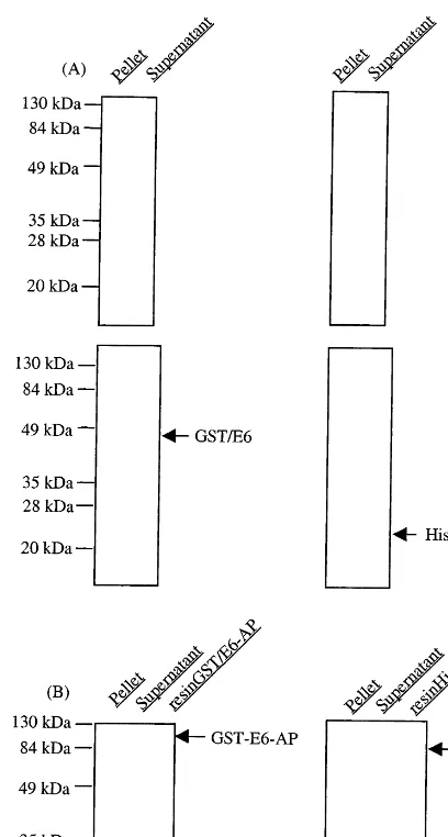
Fig. 2. Overexpression of recombinant E6 and E6AP proteins. (A) GST-tagged E6 and 6×His-tagged E6, (B) GST-tagged E6AP and 6×His-tagged E6AP. The bacterial cell lysates after sonication were separated into pellets and supernatants by centrifugation at 12 000 rpm for 30 min. Diluted aliquots (1:10) of each fraction were analyzed on SDS-PAGE. The upper and lower panels of (A) represent the Coomassie blue staining and immunoblotting, respectively. For fused E6, E6 protein was identified with goat anti-E6 (N-17) antibody fol-lowed by incubation of alkaline phosphatase-conjugated anti-goat IgG. After washing, the bound enzyme was visualized using alkaline phosphatase conjugate substrate kit.
tive washing with PBS containing 0.5% Tween 20 (PBST) to remove unbound E6, goat anti-E6 was dispensed to plate at a concentration of 0.1mg/ml followed by 1-h incubation. After washing, horseradish peroxidase (HRP)-conjugated anti-goat antibody (Sigma) was applied to the plate for immunoassay. After washing twice with PBST, 100 ml of substrate (4 mg o-phenylenediamine, 5 ml 37% H2O2per 10 ml of 0.1M citrate buffer, pH 5.1) was added, and the enzyme activity was stopped 15 min later with 2.5 N sulfuric acid and detected at 490 nm using an ELISA reader (Molecular Devices, Hercules, CA).
3. Results
As a strategy for amplification of E6AP cDNA, two pairs of primers which are overlapped at nucleotides (nt) 1524 – 1553 were designed as de-picted in Fig. 1(B). Both E6 and E6AP cDNA were fused to pGEX or pET28 vector to prepare the GST- or 6×His-fused proteins appropriate for screening systems. Overexpression and immo-bilization of fusion proteins on resins for pull down assays are shown in Fig. 2. Expression levels of E6 proteins present in both supernatant and pellet after sonication were detected by Coomassie-blue staining and Western blot analy-sis. Immunoblot analysis showed that apparent molecular weights of expressed His-E6 and GST-E6 proteins were approximate 24 and 46 kD, respectively. The migration of His-tagged E6 was faster than the 20-kDa molecular weight marker as predicted, which likely to be due to the addi-tional nucleotides generated on subclone and pos-itive charges of six histidine residues linked to N-terminus of E6 (Fig. 2(A)). Also, expression plasmids for E6AP were transformed into E. coli
BL21 and DH5a, and proteins present in the bacterial lysates resolved by SDS-PAGE and Coomassie staining, proteins migrating at the pre-dicted molecular weights for E6AP were clearly visible. His-tagged E6AP and GST-tagged E6AP were over than 100 kD, molecular weight corre-sponding to authentic E6AP alone, and were im-mobilized on GSH- or Ni-NTA Sepharose, respectively, to utilize as a bait in pull down containing 3% skimmed milk. Serial dilutions of
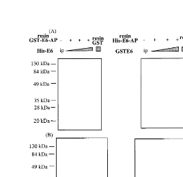
exhaus-assays. E6AP was purified to be homogeneous as bead bound form of either GST- or His-tagged (Fig. 2(B)). E6AP immobilized on resins was incu-bated with E6 fusion proteins derived from differ-ent vector systems, respectively, to avoid bindings to the same ligands on resins (Fig. 3). E6 fusion proteins were bound to His-E6AP and GST-E6AP in a dose dependent manner, respectively, whereas E6 proteins had binding to neither GST immobilized on GSH- nor Ni-NTA-resin alone, indicating that binding systems using E6AP-E6 interaction may be useful for drug screening. For practical application, pull down assays between E6AP and E6 were extended to ELISA systems (Fig. 4). However, His-E6AP was readily eluted from Ni-NTA Agarose resin by addition of 200 mM immidazole, compared with elution of
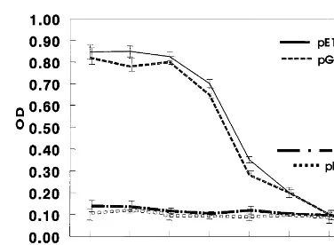
GST-E6AP from GSH-Sepharose resins by elution buffer (10 mM glutathione in PBS). His-E6AP was coated to plate at a concentration of 4mg/ml and then mixed with various dilutions of E6 lysates. E6 proteins bound to E6AP were assayed with antibody, which recognize specifi-cally E6. As shown in Fig. 4, GST-E6 or His-E6 fusion proteins were specifically bound to E6AP in a dose dependent manner while any other non-relevant lysates derived from E. coli
harboring pET28-E7 and vector alone pET28a, were not. For mass-screening potential drugs against cervical cancer caused by HPV in-fection, we determined 16× dilution of GST-E6 or His-GST-E6 lysates as optimum dose in the ELISA based on the interaction between E6AP and E6.
Fig. 4. Optimization of ELISA for screening drug targeting to HPV E6. Eluted E6AP was coated onto Maxisorb 96-well plate for overnight and blocked with PBS containing 3% skimmed milk for 2 h at room temperature. After washing with PBST, various doses of E6 supernatants derived from pET28a/E6 and pGEX/E6 were incubated, respectively, for 1 h. The E6 bound to E6AP was detected with goat anti-E6 (N-17) antibody followed by incubation of horseradish phos-phatase-conjugated anti-goat IgG. After washing, the bound enzyme activity was read at 490 nm by using ELISA reader. As non-specific controls, non-relevant agents such as E7 and supernatant of vector, pET28a, were incubated with E6AP. Results are the mean9standard deviation and are representa-tive of three experiments.
system for screening antiviral drugs. Screening system based on interaction between E6AP and E6 has been recently reported (Beerheide et al., 1999). In previous report, the zinc-ejecting in-hibitor of E6 binding to E6-associated protein (E6AP) and E6-binding protein (E6-BP) was in-vestigated using BIACORE assays. Among com-pounds ejecting zinc from E6 protein, some inhibit the interaction of E6 with E6AP and E6-BP. The E6 protein of HPV 16 consists of 158-amino acid residues and contains two hypothetical Cys-X2-Cys-X29-Cys-X2-Cys zinc fingers. The conservation of the zinc fingers in E6 and E7 among distantly related HPV types sug-gests that this zinc-binding motif is strictly re-quired for the function of the E6 and E7 oncoproteins. However, BIACORE system is very expensive and laborious compared to the ELISA system. In this study, we first adopted ELISA system for screening system for anti-HPV drugs. First, we constructed the GST- or His-fused E6AP and E6 to investigate the direct binding between E6AP and E6. Western blotting showed that His-E6 was bound to GST-E6AP immobi-lized on resins but not to GST. Similar results were obtained with the His resin pull-down assay, which showed that GST-fused E6 was efficiently retained by His-E6AP but not by resin alone. Second, we applied in vitro binding of E6 and E6AP into the development of ELISA. Elution of His-tagged E6AP by addition of imidazole was more readily achieved than that of GST-fused E6AP. Hence, E6AP protein, eluted from His-E6AP bound to resin, was coated onto the plate to which various doses of E6 derived from GST-E6 or His-GST-E6 were added. Either GST-GST-E6 or His-E6 lysates were bound to E6AP in a dose dependent manner to 32:1 dilution. E6AP-E6 binding was saturated at concentrations more than 32-fold diluted E6 lysates. These ELISA systems were first established and may be useful in the screening of drugs against cervical cancer caused by HPV infection. Moreover, the estab-lishment of these screening systems may provide a basis for the further analysis of the normal cellu-lar functions as well as development of anti-can-cer drugs.
4. Discussion
Acknowledgements
This work was supported by grant Molecular Medicine Program (98-J03-02-A-03) and I03-008 from Ministry of Science and Technology, Korea.
References
Beerheide, W., Bernard, H.U., Tan, Y.J., Ganesan, A., Rice, W.G., Ting, A.E., 1999. Potential drugs against cervical cancer: zinc-ejecting inhibitors of the human papillo-mavirus type 16 E6 oncoprotein. J. Natl. Cancer Inst. 91 (14), 1211 – 1220.
Chen, J.J., Reid, C.E., Band, V., Androphy, E.J., 1995. Inter-action of papillomavirus E6 oncoproteins with a putative calcium-binding protein. Science 269 (5223), 529 – 531. Crusius, K., Kaszkin, M., Kinzel, V., Alonso, A., 1999. The
human papillomavirus type 16 E5 protein modulates phos-pholipase C-gamma-1 activity and phosphatidyl inositol turnover in mouse fibroblasts. Oncogene 18, 6714 – 6718. de Villiers, E.M., 1989. Heterogeneity of the human
papillo-mavirus group. J. Virol. 63, 4898 – 4903.
Filatov, L., Golubovskaya, V., Hurt, J.C., Byrd, L.L., Phillips, J.M., Kaufmann, W.K., 1998. Chromosomal instability is correlated with telomere erosion and inactivation of G2 checkpoint function in human fibroblasts expressing hu-man papillomavirus type 16 E6 oncoprotein. Oncogene 16, 1825 – 1838.
Howley, P.M., 1996. Papillomavirinae: the viruses and their replication. In: Knipe, D.M., Howley, P.M. (Eds.), Field’s Virology. Lippincott-Raven, Philadelphia, PA, pp. 2045 – 2076.
Huibregtse, J.M., Scheffner, M., Howley, P.M., 1993. Local-ization of the E6AP regions that direct human
papillo-mavirus E6 binding, association with p53, and ubiquitination of associated proteins. Mol. Cell. Biol. 13, 4918 – 4927.
Marchuk, D., Drumm, M., Saulino, A., Collins, F.S., 1990. Construction of T-vectors, a rapid and general system for direct cloning of unmodified PCR products. Nucleic Acids Res. 19 (5), 1154.
Sherman, L., Jackman, A., Itzhaki, H., Stopper, M.C., Koval, D., Schlegel, R., 1997. Inhibition of serum- and calcium-in-duced differentiation of human keratinocytes by HPV16 E6 oncoprotein: role of p53 inactivation. Virology 237, 296 – 306.
Stellato, G., Nieminen, P., Aho, M., Lehtinen, T., Lehtinen, M., Paavonen, J., 1997. Type 1 cytokine response and treatment outcome of genital HPV lesions. Genitourin Med. 73, 387 – 390.
Syrjanen, S.M., Syrjanen, K.J., 1999. New concepts on the role of human papillomavirus in cell cycle regulation. Ann. Med. 31, 175 – 187.
Tong, X., Howley, P.M., 1997. The bovine papillomavirus E6 oncoprotein interacts with paxillin and disrupts the actin cytoskeleton. Proc. Natl. Acad. Sci. USA 94 (9), 4412 – 4417.
Trobs, R., Metzner, G., Friedrich, T., Pustowoit, B., Han-drick, W., Nestler, I., 1998. Papillomavirus-induced genital warts in a girl — management by surgery and im-munomodulating therapy. Pediatr. Surg. Int. 13, 301 – 303. zur Hausen, H., de Villiers, E.M., 1994. Human papilloma
viruses. Annu. Rev. Microbiol. 48, 427 – 447.
zur Hausen, H., Schneider, A., 1987. The role of papillo-maviruses in human anogenital cancers. In: Howley, P.M., Salzman, N.P. (Eds.), The Papovavirinae: The Paillo-maviruses. Plenum, New York, pp. 245 – 263.
Zwerschke, W., Jansen-Durr, P., 2000. Cell transformation by the E7 oncoprotein of human papillomavirus type 16: interactions with nuclear and cytoplasmic target proteins. Adv. Cancer Res. 78, 1 – 29.