Laporan Kasus
ICP MONITOR PLACEMENT STEPWISE EARLY DECOMPRESSIVE
CRANIECTOMY FOR THE MANAGEMENT OF SEVERE TBI PATIENTS:
A CASE REPORT
Dirga Rachmad Aprianto
1, Achmad Kurniawan
2, Andhika Tomy Permana
2,
Fadillah Putri Rusdi
2, Akbar Wido
2, Bagus Sulistyono
2, Made Gemma
Daniswara Maliawan
2, Tedy Apriawan
3, Abdul Hafid Bajamal
41
Neurosurgery Division, Surgery Department, Faculty of Medicine, Universitas Islam
Sultan Agung
2
Resident of Neurosurgery Department, dr. Soetomo General Hospital, Surabaya, East
Java, Indonesia
3
Staff of Neurosurgery Department, dr. Soetomo General Hospital & Airlangga
University Hospital Surabaya, East Java, Indonesia
4
Professor of Neurosurgery Department, dr. Soetomo General Hospital, Surabaya,
East Java, Indonesia
Correspondence author: Aprianto, E-mail: [email protected]
Abstract
Introduction. Increased intracranial pressure (ICP) is a secondary event that mostly occurs following traumatic brain injury (TBI) and it correlates with poor outcome of the patients. Several studies have suggested that early decompressive craniectomy (DC; within 48 hours after injury) is recommended for severe TBI patients requiring removal of intracranial hemorrhage and early DC was able to reduce the complications of TBI caused by increased ICP. However, even early DC has been performed, increased ICP may still progress due to massive brain edema. Methods. We herein report a case report of patient admitted with severe TBI and intracranial hemorrhage. The patients were underwent DC and ICP monitor placement after the removal of the intracranial hemorrhage. During postoperative observation in ICU, the CSF of the patients was gradually drained if the ICP was over 15mmHg.
Results. The ICP right after performed early DC was 30 cmH2O (22 mmHg). One day after surgery,
the hemodynamic of the patient was stable and the GCS was 2X5 with the ICP of the patient was about 18 cmH2O. On day 2-5, patient was hemodynamically stable with improved GCS (3X5) and decreased
of ICP (around 13-15 cmH2O). On day 6, the ICP monitor was removed and the patient discharged on
day 19 after fully recovered. Conclusion. The placement of ICP monitor and the application of gradual release of CSF after DC might be helpful to reduce increased ICP in severe TBI patients, and thus reducing the morbidity and mortality.
Keywords: Traumatic brain injury, intracranial pressure monitor, decompressive craniectomy
Pemasangan ICP Monitor Bersamaan Dengan Kraniektomi Dekompresi Awal
untuk Tatalaksana Pasien Cedera Otak Berat: Laporan Kasus
Abstrak
Pendahuluan. Peningkatan tekanan intrakranial (ICP) merupakan kejadian sekunder yang sering terjadi setelah cedera otak traumatis (TBI) dan berkorelasi dengan hasil yang buruk pada pasien. Beberapa penelitian menunjukkan bahwa kraniektomi dekompresif awal (DC) (dalam 48 jam setelah cedera) direkomendasikan untuk pasien dengan TBI berat yang membutuhkan evakuasi perdarahan intrakranial dan DC awal mampu mengurangi komplikasi TBI yang disebabkan oleh peningkatan TIK. Namun, meskipun DC awal telah dilakukan, peningkatan TIK masih dapat berlangsung karena terjadi
edema otak yang masif. Metode. Sebuah kasus pasien yang dirawat dengan TBI berat dan perdarahan
30cm H20 (22 mmHg). Hari pertama setelah operasi, hemodinamik pasien stabil dan GCS 2X5 dengan ICP pasien sekitar 18 cmH2O. Pada hari ke 2-5, pasien hemodinamik stabil dengan GCS membaik (3X5) dengan penurunan ICP (sekitar 13-15 cmH2O). Pada hari ke-6, ICP monitor dilepaskan dan
pasien dipulangkan pada hari ke 19 setelah pulih sepenuhnya. Kesimpulan. Penempatan ICP monitor
dan aplikasi pelepasan CSF secara bertahap setelah DC mungkin membantu mengurangi peningkatan ICP pada pasien dengan TBI berat, dan dengan demikian mengurangi morbiditas dan mortalitas.
Kata kunci: Cedera otak traumatik, monitor tekanan intrakranial, kraniektomi dekompresi
Introduction
Traumatic brain injury (TBI) is a
well-recognized as the leading cause of
death of the traumatic injury patients. In
the United States, from no less than 2.5
million TBI-related patients, the mortality
is accounted for more than 55.000 cases
per year.1 Beside intracranial hemorrhages,
elevated intracranial pressure (ICP) is
mostly attributed due to brain edema
formation and thus resulting unfavorable
outcomes among TBI patients. 2,3
medical treatments have failed in lowering
ICP, decompressive craniectomy (DC) is
an option to treat increased ICP following
TBI. Further, recent studies recommended
that DC should be performed within 48
hours after trauma to significantly improve
neurological functions and mortality
outcomes. 2,5-7
ICP monitors in treating increased ICP8. In
a retrospective study, 1.874 patients (out
of 10.628) who have ICP monitors had
lower odds of mortality especially in
patients under 659. Another retrospective
study also showed that the mortality in
massive brain edema even after early DC.
If uncontrollable ICP happens, early DC
will not significantly improve the
outcomes. Therefore, it is questionable
whether ICP monitor placement stepwise
early DC have favorable outcomes in
severe TBI patients after the removal of
the intracranial lesions. If the ICP remains
uncontrollable even after early DC and
medical therapies, gradual release of
cerebrospinal fluid (CSF) might be helpful
to reduce the increased ICP.
Case Presentation
A 31 years old male patient was
hours before admission. Previously, he
was brought for treatment in a local
hospital right after the accident. He was
then referred to our hospital for further
treatment. The patient had history of
decreased consciousness since the
accident, vomited 3 times, bloody otorrhea
of the left ear, and no seizure. Upon arrival
at our hospital the GCS score was 8 with
unequal pupil diameter, decreased light
reflex of the left eye and right
parieto-occipital cephalohematoma. The latest
head CT scan, 15 hours after accident,
showed multiple contusions at left-right
frontal region and enlargement of
intracerebral hemorrhage (ICH) at left
frontal region with volume around + 25 cc
compared to previous head CT Scan 4
hours after accident (+ 7,5cc), and thus
causing midline shift > 0,5 cm to the
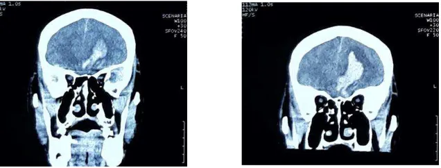
contralateral side (fig.1&2).
Figure 1. an axial pre-operative CT scan show multiple contusions at left-right frontal region and enlargement of intracerebral hemorrhage (ICH) at left frontal region, causing midline shift > 0,5 cm to the contralateral side)
Source: Database Neurosurgery Department, Dr. Soetomo General Hospital
Figure 2. A coronal pre-operative CT scan show multiple contusions at left-right frontal region and enlargement of intracerebral hemorrhage (ICH) at left frontal region.
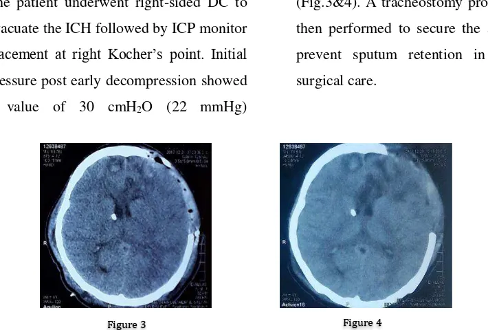
The patient underwent right-sided DC to
evacuate the ICH followed by ICP monitor placement at right Kocher’s point. Initial pressure post early decompression showed
a value of 30 cmH2O (22 mmHg)
(Fig.3&4). A tracheostomy procedure was
then performed to secure the airway and
prevent sputum retention in the
post-surgical care.
Following surgery, the patient was
treated and intensively observed in the
intensive care unit (ICU). The ICP was
monitored periodically and the CSF was
drained if the ICP above 15 cmH2O (11
mmHg), with a maximum 100cc in a day.
One day after surgery, the hemodynamic
of the patient was stable and the GCS was
2X5 with the ICP of the patient was about
18 cmH2O (100cc of CSF drained). On
day 2, the GCS was improved to 3X5 with
the ICP of 15 cmH2O (100cc of CSF
drained). Until day 5 post surgery, patient
was hemodynamically stable with GCS
3X5 and ICP around 13 – 15 cmH2O (less
than 100cc of CSF drained per day). The
ICP monitor was then removed on day 6
after surgery and the patient was
transferred from ICU to our high care unit
(HCU). During treatment in HCU, the
patient was also treated by pulmonologist
due to pneumonia complications. The
patient was discharged from hospital on
day 19 after fully recovered with GCS
4X6 and pneumonia treatment was
finished.
Discussion
The high number of mortality and
morbidity in TBI is often attributed to the
increased ICP as a result of brain
edema11,12. Therefore, managing brain
Figure 3. 2 days post-operative axial CT scan showed condition after DC, note that the ICH had been removed and ICP monitor had placed. Figure 4. 6 days post-operative CT scan, notice the ICP monitor still exist and brain became swollen)
Source: Database Neurosurgery Department, Dr. Soetomo General Hospital
edema in order to prevent increased ICP is
one of the focuses of medical management
in TBI. However, progressive brain
edema refractory to medical management
is likely occurred in severe TBI, thus
resulting high mortality rate13 (close to
100%; Miller, 1977). In managing
refractory intracranial hypertension DC is
considered as a salvage procedure and has
been widely used in severe TBI. Recently,
many studies showed that early DC, within
48 hours of injury, in severe TBI improves
the outcome and reduces mortality rate. In
a study conducted by Grindlinger et al3
ICP post early DC was significantly
decreased, thus resulting in good outcome
and decreased mortality rate3.
In our case we performed early
decompressive craniectomy due to
enlargement of intracerebral haemorrhage
at left frontal region, previous head CT 4
hours after accident showed (+¬ 7,5cc)
while 15 hours after accident showed + 25
cc of hematoma, thus causing > 0,5 cm
midline shift. After DC was performed,
the patient GCS was 2X5, and improved to
3x5 at Day 2, corresponds as Kim said that
DC performed within 48 hours after
trauma, significantly improve neurological
functions and mortality outcomes5. Not
only DC was performed, ICP monitor was
also placed and drained the CSF if the ICP
above 15 cmH2O (11 mmHg), this aimed
to treat increased ICP so that cerebral
perfusion and oxygenation can be
maintained, as recommended by brain
trauma foundation in the management of
severe TBI8. Alali also support this
guidelines in his retrospective study which
showed that mortality in severe TBI
patients was lower in group of patients
treated with ICP monitor9.
Until 5th day after surgery, the
patient's haemodynamic and neurological
functions was stable, which is showed a
favorable outcome as Grindlinger stated in
his study3. However this case found that
enough to control refractory increased ICP
Conclusions
The placement of ICP monitor and
the application of gradual release of CSF
after DC might be helpful to reduce the
increased ICP of severe TBI patients, and
thus reducing the morbidity and mortality.
However, in this case report
showed that that early DC might not
enough to control refractory increased
ICP.
Reference
1. Taylor CA, Bell JM, Breiding MJ, Xu
L. Traumatic Brain Injury-Related
Emergency Department Visits,
Surveill Summ. 2017 Mar 17;66(9):1-16.
2. Plesnila N. Decompression
craniectomy after traumatic brain injury: recent experimental results. Prog Brain Res. 2007;161:393-400.
3. Grindlinger GA, Skavdahl DH, Ecker
RD, Sanborn MR. Decompressive craniectomy for severe traumatic brain injury: clinical study, literature review and meta-analysis. Springerplus. 2016 Sep 20;5(1):1605.
4. Chibbaro S, Tacconi L. Role of
decompressive craniectomy in the management of severe head injury with refractory cerebral edema and intractable intracranial pressure. Our experience with 48 cases. Surg Neurol. 2007 Dec;68(6):632-638
5. Kim DR, Yang SH, Sung JH, Lee SW,
Son BC. Significance of Intracranial
Pressure Monitoring after Early
Decompressive Craniectomy in
Patients with Severe Traumatic Brain Injury. J Korean Neurosurg Soc. 2014 Jan;55(1):26-31.
6. Alvis-Miranda H, Castellar-Leones
SM, Moscote-Salazar LR.
Decompressive Craniectomy and
Traumatic Brain Injury: A Review. Bull Emerg Trauma. 2013;1(2):60-68.
7. Shutter LA, Timmons SD. Intracranial
Pressure Rescued by Decompressive Surgery after Traumatic Brain Injury.
N Engl J Med. 2016 Sep
22;375(12):1183-4.
8. Brain Trauma Foundation.
“Guidelines for the Management of Severe Traumatic Brain Injury 4th Edition”. Neurosurgery. 2017 Jan 1;80(1):6-15.
9. Alali AS, Fowler RA, Mainprize TG,
et al. Intracranial pressure monitoring in severe traumatic brain injury: results from the American College of
Surgeons Trauma Quality
Improvement Program. J
Neurotrauma. Oct 15
2013;30(20):1737-1746. PMID:
23731257
10. Farahvar A, Gerber LM, Chiu YL,
Carney N, Hartl R, Ghajar J. Increased mortality in patients with severe traumatic brain injury treated without intracranial pressure monitoring. J Neurosurg. Oct 2012;117(4):729-734. PMID: 22900846.
11. Nirula R, Millar D, Greene T,
McFadden M, Shah L, Scalea TM, Stein DM, Magnotti LJ, Jurkovich GJ, Vercruysse G, Demetriades D, Scherer LA, Peitzman A, Sperry J, Beauchamp K, Bell S, Feiz-Erfan I, O'Neill P,
Coimbra R. Decompressive
craniectomy or medical management
for refractory intracranial
hypertension: an AAST-MIT
propensity score analysis. J Trauma Acute Care Surg. 2014 Apr;76(4):944-52; discussion 952-5
12. Gouello G, Hamel O, Asehnoune K,
Bord E, Robert R, and Buffenoir K. Study of the Long-Term Results of
Decompressive Craniectomy after
Severe Traumatic Brain Injury Based on a Series of 60 Consecutive Cases.
ScientificWorldJournal. 2014 Feb
24;2014:207585.
13. Miller JD, Becker DP, Ward JD, Sullivan HG, Adams WE, Rosner MJ: Significance of intracranial hypertension in severe head injury. J