LAPORAN KASUS
Foreign body a fish in orohypopharynx with complication
vocal cord paralysis
Ade Asyari, Novialdi, Nur Azizah
Bagian Telinga Hidung Tenggorok Bedah Kepala & Leher, Fakultas Kedokteran, Universitas Andalas
Korespondensi: Ade Asyari, email: [email protected]
Abstract
Foreign body a fish in orohypopharynx is a rare case and require rapid diagnosis and immediate treatment to prevent complication. There are some complications that can occur, such as upper airway obstruction, perforation of the pharyngeal wall, vocal cord paralysis, pneumomediastinum and emphysema. Vocal cord paralysis is rare complication, may be due to direct trauma to the recurrent laryngeal nerve by foreign body or surgical procedure and or due to recurrent nerve palsy secondary to inflammation. The management for pharyngeal foreign bodies is extraction of foreign body with Magill forceps, direct laryngoscope and rigid endoscopy. Tracheostomy should be performed if endotracheal intubation could not be done or failed to be performed. Objective: Understanding diagnosis and management of patient foreign body a fish in an orohypopharynx, as well as to know the possible complications. Case report: Reported a case, male 40 years old, with diagnosis foreign body a fish in orohypopharynx with complication unilateral vocal cord paralysis. Foreign body was extracted using Magill forceps and rigid esophagoscopy with tracheostomy preparation if endotracheal intubation was failed to perform. Conclusion: Foreign body a fish in orohypopharynx is a rare case. Precise diagnosis and treatment are very important to prevent complication. Vocal cord paralysis is a rare complication caused by foreign body in orohypopharynx.
Keywords: foreign body a fish; vocal cord paralysis; Magill forceps; rigid esophagoscopy
Abstrak
Benda asing ikan di orohipofaring merupakan kasus yang jarang terjadi dan memerlukan diagnosis serta penatalaksanaan yang cepat dan tepat untuk mencegah komplikasi. Komplikasi yang dapat terjadi berupa sumbatan jalan nafas atas, perforasi dinding faring, paralisis pita suara, pneumomediastinum dan emfisema. Paralisis pita suara merupakan komplikasi yang jarang, penyebabnya bisa karena trauma langsung pada nervus laringeal rekuren oleh benda asing atau karena tindakan ekstraksi benda asing itu sendiri, bisa juga karena inflamasi pada nervus laringeal rekuren sekunder karena benda asing. Penatalaksanaan benda asing ikan di orohipofaring dapat menggunakan ekstraksi dengan forsep Magill, laringoskop langsung dan penggunaan endoskopi kaku. Tindakan trakeostomi sangat diperlukan bila intubasi endotrakea tidak dapat atau gagal dilakukan. Tujuan: mengetahui dan memahami diagnosis dan penatalaksanaan yang cepat dan tepat pada kasus benda asing ikan di orohipofaring serta mengetahui komplikasi yang mungkin terjadi. Laporan Kasus: Satu kasus dilaporkan, seorang laki-laki 40 tahun dengan diagnosis benda asing ikan di orohipofaring dengan komplikasi paralisis pita suara unilateral. Benda asing ditatalaksana dengan ekstraksi menggunakan forsep Magill dan esofagoskop kaku. Dilakukan persiapan trakeostomi bila gagal intubasi. Kesimpulan: Benda asing ikan di orohipofaring merupakan kasus yang jarang. Diagnosis dan penatalaksanaan yang cepat dan tepat dapat mencegah komplikasi. Salah satu komplikasi benda asing ikan di orohipofaring berupa paralisis pita suara.
INTRODUCTION
Various foreign bodies that impaction in the pharynx and upper oesophagus is common in otolaryngological practices. It may impaction in tonsil, base of tongue, hypopharynx, piriform fossae and oesophagus or sometimes in the larynx or lower down in the respiratory tract that may be life threatening and need immediate removal because it can lead medical or surgical emergencies which are
often challenging for an
otorhinolaryngologist.1–5
Accidental ingestion of foreign bodies and their impaction is more common in children than adult. In children exploring their environment with their mouth, uncoordinated swallowing, absence of molars for proper chewing are at risk for the ingestion and aspiration of small edible object. Foreign bodies like coin, marble, button battery, bottle top, bean, grain and seed have been mostly reported in infant and children.4,6
Foreign body ingestion, either intentional or unintentional, appears more often in the elderly population in patient with psychiatric disorders, developmental delay or alcohol intoxication and in prisoners seeking secondary gain.7,8 Foreign body
bone, denture and metallic pin or wire have been reported in adult. While fish bone most common foreign body in the pharynx in adult, live fish impacted in the throat too has been reported.5,1,9–12
The diagnosis is based on anamnesis or clinical presentation, physical, radiological and or endoscopy examination.4,6,13 The
use of rigid endoscopy under general anaesthesia has long been used by
otorhinolaryngologist for the diagnosis and extraction of foreign bodies in aerodigestive tract. Extraction with Magill forceps and direct laryngoscope have been reported as an immediately procedure that should be performed to a foreign body in the pharynx and proximal esophagus.3,5,6,14
Various studies have been reported the extraction of foreign bodies in pharynx with tracheostomy procedure to reduce the risk of morbidity and mortality. Preparation for performed tracheostomy in cases with a foreign body or virtually lodged the airway should be done to save the airway.5,12,15
Complications in cases foreign body are mucosal injury, upper airway obstruction, perforation the wall of pharynx, pneumomediastinum, emphysema and vocal cord paralysis.5,15 Vocal cord paralysis
is rare complication, that can cause by mechanical obstruction of vocal cord
movement, recurrent nerve palsy
secondary to inflammation, trauma and nerve damage caused by foreign body or surgical prosedure.16–19
CASE REPORT
patient felt pain and difficult in swallowing. There was saliva mixed with blood, there was drooling. There was nausea, but no vomitus. There wasn’t any pain in neck and chest. The patient felt quite hard to breath, no cough and there wasn’t cyanosis after the incident. The patient’s voice sounded like mumbling. There was no history of hoarseness before the incident.
On general examination, general appearance was moderately ill, blood pressure 110/70 mmHg, pulse rate 88x/minute, respiration rate 19x/minute,
temperature 36,8˚C. On thorax
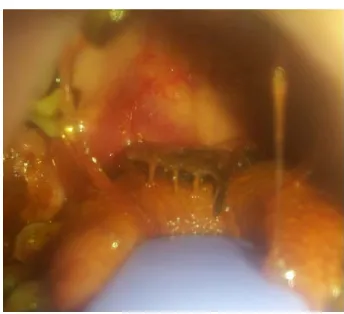
examination, thorax movement was symmetric, no retraction, no abnormal breath sound, no stridor, no wheezing and no crackles. On ear and nose examination in normal limit. In oral cavity, there was saliva mixed with blood, there was fish tail (1 cm in length), no movement, erosion on soft palate and hard palate and no laceration. Throat examination was difficult to evaluated because of the fish (figure 1).
Figure 1. The fish tail was seen on oral cavity examination
On plain lateral cervical X-ray examination,
shown radiopaque appearance that
resemble the shape of a fish skeleton as
high as cervical 1 to cervical 5 (figure 2). The patient was diagnosed with foreign body “a fish” in Oro-hypopharynx and prepared to perform extraction of foreign body in general anaesthesia, also was prepared to perform tracheostomy if the intubation failed.
Figure 2. On plain lateral cervical X-ray
The therapy was IVFD RL, ceftriaxone injection 2x1 gr, dexamethasone injection 3x5 mg, ranitidine injection 2x50 mg and tramadol drip 50mg/500cc RL. Blood test was performed with result leucocytosis (leucocyte 11.500/mm3) and other blood test the result was in normal limit. On thorax X-ray was no abnormalities found.
began. The mouth and head of the fish were almost in oesophagus introitus, by holding the body of the fish slowly. The remain part of the fish was cleaned up with NaCl 0,9%. Evaluation there was a lacerating on right lateral hypopharynx that the place where remain fin of the fish was stuck.
Esophagoscopy was performed to evaluate the oesophagus area by using the rigid esophagoscope (size 12x16x40). Esophagoscope was inserted vertically into the mouth along pharyngeal posterior wall until introitus of oesophagus. There was no fish fin on mucosa of oesophagus (crycopharynx), no erosion on mucosa of crycopharynx and oesophagus, no clotting and no active bleeding. Esophagoscope pushed down until as level as 18 cm from upper incisivus. There was liquid and blackish discharge fulfilled oesophagus. Discharge was suction while evaluates mucosa of oesophagus. Esophagoscopy inserted out smoothly. Applied nasogastric tube number 18 and operation was finished.
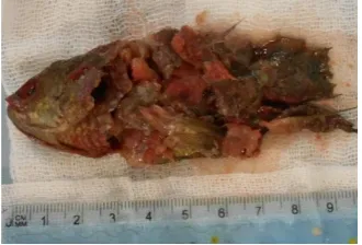
Figure 3. Foreign body a fish after extraction.
Patient diagnosed as post extraction foreign body “fish” in oro hypopharyngeal. Patient hospitalized in ORL Departement, therapy was continued. Post operation instruction, monitoring vital sign, bleeding from the mouth, sign of airway
obstruction. Diet liquid meal via NGT.
The first day postoperative follow-up, July 29th 2016, patient still felt pain in the throat, there was no bloody saliva, no difficulty in breathing, no fever, there was hoarseness. On physical examination, on throat, posterior pharyngeal wall was hyperaemic. Indirect laryngoscopy hypopharynx mucosa was hyperaemic, epiglottic and arytenoid hyperaemic, oedema, ventricularis fold and vocal cords was not hyperaemic, left ventricular and vocal fold limited movement adduction type in paramedian position, glottic rim was open, pyriformis sinus there was standing secretions, there was NGT at the left side of piriformis sinus. Patient diagnosed with post-extraction of foreign bodies “fish” in the oro-hypopharynx with complication left vocal cord paralysis adduction type in paramedian position the first day, therapy was continued.
All of therapy was continued except dexamethasone that tapered off into 2x5 mg of intravenous.
On the sixth day August 2nd, 2016 the patient asking to go home and has been explained about the risks and possibilities that will happen, but the patient still wants to go home. Patients discharged with NGT still inserted and had given therapy cefixime syrup 2 x 100 mg, ibuprofen syrup 3x100 mg, suggest controlling to the ORL outpatient clinic Dr. M. Djamil Hospital Padang 3 days later.
On August 10th, 2016 (the 13th day after the operation) the patient control with hoarseness, but there was no sorer throat and no fever. NGT is still attached. On nose, ear and throat in normal limit. Indirect laryngoscopy, epiglottic and arytenoid was not hyperaemic, left ventricularis fold and vocal cords, there were limited movement adduction type in paramedian position, glottic rim was open, piriformis sinus there was no standing secretions, NGT was sited at the left side of piriformis sinus. The patient was taking drinking test and there were no choking and coughing. The NGT was excluded. The patient diagnosed with post extraction of foreign bodies “fish” in oro-hypopharynx with complication left vocal cord paralysis adduction type in paramedian position. No given therapy. Patients has been asked to control in 2 weeks. But patients didn’t control anymore. We did a follow up the patient via telephone (a month after the incident) the patients suggest controlling but has no more complaints of hoarseness, pain in swallowing and no fever, there was no choking when eat and drink.
On February 24th, 2017 (seven months after incident), the patient control with no hoarseness, no sore throat, no chocking and no difficulty in breathing. Indirect laryngoscopy, epiglottic and arytenoid was not hyperaemic, ventricularis fold and vocal cords, there was symmetric movement, glottic rim was open, piriformis sinus there was no standing secretions. The patient diagnosed with post extraction of foreign bodies “fish” in oro-hypopharynx with complication left vocal cord paralysis in improvement. The patient not given any medication.
DISCUSSIONS
Reported one case a foreign body a fish lodged in the orohypopharynx with complication paralysis of left vocal cord in a man, 40-year-old. This is a rare case. At the ORL-HNS Dr. M Djamil Padang recorded of the same cases from the year 2008-2015 as many as four cases which was treated by extraction of the foreign body under general anaesthesia with and without tracheostomy. The same case was reported by Sangma R1 and Tang LM12
a live fish in the throat, management with tracheostomy procedure than removal foreign body with under general anaesthesia. Parida KP and Surianarayanan5 also reported a case
accidentally entry of a live fish into his throat. They remove the fish under topical anaesthesia without tracheostomy.
seven hours. It was very challenging as the fish was alive and struggled to move deeper into the pharynx and at the same time. Due to the presence of sharp stiff spines on both the dorsum and ventral surfaces of the fish, it moved further in rather than out of the pharynx.12
The most commonly symptoms of impacted foreign body in the throat are odynophagia or dysphagia, followed by cough, vomiting, hypersalivation, saliva mixed with blood until breathless. Odynophagia which is accompanied with hypersalivation and mixed with blood in this case is caused by a foreign body that causes difficulty in swallowing saliva of patients themselves and injuries at hypopharynx area because of the fins and results into bleeding.4,11
Orohypopharynx foreign bodies can be detected by direct visualization, indirect laryngoscopy and endoscopy examination. Soft tissue radiography of the neck is important for diagnose, determine the location and general shape of foreign body and determine complication.14,20
In this patient, tracheostomy was prepared if intubation failed. This is because of the foreign body a fish is expected to close the larynx, making it difficult intubation. Panigrahi reported one case of fish in the throat performed a tracheostomy because intubation cannot
be performed.15 Preparation
tracheostomy is an important procedure to reduce the risk of morbidity and mortality if endotracheal intubation
cannot be performed. Meanwhile,
according to Parida, tracheostomy
procedure is required if upper airway obstruction is present.5
Management in this case is with extraction of foreign body using Magill forceps. Senthilkumaran also reported successfully using Magill forceps to extract the fish live in the throat.11
Esophagoscopy actions is carried out because the head of the fish is already visible in the crycopharynx area so the evaluation into the oesophagus needs to be done to see whether there is any remaining foreign body like fish scales and the possibility of damage to the oesophageal mucosa.
Patients after extraction of foreign body, NGT installation is done with the aim that wound caused by foreign body can be recovered perfectly. Tang LM reported case foreign body a fish in the throat, after extraction there was lacerations of the mucosa of the throat, the aim of performing NGT is allow time for the wound to heal and avoid infection or food bolus that can inhibit mucosal repair.12
NGT can be released on the 3rd day until the seventh day. The period of wound healing in mucosal areas can occur about 3 until 10 days, depending on the area and depth of laceration wound.4,6,21,22
in the hypopharynx, vocal cord paralysis occurred can be due to mechanical obstruction of vocal cord movement, recurrent nerve palsy secondary to inflammation or by direct damage to the nerve.17
In this case, vocal cord paralysis may be due to direct trauma to the recurrent laryngeal nerve by foreign body or due to the act of extraction. Endotracheal intubation can lead to pathological changes, trauma and nerve damage. It seems that when extraction foreign body the cuff shifts and the pressure of cuff is related with nerve paralysis caused by nerve pressure and neuropraxia.19 The proposed mechanism is entrapment of the anterior (adductor) ramus of the recurrent nerve 6 to 10 mm below the level of the cords among the thyroid lamina, the superiorly located arytenoid and the inflated cuff of the endotracheal tube presses the nerve that causes nerve paralysis and neuropraxia. Therefore, the cuff’s position of the endotracheal tube should be located at more than 15 mm below vocal cord.19,23,24 Poncz and Cavo as
quoted by Virgilis reported cases of paralysis of the recurrent nerves because of foreign body impaction are because of endotracheal intubations.23
In this case vocal cord paralysis improvement expected in one month. Because when followed up via telephone in one month there was no voice harness. In seven months after the incident patient control with no complain and in indirect laryngoscopy examination epiglottic and arytenoid was not hyperaemic, ventricularis fold and vocal cords, there was symmetric movement, glottic rim was open, piriformis sinus there was no standing secretions.
CONCLUSIONS
Foreign body a fish in orohypopharynx is a rare case. Precise diagnosis and treatment are very important to prevent complication. Vocal cord paralysis is a rare complication, may be due to direct trauma to the recurrent laryngeal nerve by foreign body or surgical procedure and or due to recurrent nerve palsy secondary to inflammation.
REFERENCES
1. Sangma R ranjan K. An unusual case of foreign body whole fish in throat – A case report. J Dent Med Sci. 2015;14(6):57-59.
2. Sharma RC, Dogra SS, Mahajan VK. Oro-pharyngolaryngeal foreign bodies : some interesting cases.
Indian J Otolaryngol Head Neck Surg. 2012;64(6):197-200.
3. Gupta R PVK. Incidence of foreign bodies in aerodigestive tract in vindhya region : our experience.
Indian J Otolaryngol Head Neck Surg. 2014;66(2):135-41.
4. Hariga Ines, Khamassi khaled, zribi sarra, Amor Ben M, Gamra Ben Olfa, Mbarek C et al. Management of foreign bodies in the aerodigestive tract. Indian J Otolaryngol Head Neck Surg. 2014;66(1):220-24.
6. Sharma RC, Dogra SS, Mahajan VK. Oro-pharyngo-laryngeal foreign bodies : some interesting cases. 2012;64(6):197-200.
7. Birk Michael, Bauerfeind Peter, Deprez Pierre H, Häfner Michael, Hartmann Dirk HC et al. Removal of foreign bodies in the upper gastrointestinal tract in adults : European Society of Gastrointestinal Endoscopy ( ESGE ) Clinical Guideline. Euran Soc Gastrointest Endosc. 2016;48:1-8.
8. Ambe Peter, Weber Sebastian A, Schauer Mathias KWT. Swallowed foreign bodies in adults. Dtsch Arztebl Int. 2012;109:869-76.
9. Mittal M, Yadav R, Mody N DP V. Live fish: unique foreign body throat. Bombay Hosp J. 2011;53(4):804-6.
10. Panigrahi, R, Sarangi, T R, Behera SK BR. Unusual foreign body in throat. Indian J Otolaryngol Head Neck Surg. 2007;59(12):384-5.
11. Senthilkumaran S, Sweni S, Ganapathysubramanian, Ponuswamy S,
Thirumalaikolundusubramanian P. Live fish impaction in hypopharynx in an elderly patient. Int J Gerontol. 2012;5(4):225-6.
12. Tang ML, Ching Ls, mutunayagam SB RG. Fish in throat : An unusual foreign body. Med J Malaysia. 2013;68(6):469-70.
13. Kirfi AM, Labaran AS, Samdi MT. Clinical profile and management of aerodigestive foreign bodies in North-western Nigeria. Sudan Med Monit |. 2017;9(1):39-43.
14. Guelfguat M, Kaplinskiy V, Reddy sh DJ. Clinical guidelines for imaging and reporting ingested foreign bodies. Am J Roentgenol. 2014;(7):37-53.
15. Panigrahi R, Sarangi TR. Unusual foreign body in throat. 2007;(December):384-5.
16. Honda K, Tanaka S, Tamura Y, Asato R, Hirano S IJ. Vocal cord fixation caused by an impacted fish bone in hypopharynx : report of a rare case. Am J Otolaryngol Neck Med Surg. 2007;28(8):257-9. 17. Hung CC, Lee JC, Hsiao LC LY. Vocal cord immobility caused by the long-standing impaction of a
fishbone in the hypopharynx. Laryngoscope. 2009;(1):228-30.
18. Wu CK, Wang CH, Lee JC CH. ScienceDirect Outcome of vocal fold palsy caused by an impacted fish bone in hypopharynx : Case report and literature review. Eur Geriatr Med. 2014:3-6.
19. Javad S, Toutounchi S, Eydi M, Golzari SEJ, Ghaffari MR. Vocal cord paralysis and its etiologies : A prospective study. J Cardiovasc Thorac Res. 2014;6(1):47-50.
20. Pinto A Muzj C, Gagliardi N, pinto F, Setola R, scaglione m et al. Role of imaging in the assessment of impacted foreign bodies in the hypopharynx and cervical esophagus. Semin Ultrasound CT MRI. 2012;33(5):463-70.
21. Liu J, Zhang X, Xie D, Peng A, Yang X, Yu F et al. Acute mediastinitis associated with foreign body erosion from the hypopharynx and esophagus. Gen Otolaryngol Acute. 2012;146(1):58-62. 22. Yunker WK, Friedman EM. Ingestion injuries and foreign bodies in the aerodigestive tract. In:
Johnson Jonas T, Rosen Clark A, editors. Bailey’s head and neck surgery otolaryngology. fifth ed. Philadelphia Lippincott Inc ; 2014. p. 1399-1407.
23. Virgilis D, Weinberger JM, Fisher D, Goldberg S, picard E kerem E. Vocal Cord Paralysis Secondary to Impacted Esophageal Foreign Bodies. Pediatrics. 2001;107(6):1-3.