The specific PKR inhibitor C16 prevents apoptosis and IL-1
b
production
in an acute excitotoxic rat model with a neuroinflammatory component
C. Tronel
a,b,⇑
, G. Page
c, S. Bodard
a,b, S. Chalon
a,b, D. Antier
a,b aINSERM U930, Tours, FrancebUniversité François Rabelais de Tours, UMR U930, Tours, France
cEA 3808, University of Poitiers, ‘‘Molecular Targets and Therapeutics of Alzheimer’s Disease (CiMoTheMA)’’, 6 rue de la Milétrie, BP 199, 86034 Poitiers, France
a r t i c l e
i n f o
Article history: Received 11 June 2013
Received in revised form 10 October 2013 Accepted 22 October 2013
Available online 6 November 2013
Keywords: Neuroinflammation PKR
Caspase-3
Oxindole/imidazole C16 Cytokines
Neuroprotection
a b s t r a c t
The double-stranded RNA-dependent protein kinase (PKR), an apoptotic inducer, regulates much pro-inflammatory cytokine production. The purpose of this study was to evaluatein vivothe effects of the specific PKR inhibitor C16 in the striatum in an acute excitotoxic rat model with an important neuroin-flammatory component. Inflammation was induced by unilateral striatal injection of quinolinic acid (QA) in 10-week-old normotensive rats. Animals were separated into groups receiving either vehicle or C16 for both sham and QA rats. The effects were assessed in ipsi- and contralateral striata by immunoblotting for PKR activation, by Luminex assay for cytokine levels and by immunofluorescent staining for cleaved cas-pase-3 to detect neuronal apoptosis. The highest dose of C16 (600
l
g/kg; C16-2) in QA rats reduced expression of the active catalytic domain of the PKRvs. that in vehicle-injected QA rats. A robust increase of IL-1blevels on the contralateral side of QA rats was prevented by C16-2 (97% inhibition). Macroscopic and microscopic observation of cerebral tissue (Hematoxylin & Eosin staining) revealed that tissue integ-rity was more preserved with C16-2 treatment than its vehicle in QA rats. Furthermore, C16-2 treatment decreased by 47% the neuronal loss and by 37% the number of positive cleaved caspase-3 neurons induced by QA injection.In conclusion, C16 prevented not only the PKR-induced neuronal loss but also the inflammatory response in this acute excitotoxicin vivomodel, highlighting its promising neuroprotective properties to rescue acute brain lesions.
Ó2013 Elsevier Ltd. All rights reserved.
1. Background
Neuroinflammation is known as an important element of
neurodegenerative diseases (Glass et al., 2010) and is considered
as a consequence of microglia activation responsible for a
pro-inflammatory cascade that results in the release of chemokines,
cytokines, reactive oxygen species and proteolytic enzymes
(Men-nicken et al., 1999; Kalaria, 1999). Amongst investigated treatment
strategies to down-regulate the inflammation in brain, we can
mention the immunotherapy approach which aims to control the
immune system and direct it against the pathological features of
Alzheimer’s (AD) and Parkinson’s (PD) diseases, amyotrophic
lat-eral sclerosis (Zotova et al., 2010; Panza et al., 2012; Gendelman
and Appel, 2011; Schneeberger et al., 2012) and of course
anti-inflammatory drugs (Hoozemans et al., 2011; Whitton, 2010; Chen
et al., 2011). Many experimental studies have been carried out in
animal models to explore the effects of anti-inflammatory
ments against neurodegeneration, some demonstrating that
treat-ment with non-steroidal anti-inflammatory drugs (NSAIDs)
(McGeer and McGeer, 2007; Gupta et al., 2011) or nitric
oxide-releasing NSAIDs (Doherty, 2011; Jantzen et al., 2002) could have
a beneficial effect. However, prospective anti-inflammatory
strate-gies against disease progression in human subjects with
neurode-generative pathologies have failed to show significant positive
results (Gagne and Power, 2010).
Highlighting the molecular mechanisms involved in
inflamma-tion has led to new targeted therapeutic strategies such as
anti-Nuclear Factor-
j
B, anti-tumor necrosis factor alpha (TNF-
a
) and
peroxisome proliferator-activated receptor (PPAR) agonists for
neurodegenerative disorders (Paris et al., 2010; Tobinick and Gross,
2008; Martin et al., 2012; Chiang et al., 2012). Among the
molecu-lar mechanisms, it was also demonstrated that the
double-stranded RNA-dependent protein kinase (PKR) interacts with the
signaling pathway of NF-
j
B (Gil et al., 2004) and is involved in
degenerative diseases (Bando et al., 2005).
PKR is a serine/threonine stress kinase, which is activated by
several agents like interferon alpha or calcium leading to its
0197-0186/$ - see front matterÓ2013 Elsevier Ltd. All rights reserved. http://dx.doi.org/10.1016/j.neuint.2013.10.012
⇑
Corresponding author at: UMR INSERM U930, Université François Rabelais – UFR Pharmacie, 31 Avenue Monge, Tours, France. Tel.: +33 (0) 247 367 243; fax: +33 (0) 247 367 224.E-mail address:[email protected](C. Tronel).
Contents lists available at
ScienceDirect
Neurochemistry International
autophosphorylation (
Peel and Bredesen, 2003
). PKR is involved in
the regulation of the expression of a wide range of cytokines as PKR
inhibition greatly decreased TNF
a
and interleukin (IL)-1
b
, IL-6
pro-duction in various models (
Couturier et al., 2010a, 2011; Lu et al.,
2012
). On the contrary, activated PKR has been associated with
the pathogenesis of conditions characterized by a massive
neuro-nal loss and PKR is known for its property to induce apoptosis (
Bal-achandran et al., 1998, 2000; Morel et al., 2009
) with up-regulation
of Fas and Bax and activation of caspase-3 involved (
Field et al.,
2010
). We showed that specific inhibition of PKR at the peripheral
level could decrease the inflammatory response in peripheral
blood mononuclear cells from AD patients, making the use of
phar-macological PKR inhibition a potential strategy against cerebral
inflammation (
Couturier et al., 2010b
).
Several compounds have already been used to inhibit PKR. A
cell-permeable peptide was reported to protect SH-SY5Y cells from
b
-amyloid peptide-induced apoptosis (
Page et al., 2006
).
Further-more, in this cellular model the oxindole/imidazole derivative
C16 prevented not only PKR-phosphorylation but also the
activa-tion of caspase-3 induced by A
b
(
Couturier et al., 2010a
),
confirm-ing published data reportconfirm-ing the protective effect of C16 treatment
against the neuronal cell death induced by endoplasmic
reticulum-stress in SH-SY5Y cells (
Shimazawa and Hara, 2006
). The C16 is
able to inhibit the autophosphorylation of PKR and to unlock the
translation blockade induced by PKR in primary neuronal cultures
(
Morel et al., 2009
).
In vivo
approaches allow an evaluation of the
brain response while maintaining the connections between
cellu-lar populations like neurons known for producing inflammatory
mediators (
Griffin et al., 2006; Murphy et al., 1999; Botchkina
et al., 1997
). However, only a few experimental results relating
to
in vivo
investigations of C16 effects are available, one in rat pups
and another in an APPswePs1dE9 mouse model of AD, showing
that an intraperitoneal C16 treatment prevents cerebral activation
of PKR and neuroinflammation in early stages of disease (
Ingrand
et al., 2007; Couturier et al., 2012
) but no acute cerebral
inflamma-tion model has been used in that purpose so far.
The acute excitotoxic rat model with an important
neuroinflam-matory component obtained by unilateral striatal injection of
quinolinic acid (QA) is a described model of Huntington’s disease
(
Schwarcz and Köhler, 1983
). It has recently been shown to be
use-ful to study the over expression of the translocator protein (TSPO)
as a relevant marker of neuroinflammation (
Arlicot et al., 2008
). QA
is a strong agonist of glutamate NMDA (
N
-methyl-D-aspartate)
receptors. Over activation of NMDA receptors causes a massive
intracellular influx of calcium that leads to neuronal death by
activation of various enzymes (lipases, proteases, endonucleases)
triggering different cell components then leading to neuronal
death (
Estrada Sánchez et al., 2008
). Factors released during the
death of neurons rapidly lead to massive microglial activation.
Therefore, the purpose of the present study was to evaluate
in vivo
the effects of the specific PKR inhibitor C16 on PKR
activa-tion and cleaved caspase-3 expression, on neuron survival and on
pro- and anti inflammatory cytokine production in the striatum
of this acute neuroinflammatory rat model.
2. Methods
2.1. Chemical products
Sodium fluoride (NaF), phenylmethylsulfonyl fluoride (PMSF),
protease and phosphatase inhibitor cocktails, dithiothreitol (DTT),
paraformaldehyde (PFA), DAPI, Eukitt
Òquick-hardening mounting
medium and all reagent-grade chemicals for buffers were
purchased from Sigma (St Quentin Fallavier, France); NuPAGE
ÒNovex
ÒBis–Tris Mini Gels, NuPAGE
ÒLDS 4
LDS Sample Buffer,
NuPAGE
ÒSample Reducing Agent (10
), NuPAGE
ÒMES SDS
Run-ning Buffer, Seeblue
ÒPlus2 pre-stained standard and NuPAGE
ÒAntioxidant, iBlot
ÒGel Transfer Device (EU) from Gibco-Invitrogen
(Fisher Bioblock Scientific distributor, Illkirch, France); the
imidaz-olo-oxindole compound C16 from Merck Chemicals Calbiochem
Ò(Nottingham, UK). For Western blot, primary antibodies and
sec-ondary anti-rabbit IgG antibody conjugated with Horseradish
Per-oxydase were purchased from Cell Signalling (Ozyme, St Quentin
Yvelines, France) except the anti-P
T451-PKR which was obtained
from Eurogentec (Seraing, Belgium), anti-
b
tubulin from Sigma
(St Quentin Fallavier, France), peroxidase-conjugated anti-mouse
IgG from Amersham Biosciences (Orsay, France), IgG- and
prote-ase-Free Bovine Serum Albumin (BSA) from Jackson
ImmunoRe-search. Rat cytokine/chemokineLuminex custom 4-plex kits (for
TNF-
a
, IL-1-
b
, IL-4 and IL-10), Mayer’s hemalum solution and eosin
Y solution 0.5% aqueous were purchased from Millipore (St
Quen-tinYvelines, France). For immunofluorescence and
immunohisto-chemistry, primary antibodies were purchased from Millipore
(Molsheim, France) for anti-NeuN, Cell Signalling (Ozyme, St
Quentin Yvelines, France) for anti-cleaved caspase-3. Secondary
antibody TRITC and fluorescent mounting medium are from
Dako-Cytomation (Trappes, France). Secondary antibody FITC was from
Rockland (Tebu-Bio, Le-Perray-en-Yvelines, France).
2.2. Animals
Normotensive male Wistar rats (Animal housing René Janvier,
France), weighing
350 g were used for this study. The
experi-ments were performed in accordance with the requireexperi-ments of
the European Community Directive 2010/63/EU and approved by
the local ethical committee (agreement no. 2012-03-1). Rats were
kept in a temperature- (22 ± 1
°
C) and hygrometry- (40 ± 7%)
con-trolled environment under a 12-h light–dark cycle with food and
water available
ad libitum
. A total of 62 animals were used for
the experiments described below. All efforts were made to
mini-mize animal suffering and discomfort.
2.3. Excitotoxic neuroinflammatory model
We used an
in vivo
model of unilateral striatal excitotoxic lesion
in 10-week-old male normotensive rats displaying a high
activa-tion of astroglial cells (
Gil et al., 2004
). Rats were anaesthetized
with isoflurane (4% for induction and 2% for maintenance) and
placed in a stereotaxic David Kopf apparatus (tooth bar:
3.3 mm). The animals were unilaterally injected with 150 nmol
of quinolinic acid (QA; Sigma; dissolved in 0.1 M PBS, pH = 7.4) into
the right striatum (injection rate: 0.5
l
L/min) using a 25-
l
L
micro-syringe (Hamilton) and a micropump (KD Scientific). QA (2
l
L) was
injected at the following coordinates: AP: +0.7 mm; ML:
3 mm;
DV:
5.5 mm from bregma according to
Paxinos and Watson
(1982)
. The animals body temperature (36.9 ± 0.6
°
C) was
moni-tored during the surgery by using a thermal probe. The injection
syringe was left in place for an additional 4 min to avoid QA
back-flow, and then slowly removed. The scalp was sutured and animals
replaced into their cages and examined daily until sacrifice.
2.4. Treatment with the PKR inhibitor: the compound C16
C16 at 600
lg/kg i.p.); QA (2
lL QA 150 nmol; DMSO i.p.); QA-C16-1
(2
lL QA 150 nmol; C16 at 60
lg/kg i.p.) and; QA-C16-2 (2
lL QA
150 nmol; C16 at 600
lg/kg i.p.).
2.5. Immunoblotting
Rats were killed by decapitation following deep isoflurane
anes-thesia (2 min; 4%) and both ipsi- and contra-lateral striatum were
dissected from brain tissue. Striatum have been homogenized in 20
volumes of lysis buffer (25 mM Tris–HCl, 150 mM NaCl, 1 mM
EDTA, pH 7.4) and supplemented with 50 mM NaF, 1 mM PMSF,
protease and phosphatase inhibitor cocktails (50
lL/g of tissue
and 10
lL/mL of lysis buffer, respectively) as previously described
(Couturier et al., 2012). Lysates were sonicated and centrifuged at
15,000g
for 15 min at 4
°
C. The resulting supernatants (dilution 1/
200) were collected to measure the quantity of total protein with
the Qubit
Òfluorescence detector (Life Technologies, Saint Aubin,
France). Samples were stored at
80
°
C until Western blot (WB)
analysis and Luminex assay as described below.
Samples (30
lg proteins of striatal lysates) were prepared for
electrophoresis by adding NuPAGE
ÒLDS 4
LDS sample buffer
and NuPAGE
ÒSample Reducing Agent (10
). The samples were
heated up to 100
°
C for 5 min, loaded into 4–12% NuPAGE
ÒNovex
ÒBis–Tris Mini Gels, and run at 200 V for 35 min in NuPAGE
ÒMES
SDS running buffer containing 0.5% NuPAGE
Òantioxidant. Proteins
were transferred to nitrocellulose membranes using the iBlot
ÒDry
blotting system (Life Technologies, Saint Aubin, France) set to
pro-gram 20 V for 7 min. Membranes were washed for 10 min in
Tris-buffered saline/Tween (TBST: 20 mM Tris–HCl, 150 mM NaCl, pH
7.5, 0.05% Tween 20) and blocked 2 h in TBST containing 5% bovine
serum albumin and 0.21% of NaF. Blots were incubated with
pri-mary antibody in blocking buffer overnight at 4
°
C. The antibodies
used were rabbit anti-P
T451-PKR (1:100) which detect threonine
451 phosphorylated from both full-length (74 kDa) and catalytic
fragments of PKR (35 kDa) and rabbit anti-total PKR (1:500), rabbit
anti-P
S51-eIF2a
and rabbit anti-total eIF2a
(38 kDa). eIF2a
is a
translation factor which can be phosphorylated by PKR. The
mem-branes were washed twice with TBST and incubated with the
peroxidase-conjugated secondary antibody either anti-rabbit or
mouse IgG (1:1000) depending on the origin of primary
anti-body for 1 h at room temperature (RT). The membranes were
washed again and exposed to the chemiluminescence ECL luminol
plus WB system (Amersham Biosciences, Orsay, France) followed
by signal capture with the Gbox system (GeneSnap™, Syngene,
Ozyme distributor, Montigny-Le-Bretonneux, France). After 2
washes in TBST, the membranes were probed with mouse antibody
against tubulin (1:10,000) overnight at 4
°
C. They were washed
with TBST, incubated with peroxidase-conjugated secondary
anti-body anti-mouse (1:1000) for 1 h, exposed to the
chemilumines-cence ECL luminol WB system and the signals were captured.
Automatic image analysis software was supplied with GeneTools™
(Syngene, Ozyme distributor, Montigny-Le-Bretonneux, France).
Protein/tubulin (housekeeping protein)
ratios
were calculated and
are displayed in the corresponding figures.
2.6. Luminex assay
We quantified the expression levels of the pro-inflammatory
cytokines IL-1
b
and TNF-a
as well as the anti-inflammatory
cyto-kines IL-4 and IL-10 in both contra- and ipsilateral striatum of all
groups of rats (sham; C16-1; C16-2; QA; QA-C16-1; QA-C16-2;
n
= 8 for each group).
Striatum tissues were prepared and the supernatant protein
concentration measured as previously described (see Section
2.5).
The Rat cytokine Luminex custom 4-Plex kit reagents were used
at 25
°
C according to the user manual to evaluate interleukins for
TNF-alpha (sensitivity: 2 pg/mL), IL-1
b
(sensitivity: 15 pg/mL),
IL-4 (sensitivity: 2 pg/mL) and IL-10 (sensitivity: 2 pg/mL) expression
patterns. The assay was performed in 96-well plates and all
re-agents were prepared according to the manufacturers’ instructions.
The wells were pre-wet with 200
lL of assay buffer for 10 min at
RT on a plate shaker set at 500 rpm and the plate blotted on paper
towels to remove excess fluid. Each standard (25
lL) from a range
of concentrations (20,000–4.9 pg/mL) (assay buffer was used as a
blank); quality control preparations, lysis buffer (as background)
and samples were added to the relevant wells. Then 25
lL of assay
buffer were added to each well. The mixed magnetic bead solution
was sonicated and vortexed prior to adding 25
lL into each well.
The plates were sealed and incubated with agitation on a plate
sha-ker (at 800 rpm) overnight at 4
°
C in a darkroom.
After incubation, the magnetic beads were washed twice with
200
lL/well wash buffer and wash buffer was removed after
con-necting the 96-well plate to the magnetic stand to hold the beads
followed by the addition of 25
lL biotinylated detection antibodies
in each well and incubation for 1 h at RT on the plate shaker (at
800 rpm) in a darkroom. Without washing, 25
lL/well
streptavi-din–phycoerythrin solution was added, and the plates were
incu-bated for another 30 min at RT on a plate shaker, protected from
direct light.
After staining was complete, the microbeads were washed
twice with 200
lL/well wash buffer and were resuspended in
150
lL/well of Luminex sheath fluid on a plate shaker for 5 min
at RT before analyzing. The assay was carried out on a Luminex
200™ instrument using Exponent software (Luminex Corp., Austin,
TX). Fifty events (beads) were collected for each cytokine/sample
and median fluorescence intensities (MFIs) were obtained and
con-verted to concentrations using results from a standard cytokine
preparation. The concentration of each analyte for each sample
(pg/mL) was calculated using a five parameter logistic fit curve
generated for each analyte from the seven standards using
Expo-nent™ software. The concentrations of each analyte in samples
were expressed in pg/mg protein.
2.7. H&E staining and immunofluorescence procedure
Fourteen rats were separated into 4 groups as follows: sham
(PBS intrastriatal; DMSO i.p.;
n
= 3); C16-2 (PBS intrastriatal; C16
at 600
lg/kg i.p.;
n
= 3); QA (QA 150 nmol/2
lL intrastriatal; DMSO
i.p.;
n
= 4); QA-C16-2 (2
lL QA 150 nmol; C16 at 600
lg/kg i.p.;
n
= 4). Rats were euthanized 24 h post-surgery by intracardiac
per-fusion to avoid signal noise caused by blood auto fluorescence.
Ani-mals were deeply anesthetized by i.p. injection of pentobarbital
(50 mg/kg; Céva Santé Animale, Paris, France), perfused through
the heart with 250 mL of heparinized saline (1 UI/mL of saline)
be-fore the brains were removed and snap-frozen at
80
°
C.
Briefly, coronal sections (8
lm thickness) of striatum were
made for hematoxylin and eosin (H&E) staining (Millipore, St
Quentin Yvelines, France). Slices were immersed in hemalum for
5 min, and then rinsed with tap water. The nuclear staining
achieved in eosin aqueous solution during 2 min. Slides were
washed in water and dehydrated in ascending alcohol solution
and xylene before mounting on glass side with Eukitt™ mounting
medium (Sigma, St Quentin Fallavier, France).
for 30 min, staining 1 h at RT in a buffer to enhance cell
permeabil-ity and to block non-specific sites (PBS 0.1 M/0.3% Triton X-100/5%
horse serum) before incubation with both primary antibody NeuN
(1:500) and cleaved caspase-3 (1:50) diluted in PBS/0.3% triton
X-100/1% horse serum) overnight at 4
°
C. The slices were incubated
1 h at RT with the mix containing secondary antibodies: swine
anti-mouse fluorescein isothyocyanate isomer 1 (FITC) (1:200),
and goat anti-rabbit conjugated with tetramethyl rhodamine
iso-mer R (TRITC) (1:200) diluted in PBS/0.3% triton X-100/1% horse
serum. The slices were washed twice in distilled water and
incu-bated with DAPI (1
l
g/mL) for 15 min. After 3 washings in distilled
water, the slices were mounted with fluorescent mounting
med-ium (DakoCytomation, Trappes, France) and kept at 4
°
C.
Multiple labeled samples were examined with a spectral
confo-cal FV-1000 station installed on an inverted microscope IX-81
(Olympus, Tokyo, Japan) with Olympus Uplansapo
60 water, 1.2
NA, objective lens. Fluorescence signal collection and image
con-struction were performed using the control software (Fluoview
FV-AS10, Olympus). Multiple fluorescence signals were acquired
sequentially to avoid cross-talk between image channels.
Fluoro-phores were excited with 405 nm line of a diode (DAPI), 488 nm
line of an argon laser (FITC), 543 nm line of an HeNe laser (TRITC).
Emitted fluorescence was detected through spectral detection
channels between 425–475 nm and 500–530 nm, for blue and
green fluorescence, respectively and through a 560 long pass filters
for red fluorescence. The images were merged as an RGB image.
Neurons and activated caspase-3 cell counting and
correspond-ing merges were performed with Image J software (Rasband, WS,
Image J, US National Institute of Health, Bethesda, MD, USA) in 3
areas per hemisphere for each slice and 3 slices per rat. Slices were
chosen close to the site of injury (bregma – 3 mm). Cells were
counted using a non-stereological approach. The percentage of
neurons loss in ipsi-
vs
contra-lateral hemisphere was calculated
as was the number of cells or neurons expressing cleaved
cas-pase-3 in ipsilateral hemisphere too.
2.8. Statistics
Results are expressed as means ± SEM. Data for multiple
vari-able comparisons was analyzed by a Kruskal–Wallis test followed
by a Dunn’s multiple comparison test using GraphPad™ Prism
ver-sion 5 (Graphpad Software, San Diego, CA, USA). The level of
signif-icance was
p
< 0.05.
3. Results
The influence of the acute treatment with PKR-inhibitor C16 on
PKR activation and cleaved caspase-3 expression and on pro- and
anti inflammatory cytokine production was evaluated in striatum
of contra- and ipsilateral cerebral hemispheres for all groups.
Po-tential C16 dose-dependent effect has been highlighted by
compar-ing results obtained with 60 and 600 mg/kg administered to
animals in C16-1 and C16-2 groups, respectively.
3.1. Effects of C16 treatment on striatal PKR activation
PKR consists of two functionally distinct domains: an
amino-terminal regulatory domain and a carboxyl-amino-terminal catalytic
domain (
Meurs et al., 1990
). The regulatory domain consists of
two dsRNA-binding motifs, followed by a spacer. Binding of dsRNA
exposes the catalytic site and induces dimerization of PKR.
Dimer-ization allows autophosphorylation, rendering PKR active (
Ung
et al., 2001
). However, dsRNA-independent protein activators of
PKR, such as PACT (human) and RAX (mouse) have been identified
(
Singh and Patel, 2012; Patel and Sen, 1998; Ito et al., 1999
).
Furthermore, the T
451phosphorylated site in the PKR activation
loop was required
in vitro
and
in vivo
for the high-level kinase
activity (
Romano et al., 1998
). During apoptosis, WB analysis
re-vealed that PKR is cleaved by caspase-3, releasing the
COOH-termi-nal part of the protein corresponding to the catalytic kinase
domain (KD) of PKR (
Saelens et al., 2001; Suen et al., 2003
). The
specific antibody anti-P
T451-PKR used detects both full-length and
catalytic KD of PKR at 74 kDa and 35 kDa, respectively. The
anti-body also detects two bands of full-length of PKR as previously
de-scribed in rodent brain tissue and some cell line (
Page et al., 2006;
Kalai et al., 2007; Vantelon et al., 2007
). The second band could be
due to an isoform or an alternative splicing of the PKR. The two
bands have been quantified in this work. It was demonstrated that
the released KD domain of PKR is constitutively active (
Saelens
et al., 2001
), which is well supported by structure studies (
Wu
and Kaufman, 1997
). Moreover, the caspase-generated fragments
of PKR cooperate to activate full-length PKR and amplify the
trans-lation inhibitory signal (
Kalai et al., 2007
).
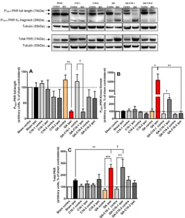
We first checked if the
in vivo
model displayed a significantly
increased level of P
T451-PKR by WB before evaluating, in a second
period, the influence of the PKR inhibitor C16 acute treatment in
both ipsi- (hemisphere of the QA injection) and contralateral
striatum.
As shown in
Fig. 1
, QA injection induced a significant decrease
of full-length P
T451-PKR levels (82% inhibition,
p
< 0.01) and on
the contrary a robust increase of catalytic KD P
T451-PKR levels
(959% increase,
p
< 0.05) in ipsilateral striatum compared to the
contralateral side. No modification of P
T451-PKR levels was
ob-served in ipsilateral side of sham rats (
Fig. 1
A and B). In addition,
QA injection induced a significant increase of total PKR in the
ipsi-lateral striatum compared to the contraipsi-lateral side (253% increase,
p
< 0.001) while in sham rats this increase was not significant.
In these conditions, we studied the effects of C16 used as a
spe-cific inhibitor of PKR. Two concentrations were i.p. injected before
and after QA injection: 60 (C16-1) and 600
l
g/kg (C16-2). Results
of PKR immunoreactivity showed that neither C16-1 nor C16-2
alone modified the levels of phosphorylated forms of PKR (
Fig. 1
A
and B). The treatment with C16-1 did not modify QA-induced PKR
immunoreactivity (82% of inhibition and 293% of activation for full
length and KD domain, respectively) but it is noteworthy that the
highest dose (C16-2) greatly reduced the KD P
T451-PKR level (93%
inhibition,
p
< 0.01). The decrease of full-length P
T451-PKR level
was not significant in QA-C16-2 rats (
Fig. 1
A).
Interestingly, C16-2 decreased the levels of total PKR observed
in QA ipsi striatum but not C16-1 (
Fig. 1
C). Compared to C16-1,
the dose C16-2 prevents the cleavage of full-length of P
T451-PKR
le-vel (
Fig. 1
A and B) and the increase of total PKR (
Fig. 1
C), therefore
we selected this dose for all other experiments.
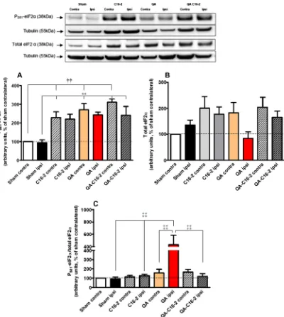
3.2. Effects of C16 treatment on striatal eIF2
a
expression
In
Fig. 2
, results showed the expression of eIF2
a
, a well-known
downstream substrate of PKR. Indeed, PKR phosphorylates eIF2
a
,
leading to the inhibition of the protein synthesis initiation (
Dalton
et al., 2012; Williams, 1999
). Rats receiving C16-2, QA or QA-C16-2
displayed an increase of P
S51-eIF2
a
in both contra- and ipsi-lateral
sides compared to sham rats (between 130% and 210% of increase).
Expression of total eIF2
a
showed variability within groups of rats
and no significant difference was seen, although the rate of total
eIF2
a
would tend to increase in the groups that received C16-2
except that in the ipsilateral striatum injured by QA which was
comparable to the control (
Fig. 2
B). However, calculated
ratio
P
S51-eIF2
a
/total eIF2
a
showed a great and significant increase
(p
< 0.05) in QA ipsilateral striatum compared to ipsilateral striata
from sham, C16-2 and QA-C16-2 rats (365%, 245% and 261%,
3.3. Effects of C16 treatment on macroscopic and microscopic striatal
tissue integrity
No difference was observed between sham and C16-2 animals
(Fig. 3). However, QA rats showed massive tissue impairment in
ipsilateral hemisphere. However, QA-C16-2 group displayed better
tissue integrity, with rescue of striosomes contrary to QA animals.
3.4. Effects of C16 treatment on pro- and anti inflammatory cytokine
production
We compared by Luminex™ assay the levels of cytokines IL-1
b
,
TNF
a
, IL-4 and IL-10 in the striatum of sham, C16-1, C16-2, QA,
QA-C16-1 and QA-C16-2 animals. A robust increase of IL-1
b
levels in
the contralateral striatum of QA rats was significantly prevented
by C16-2 (97% inhibition;
p
< 0.01). The treatment of C16-2 in
PBS-injected rats has no effect on IL-1
b
compared to sham
(Fig. 4A). For IL-4 and IL-10, no significant difference was observed
(Fig. 4C and D). All amounts of TNF-
a
were very low and close to
the limit of detection and no difference was observed between
groups (Fig. 4B).
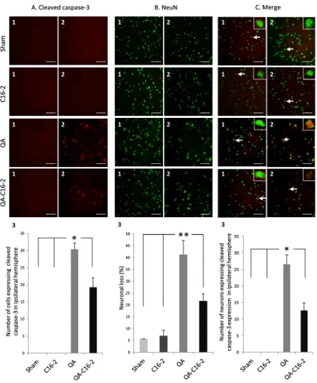
3.5. Effects of C16 treatment on neuron survival and cleaved caspase-3
expression
Neuronal loss, expression of cleaved caspase-3 and neurons
expressing cleaved caspase-3 have been analyzed in contra- and
ipsi-lateral striatum of sham (n
= 3), C16-2 (n
= 3), QA (n
= 4)
and QA-C16-2 (n
= 4) groups (Fig. 5). Immunofluorescence
showed that cleaved caspase-3 was only expressed in ipsilateral
striatum of rats exposed to QA (Fig. 5A.1
vs
A.2). Treatment with
C16 at 600
l
g/kg significantly (p
< 0.05) decreased the number of
cells expressing cleaved caspase-3 (30.3 ± 1.8
vs. 19.2 ± 2.8 cells
in QA and QA-C16-2 groups, respectively) (Fig. 5A.3). For
neu-rons, manual counting was performed in contra- and ipsi-lateral
hemispheres (Fig. 5B.1 and B.2, respectively) and percentage of
Fig. 1.Representative immunoblots and densitometric analyses of PKR immunoreactivity in the presence or absence of C16 treatment. Rats received either 60lg/kg (C16-1)
or 600lg/kg (C16-2) of C16 or DMSO as vehicle by i.p. as described in the method section. They were unilaterally injected with 150 nmol of QA or PBS. Contralateral and
ipsilateral striata were used to measure the expression of full-length PT451-PKR (panel A), kinase domain (KD) PT451-PKR (panel B) and total full-length PKR (panel C) in allneuronal loss in ipsilateral striatum was calculated (
Fig. 5
B.3).
Since cells were counted using a non-stereological approach,
the number of cells detected in the ipsilateral striatum is most
likely an over-estimate because of cell shrinkage. Neuronal loss
was significantly (
p
< 0.01) increased in both QA (41.3 ± 5.8%)
and QA-C16-2 (21.6 ± 3.0%)
vs
. sham (5.6 ± 0.1%) and C16-2
(6.9 ± 2.3%) groups but the C16 treatment significantly (
p
< 0.01)
decreased the neuronal loss induced by QA injection in
QA-C16-2 rats.
Merge images of NeuN and cleaved caspase-3 are represented
in
Fig. 5
C. Cleaved caspase-3 is only expressed in ipsilateral
stria-tum of QA and QA-C16-2 animals (
Fig. 5
C.1
vs
. C.2). Counting of
neurons expressing cleaved caspase-3 was performed in ipsilateral
striatum of these 2 groups. The number of neurons expressing
cleaved caspase-3 in ipsilateral striatum is significantly (
p
< 0.05)
decreased by C16-2 treatment (
Fig. 5
C.3).
4. Discussion
We report here the effects of treatment with the specific stress
kinase PKR inhibitor oxindole/imidazole derivative C16 in an
in vivo
excitotoxic neuroinflammatory rat model. The model we
used is based on injection in the striatum of the quinolinic acid
(QA), an excitotoxic organic acid agonist of the
N
-methyl-D-aspar-tate (NMDA) receptors able to induce brain toxicity and which has
been implicated in dementia in old age or AD. Preliminary
experi-ments aimed to check that this model generates PKR activation.
The QA injection induced a considerable decrease of full-length
P
T451-PKR which was cleaved in an apoptotic environment and
pro-duced a significant increase of its C-terminal phosphorylated
frag-ment, the KD P
T451-PKR, known to be constitutively active (
Saelens
et al., 2001
), as is borne out by structure studies (
Wu and Kaufman,
1997
). Furthermore, the caspase-induced cleavage of active PKR
Fig. 2.Representative immunoblots and densitometric analyses of eIF2aimmunoreactivity with or without C16-2 treatment. Rats received 600lg/kg (C16-2) of C16 or
DMSO as vehicle by i.p. as described in Section2. They were unilaterally injected with 150 nmol of QA or PBS. Contralateral and ipsilateral striata were used to measure the expression of PS51-eIF2a(panel A), and total eIF2a(panel B) in all groups of rats (n= 8 rats per group: sham, C16-2, QA, QA-C16-2 animals). Theratioof PS51-eIF2a/total eIF2a were calculated and showed in panel C. Results are expressed as means (% of sham contra) ± SEM. p< 0.01 compared to contralateral side of sham;**p< 0.01 compared toleads to the release of an N-terminal unphosphorylated fragment
(ND fragment) which is not recognized by the antibody against
to-tal PKR. The caspase-generated fragments of PKR (ND and KD PKR
fragments) cooperate to activate full-length PKR. Specifically,
PKR-ND facilitates the interaction of PKR-KD with full-length PKR and
thus the activation of the kinase, and amplifies the translation
inhibitory signal (
Kalai et al., 2007
). The phosphorylated form of
PKR is cleaved predominantly in ipsilateral striatum of QA-injected
rats and thus the total form at 74 kDa could correspond especially
to the non-phosphorylated form. The increased expression of total
PKR could be related to a compensatory mechanism to maintain
PKR production for its phosphorylation.
Thereafter, the impact of the C16 administered was evaluated
on the expression levels of both PKR and cleaved caspase-3, on
neuronal survival and, on pro- and anti-inflammatory cytokines
production in striatum. The study paid particular attention to
the potential dose-related effects of C16 as animals received
either 60 or 600
l
g/kg of the compound 24-h and 2-h before
and 24 h after QA injection. The choice of studied doses was
based on the local experience in 7-day old rats which received
C16 i.p. at concentrations up to 167
l
g/kg and in a transgenic
AD model treated with C16 i.p. at 0.5 mg/kg every three days
for 3, 9 or 15 months (
Ingrand et al., 2007; Couturier et al.,
2012
). Animals’ behavior (fear, aggressiveness, suffering, and
pain) and body weight were monitored from the onset of the
C16 treatment to the sacrifice and no difference between C16-1
and C16-2 groups was noticed.
In the striatum we systematically compared data from
match-ing cerebral hemispheres. Our main result concerns the highest
dose of C16 (600
l
g/kg) which significantly reduced the KD
PT451-PKR expression in the striatum. Thus, C16-2 prevented the
caspase-cleavage of active PKR, leading to a decrease of its KD
frag-ment as shown by the decreased KD PT451-PKR in the ipsilateral
striatum compared to the contralateral side.
To validate the activation of PKR in this model, we also studied
the expression of total and phosphorylated forms of eIF2
a
, a
well-known substrate of PKR. Its phosphorylation by active PKR on
ser-ine 51 results in its inactivation and in a severe declser-ine in
de novo
protein synthesis (
Donnelly et al., 2013
). Results showed a
signifi-cant increase in the
ratio
of P
S51-eIF2
a
/total eIF2
a
in ipsilateral
striatum of QA-injected rats, suggesting great protein synthesis
impairment. These results were in accordance with the severe
destruction of the striatum integrity observed with H&E staining.
Interestingly, C16-2 treatment in QA-lesioned rats rescued the
activation of PKR/eIF2
a
signalling pathway with a more preserved
tissue quality than QA rats treated with the C16 vehicle.
By inhibiting the PKR/eIF2
a
signalling pathway, C16-2 showed
neurohistoprotective properties after systemic i.p. injection. Then
we wanted to know if it can rescue the toxicity of QA on
inflamma-tion, apoptosis and neuronal death because active PKR was also
in-volved in the cytokine production and apoptosis in infected (
Gil
and Esteban, 2000; García et al., 2006
) and in neurodegenerative
disease models (
Chang et al., 2002; Couturier et al., 2011; Page
et al., 2006
).
The
in vivo
acute excitotoxic model with a neuroinflammatory
component induces microglia activation (
Arlicot et al., 2008
) and
potentially increases cytokine production in cerebral structures.
Assays performed with the Luminex™ allowed us to compare the
levels of cytokines IL-1
b
, TNF
a
, IL-4 and IL-10 between
contra-and ipsilateral striatum in sham, QA, contra-and C16-2-treated animals.
Contrary to expected results, neuroinflammatory response has
not been observed in the ipsilateral side of QA rats but rather in
the contralateral side marked by a robust increase of IL-
b
levels.
As discussed above, the robust toxicity of QA in the ipsilateral side
by activating PKR and inhibiting its downstream substrate eIF2
a
led to metabolism alteration such as translational machinery. The
inflammatory response of the contralateral side may be related
to disruption of the blood–brain barrier (BBB) by QA, allowing
var-ious pro-inflammatory actors including free radicals, cytokines,
chemokines and proteolytic enzymes to disseminate, which can
in-duce glial reactivity. Indeed, it is known that QA inin-duces a
disrup-tion of BBB after
in vivo
injection (
Reynolds and Morton, 1998;
Vezzani et al., 1989
). Furthermore, QA is a metabolite of
kynuren-ine (KYN) which is an intermediate in the pathway of the
metabo-lism of tryptophan to nicotinic acid and can be produced by
endothelial cells and pericytes and further metabolized in QA by
perivascular macrophages and microglia (
Owe-Young et al.,
2008
). Both acute (e.g. ischemic stroke) and chronic (e.g.
Hunting-ton’s disease) brain disorders have been demonstrated to involve
multiple imbalances of KYN pathway metabolism with increase
of QA production (
Zádori et al., 2012
). Thus, QA could be also
pro-duced in the contralateral side and induce inflammation by its
excitotoxic effects.
The major observation was prominently a decrease of IL-1
b
in
the contralateral side of QA-C16-2 groups
vs
the contralateral side
of QA rats. IL-1
b
plays a central role in the neuroinflammatory
process which has been demonstrated for example in IL-1
recep-tor-knockout mice where lack of IL-1 signalling was associated to
attenuation in microglial activation and pro-inflammatory
cyto-kine expression (
Lin et al., 2006
). In a previously published study,
we showed that the specific inhibitor of PKR C16 was responsible
for decreasing pro-inflammatory cytokine (IL-1, IL-6 and TNF
a
)
production in peripheral blood mononuclear cells from AD patients
and in an APPswePS1dE9 transgenic model of AD at 12-months of
age (
Couturier et al., 2010a, 2012
). Furthermore, a recent paper
underlines the crucial role of PKR in inflammasome activation
in vitro
and indicates that it should be possible to
pharmacologi-cally target this molecule to treat inflammation (
Lu et al., 2012
).
Here, we showed that PKR inhibitor C16 prevented
in vivo
the
pro-duction of IL-1
b
induced in an acute excitotoxic
in vivo
model.
However, in QA-injected rats, we showed that C16 prevented PKR
activation in the side without detectable inflammatory factors in
this damaged tissue and prevented inflammation in the
contralat-eral side where the PKR activation was not observed. The C16
compound could have other molecular targets controlling
inflammatory response than the PKR pathway but determining
these new targets would require a broad screening of the kinome
cell. Recently, we observed that C16 prevented inflammatory stress
by inhibiting the autophagosome accumulation induced by IL-1
b
in vitro
(data not published). It is well established now that active
Fig. 5.Immunofluorescence of cleaved caspase-3 and NeuN in rat striatum. Panels A–C represent the same areas of contra- and ipsi-lateral striatum (1 and 2, respectively) of sham (n= 3), C16-2 (n= 3), QA (n= 4) and QA-C16-2 (n= 4) groups. Magnification was20 and120 for inserts. Data are means ± SEM and analyzed by using a Kruskal– Wallis test followed by a Dunn multiple comparison test. Scale: 20l
m. Panel A: cleaved caspase-3 (red channel). Cells expressing cleaved caspase-3 in contra- and ipsi-lateral striatum (1 and 2) for each group. The number of positive cells in ipsilateral striatum of animal has been counted (3).*p< 0.05 panel B:NeuN (green channel). Neuronsstaining in contra- and ipsi-lateral striatum (1 and 2) for each group. The percentage of neuronal loss in ipsilateral striatum, reported to contralateral one, has been performed (3).**p< 0.01 compared to respective contralateral side. Panel C: merge representing neurons and cleaved caspase-3 in contra- and ipsi-lateral striatum (1 and 2) for each
group. Neurons expressing cleaved caspase-3 in both striatum have been counted (3).*p< 0.05 compared to respective contralateral side. (For interpretation of the references
autophagy flux limits the production of IL-1
b
by degradation of
inflammasome proteins (
Shi et al., 2012; Dupont et al., 2011;
Har-ris et al., 2011
).
Contrary to IL-1
b
, no significant difference for the other
cyto-kines TNF
a
, IL-4 and IL-10 was noticed between groups in the
pres-ent work. Considering TNF
a
, studies have shown that the
administration of IL-1 receptor antagonist resulted in reduction
in TNF
a
and limited the loss of dopaminergic neurons in a rat
model of Parkinson’s (
Koprich et al., 2008
). Therefore we were
expecting an influence of C16 treatment on this cytokine too but
Luminex™ measurements were mostly down off the scale
(
Fig. 2
B) and did not allow us to quantify the cytokine production
accurately. It has been reported the very low basal level of TNF-
a
and other cytokines in the cortex and hippocampus of adult rats
at 18 h post-lesion (
Taupin et al., 1993
). At 24 h-post lesion, IL-1
was the most relevant cytokine among those studied and C16
remarkably prevented its production in contralateral side parallel
to partially rescue neuronal death in ipsilateral side.
The immunofluorescence methodology allowed the visualizing
and measuring of cleaved caspase-3 expression associated to living
neurons in ipsi- and contralateral striatum. The QA cerebral
expo-sure model we used was characterized by a massive neuronal loss
with associated apoptosis but the C16 treatment was able to
signif-icantly limit this neuronal loss. As the C16 compound administered
at 600
l
g/kg reduced the number of neurons expressing the
cleaved caspase-3 in the ipsilateral striatum, this suggests that
the neuronal protection induced by the C16 could be linked to an
anti-apoptotic effect of the compound in our QA rat model.
5. Conclusions
This study demonstrated for the first time the involvement of
PKR in an
in vivo
rat model of brain acute excitotoxicity with a
neuroinflammatory component. Data collected from animals
receiving the inhibitor of PKR C16 showed first the ability of the
compound to cross the blood–encephalic barrier and second, the
importance of the dose on its pharmacological effects. The main
result we report here is the ability of the oxindole/imidazole
derivative C16 to massively decrease the IL-1
b
expression and to
preserve
in vivo
neurons from apoptosis in the model we used.
Although the C16 could be considered as a potentially
advanta-geous strategy for patients with neurodegenerative disease, we
note how important it is to optimize the drug administration
sche-dule of the drug as well. The compound was administered 24-h and
2-h before and then 24-h after QA injection in our experiments but
clearly this has to be re-evaluated. C16 blood concentration
mea-surements would also provide important pharmacokinetics data
as well as toxicity parameters.
Acknowledgements
The authors thank the financial support of Region Centre,
Vin-cent Courdavault for technical help and Susan Walters for English
corrections. The authors declare no conflict of interest.
References
Arlicot, N., Katsifis, A., Garreau, L., Mattner, F., Vergote, J., Duval, S., Kousignian, I., Bodard, S., Guilloteau, D., Chalon, S., 2008. Evaluation of CLINDE as potent translocator protein (18 kDa) SPECT radiotracer reflecting the degree of neuroinflammation in a rat model of microglial activation. Eur. J. Nucl. Med. Mol. Imaging 35, 2203–2211.
Balachandran, S., Kim, C.N., Yeh, W.C., Mak, T.W., Bhalla, K., Barber, G.N., 1998. Activation of the dsRNA-dependent protein kinase, PKR, induces apoptosis through FADD-mediated death signaling. EMBO J. 17, 6888–6902.
Balachandran, S., Roberts, P.C., Kipperman, T., Bhalla, K.N., Compans, R.W., Archer, D.R., Barber, G.N., 2000. Alpha/beta interferons potentiate virus-induced
apoptosis through activation of the FADD/Caspase-8 death signaling pathway. J. Virol. 74, 1513–1523.
Bando, Y., Onuki, R., Katayama, T., Manabe, T., Kudo, T., Taira, K., Tohyama, M., 2005. Double-strand RNA dependent protein kinase (PKR) is involved in the extrastriatal degeneration in Parkinson’s disease and Huntington’s disease. Neurochem. Int. 46, 11–18.
Botchkina, G.I., Meistrell III, M.E., Botchkina, I.L., Tracey, K.J., 1997. Expression of TNF and TNF receptors (p55 and p75) in the rat brain after focal cerebral ischemia. Mol. Med. 3, 765–781.
Chang, R.C., Suen, K.C., Ma, C.H., Elyaman, W., Ng, H.K., Hugon, J., 2002. Involvement of double-stranded RNA-dependent protein kinase and phosphorylation of eukaryotic initiation factor-2alpha in neuronal degeneration. J. Neurochem. 83, 1215–1225.
Chen, X., Ma, X., Jiang, Y., Pi, R., Liu, Y., Ma, L., 2011. The prospects of minocycline in multiple sclerosis. J. Neuroimmunol. 235, 1–8.
Chiang, M.C., Chern, Y., Huang, R.N., 2012. PPARgamma rescue of the mitochondrial dysfunction in Huntington’s disease. Neurobiol. Dis. 45, 322–328.
Couturier, J., Morel, M., Pontcharraud, R., Gontier, V., Fauconneau, B., Paccalin, M., Page, G., 2010a. Interaction of double-stranded RNA-dependent protein kinase (PKR) with the death receptor signaling pathway in amyloid beta (Abeta)-treated cells and in APPSLPS1 knock-in mice. J. Biol. Chem. 285, 1272–1282.
Couturier, J., Page, G., Morel, M., Gontier, C., Claude, J., Pontcharraud, R., Fauconneau, B., Paccalin, M., 2010b. Inhibition of double-stranded RNA-dependent protein kinase strongly decreases cytokine production and release in peripheral blood mononuclear cells from patients with Alzheimer’s disease. J. Alzheimers Dis. 21, 1217–1231.
Couturier, J., Paccalin, M., Morel, M., Terro, F., Milin, S., Pontcharraud, R., Fauconneau, B., Page, G., 2011. Prevention of theb-amyloid peptide-induced inflammatory process by inhibition of double-stranded RNA-dependent protein kinase in primary murine mixed co-cultures. J. Neuroinflammation 8.
Couturier, J., Paccalin, M., Lafay-Chebassier, C., Chalon, S., Ingrand, I., Pinguet, J., Pontcharraud, R., Guillard, O., Fauconneau, B., Page, G., 2012. Pharmacological inhibition of PKR in APPswePS1dE9 mice transiently prevents inflammation at 12 months of age but increases Ab42 levels in the late stages of the Alzheimer’s disease. Curr. Alzheimer Res. 9, 344–360.
Dalton, L.E., Healey, E., Irving, J., Marciniak, S.J., 2012. Phosphoproteins in stress-induced disease. Prog. Mol. Biol. Transl. Sci. 106, 189–221.
Doherty, G.H., 2011. Nitric oxide in neurodegeneration: potential benefits of non-steroidal anti-inflammatories. Neurosci. Bull. 27, 366–382.
Donnelly, N., Gorman, A.M., Gupta, S., Samali, A., 2013. The eIF2akinases: their structures and functions. Cell. Mol. Life Sci. 70, 3493–3511.
Dupont, N., Jiang, S., Pilli, M., Ornatowski, W., Bhattacharya, D., Deretic, V., 2011. Autophagy-based unconventional secretory pathway for extracellular delivery of IL-1beta. EMBO J. 30, 4701–4711.
Estrada Sánchez, A.M., Mejía-Toiber, J., Massieu, L., 2008. Excitotoxic neuronal death and the pathogenesis of Huntington’s disease. Arch. Med. Res. 39, 265–276.
Field, R., Campion, S., Warren, C., Murray, C., Cunningham, C., 2010. Systemic challenge with the TLR3 agonist poly I:C induces amplified IFNalpha/beta and IL-1beta responses in the diseased brain and exacerbates chronic neurodegeneration. Brain Behav. Immun. 24, 996–1007.
Gagne, J.J., Power, M.C., 2010. Anti-inflammatory drugs and risk of Parkinson disease: a meta-analysis. Neurology 74, 995–1002.
García, M.A., Gil, J., Ventoso, I., Guerra, S., Domingo, E., Rivas, C., Esteban, M., 2006. Impact of protein kinase PKR in cell biology: from antiviral to antiproliferative action. Microbiol. Mol. Biol. Rev. 70, 1032–1060.
Gendelman, H.E., Appel, S.H., 2011. Neuroprotective activities of regulatory T cells. Trends Mol. Med. 17, 687–688.
Gil, J., Esteban, M., 2000. Induction of apoptosis by the dsRNA-dependent protein kinase (PKR): mechanism of action. Apoptosis 5, 107–114.
Gil, J., García, M.A., Gomez-Puertas, P., Guerra, S., Rullas, J., Nakano, H., Alcamí, J., Esteban, M., 2004. TRAF family proteins link PKR with NF-kappa B activation. Mol. Cell. Biol. 24, 4502–4512.
Glass, C.K., Saijo, K., Winner, B., Marchetto, M.C., Gage, F.H., 2010. Mechanisms underlying inflammation in neurodegeneration. Cell 140, 918–934.
Griffin, W.S., Liu, L., Li, Y., Mrak, R.E., Barger, S.W., 2006. Interleukin-1 mediates Alzheimer and Lewy body pathologies. J. Neuroinflammation, 3–5.
Gupta, A., Kumar, A., Kulkarni, S.K., 2011. Targeting oxidative stress, mitochondrial dysfunction and neuroinflammatory signaling by selective cyclooxygenase (COX)-2 inhibitors mitigates MPTP-induced neurotoxicity in mice. Prog. Neuropsychopharmacol. Biol. Psychiatry 35, 974–981.
Harris, J., Hartman, M., Roche, C., Zeng, S.G., O’Shea, A., Sharp, F.A., Lambe, E.M., Creagh, E.M., Golenbock, D.T., Tschopp, J., Kornfeld, H., Fitzgerald, K.A., Lavelle, E.C., 2011. Autophagy controls IL-1beta secretion by targeting pro-IL-1beta for degradation. J. Biol. Chem. 286, 9587–9597.
Hoozemans, J.J., Veerhuis, R., Rozemuller, J.M., Eikelenboom, P., 2011. Soothing the inflamed brain: effect of non-steroidal anti-inflammatory drugs on Alzheimer’s disease pathology. CNS Neurol. Disord.: Drug Targets 10, 57–67.
Ingrand, S., Barrier, L., Lafay-Chebassier, C., Fauconneau, B., Page, G., Hugon, J., 2007. The oxindole/imidazole derivative C16 reduces in vivo brain PKR activation. FEBS Lett. 581, 4473–4478.
Ito, T., Yang, M., May, W.S., 1999. RAX: a cellular activator for double-stranded RNA-dependent protein kinase during stress signaling. J. Biol. Chem. 274, 15427– 15432.
anti-inflammatory drug in amyloid precursor protein plus presenilin-1 transgenic mice. J. Neurosci. 22, 2246–2254.
Kalai, M., Suin, V., Festjens, N., Meeus, A., Bernis, A., Wang, X.M., Saelens, X., Vandenabeele, P., 2007. The caspase-generated fragments of PKR cooperate to activate full-length PKR and inhibit translation. Cell Death Differ. 14, 1050– 1059.
Kalaria, R.N., 1999. Microglia and Alzheimer’s disease. Curr. Opin. Hematol. 6, 15– 24.
Koprich, J.B., Reske-Nielsen, C., Mithal, P., Isacson, O., 2008. Neuroinflammation mediated by IL-1b increases susceptibility of dopamine neurons to degeneration in an animal model of Parkinson’s disease. J. Neuroinflammation 27, 5–8.
Lin, H.W., Basu, A., Druckman, C., Cicchese, M., Krady, J.K., Levison, S.W., 2006. Astrogliosis is delayed in type 1 interleukin-1 receptor-null mice following a penetrating brain injury. J. Neuroinflammation, 3–15.
Lu, B., Nakamura, T., Inouye, K., Li, J., Tang, Y., Lundbäck, P., Valdes-Ferrer, S.I., Olofsson, P.S., Kalb, T., Roth, J., Zou, Y., Erlandsson-Harris, H., Yang, H., Ting, J.P., Wang, H., Andersson, U., Antoine, D.J., Chavan, S.S., Hotamisligil, G.S., Tracey, K.J., 2012. Novel role of PKR in inflammasome activation and HMGB1 release. Nature 488, 670–674.
Martin, H.L., Mounsey, R.B., Mustafa, S., Sathe, K., Teismann, P., 2012. Pharmacological manipulation of peroxisome proliferator-activated receptor
c
(PPARc
) reveals a role for anti-oxidant protection in a model of Parkinson’s disease. Exp. Neurol. 235, 528–538.McGeer, P.L., McGeer, E.G., 2007. NSAIDs and Alzheimer disease: epidemiological, animal model and clinical studies. Neurobiol. Aging 28, 639–647.
Mennicken, F., Maki, R., de Souza, E.B., Quirion, R., 1999. Chemokines and chemokine receptors in the CNS: a possible role in neuroinflammation and patterning. Trends Pharmacol. Sci. 20, 73–78.
Meurs, E., Chong, K., Galabru, J., Thomas, N.S., Kerr, I.M., Williams, B.R., Hovanessian, A.G., 1990. Molecular cloning and characterization of the human double-stranded RNA-activated protein kinase induced by interferon. Cell 62, 379–390. Morel, M., Couturier, J., Pontcharraud, R., Gil, R., Fauconneau, B., Paccalin, M., Page, G., 2009. Evidence of molecular links between PKR and mTOR signalling pathways in Abeta neurotoxicity: role of p53, Redd1 and TSC2. Neurobiol. Dis. 36, 151–161.
Murphy, P.G., Borthwick, L.S., Johnston, R.S., Kuchel, G., Richardson, P.M., 1999. Nature of the retrograde signal from injured nerves that induces interleukin-6 mRNA in neurons. J. Neurosci. 19, 3791–3800.
Owe-Young, R., Webster, N.L., Mukhtar, M., Pomerantz, R.J., Smythe, G., Walker, D., Armati, P.J., Crowe, S.M., Brew, B.J., 2008. Kynurenine pathway metabolism in human blood–brain-barrier cells: implications for immune tolerance and neurotoxicity. J. Neurochem. 105, 1346–1357.
Page, G., Rioux, Bilan.A., Ingrand, S., Lafay-Chebassier, C., Pain, S., Perault Pochat, M.C., Bouras, C., Bayer, T., Hugon, J., 2006. Activated double-stranded RNA-dependent protein kinase and neuronal death in models of Alzheimer’s disease. Neuroscience 139, 1343–1354.
Panza, F., Frisardi, V., Solfrizzi, V., Imbimbo, B.P., Logroscino, G., Santamato, A., Greco, A., Seripa, D., Pilotto, A., 2012. Immunotherapy for Alzheimer’s disease: from anti-b-amyloid to tau-based immunization strategies. Immunotherapy 4, 213–238.
Paris, D., Ganey, N.J., Laporte, V., Patel, N.S., Beaulieu-Abdelahad, D., Bachmeier, C., March, A., Ait-Ghezala, G., Mullan, M.J., 2010. Reduction of beta-amyloid pathology by celastrol in a transgenic mouse model of Alzheimer’s disease. J. Neuroinflammation 7.
Patel, R.C., Sen, G.C., 1998. PACT, a protein activator of the interferon-induced protein kinase, PKR. EMBO J. 17, 4379–4390.
Paxinos, G., Watson, C., 1982. The Rat Brain in Stereotaxic Coordinates. Academic Press, San Diego.
Peel, A.L., Bredesen, D.E., 2003. Activation of the cell stress kinase PKR in Alzheimer’s disease and human amyloid precursor protein transgenic mice. Neurobiol. Dis. 14, 52–62.
Reynolds, D.S., Morton, A.J., 1998. Changes in blood–brain barrier permeability following neurotoxic lesions of rat brain can be visualised with trypan blue. J. Neurosci. Methods 79, 115–121.
Romano, P.R., Garcia-Barrio, M.T., Zhang, X., Wang, Q., Taylor, D.R., Zhang, F., Herring, C., Mathews, M.B., Qin, J., Hinnebusch, A.G., 1998. Autophosphorylation in the activation loop is required for full kinase activity in vivo of human and yeast eukaryotic initiation factor 2alpha kinases PKR and GCN2. Mol. Cell. Biol. 18, 2282–2297.
Saelens, X., Kalai, M., Vandenabeele, P., 2001. Translation inhibition in apoptosis: caspase-dependent PKR activation and eIF2-alpha phosphorylation. J. Biol. Chem. 276, 41620–41628.
Schneeberger, A., Mandler, M., Mattner, F., Schmidt, W., 2012. Vaccination for Parkinson’s disease. Parkinsonism Relat. Disord. (Suppl. 1), 11–13.
Schwarcz, R., Köhler, C., 1983. Differential vulnerability of central neurons of the rat to quinolinic acid. Neurosci. Lett. 38, 85–90.
Shi, C.S., Shenderov, K., Huang, N.N., Kabat, J., Abu-Asab, M., Fitzgerald, K.A., Sher, A., Kehrl, J.H., 2012. Activation of autophagy by inflammatory signals limits IL-1beta production by targeting ubiquitinated inflammasomes for destruction. Nat. Immunol. 13, 255–263.
Shimazawa, M., Hara, H., 2006. Inhibitor of double stranded RNA-dependent protein kinase protects against cell damage induced by ER stress. Neurosci. Lett. 409, 192–195.
Singh, M., Patel, R.C., 2012. Increased interaction between PACT molecules in response to stress signals is required for PKR activation. J. Cell. Biochem. 113, 2754–2764.
Suen, K.C., Yu, M.S., So, K.F., Chang, R.C., Hugon, J., 2003. Upstream signaling pathways leading to the activation of double-stranded RNA-dependent serine/ threonine protein kinase in beta-amyloid peptide neurotoxicity. J. Biol. Chem. 278, 49819–49827.
Taupin, V., Toulmond, S., Serrano, A., Benavides, J., Zavala, F., 1993. Increase in IL-6, Il-1 and TNF levels in rat brain following traumatic lesion. Influence of pre- and post-traumatic treatment with Ro5 4864, a peripheral-type (p site) benzodiazepine ligand. J. Neuroimmunol. 42, 177–185.
Tobinick, E.L., Gross, H., 2008. Rapid improvement in verbal fluency and aphasia following perispinal etanercept in Alzheimer’s disease. BMC Neurol. 8. Ung, T.L., Cao, C., Lu, J., Ozato, K., Dever, T.E., 2001. Heterologous dimerization
domains functionally substitute for the double-stranded RNA binding domains of the kinase PKR. EMBO J. 20, 3728–3737.
Vantelon, N., Rioux-Bilan, A., Ingrand, S., Pain, S., Page, G., Guillard, O., Barrier, L., Piriou, A., Fauconneau, B., 2007. Regulation of initiation factors controlling protein synthesis on cultured astrocytes in lactic acid-induced stress. Eur. J. Neurosci. 26, 689–700.
Vezzani, A., Stasi, M.A., Wu, H.Q., Castiglioni, M., Weckermann, B., Samanin, R., 1989. Studies on the potential neurotoxic and convulsant effects of increased blood levels of quinolinic acid in rats with altered blood–brain barrier permeability. Exp. Neurol. 106, 90–98.
Whitton, P.S., 2010. Neuroinflammation and the prospects for anti-inflammatory treatment of Parkinson’s disease. Curr. Opin. Investig. Drugs 11, 788–794. Williams, B.R., 1999. PKR; a sentinel kinase for cellular stress. Oncogene 18 (45),
6112–6120.
Wu, S., Kaufman, R.J., 1997. A model for the double-stranded RNA (dsRNA)-dependent dimerization and activation of the dsRNA-activated protein kinase PKR. J. Biol. Chem. 272, 1291–1296.
Zádori, D., Klivényi, P., Szalárdy, L., Fülöp, F., Toldi, J., Vécsei, L., 2012. Mitochondrial disturbances, excitotoxicity, neuroinflammation and kynurenines: novel therapeutic strategies for neurodegenerative disorders. J. Neurol. Sci. 15, 187– 191.