A NEW CYTOTOXIC DOLABELLANE FROM THE INDONESIAN SOFT CORAL
Anthelia
sp.
Anggia Murni
1,3, Novriyandi Hanif
2,3,*, and Junichi Tanaka
31
Biopharmaca Research Center, Bogor Agricultural University
Kampus IPB Taman Kencana, Jl. Taman Kencana No. 3, Bogor 16128, Indonesia
2
Department of Chemistry, Faculty of Mathematic and Natural Sciences, Bogor Agricultural University Jl. Agatis Wing 2 Level 4, Bogor 16144, Indonesia
3
Department of Chemistry, Biology and Marine Science, University of the Ryukyus Nishihara, Okinawa 903-0213, Japan
Received June 10, 2013; Accepted August 29, 2013
ABSTRACT
One new dolabellane (1) and two known diterpenoids stolonidiol (2) and clavinflol B (3) have been isolated from the ethyl acetate extract of the Indonesian soft coral Anthelia sp. A new compound 1 exhibited a moderate cytotoxicity against NBT-T2 cells at 10 µg/mL, while known compounds 2 and 3 showed cytotoxicity at 1 and 0.5 µg/mL, respectively. Structure of the new compound1 was elucidated by interpretation of NMR spectroscopic data (1D and 2D NMR data) and mass spectrometry (ESIMS data) as well as comparison with those of related ones. This finding should be useful for anti cancer drug development of the promising dolabellane-types compound.
Keywords:Anthelia sp.; soft coral; dolabellane; NBT-T2; cytotoxicity; marine
ABSTRAK
Senyawa baru dolabellane (1) dan dua senyawa diterpenoid, yaitu stolonidiol (2), dan clavinflol B (3) telah diisolasi dari fraksi etil asetat koral lunak Anthelia sp. yang berasal dari perairan Indonesia. Senyawa baru 1 menunjukkan aktivitas sitotoksik moderat terhadap sel tumor NBT-T2 pada konsentrasi 10 µg/mL, sedangkan senyawa2dan3 menunjukkan sitotoksik pada konsentrasi berturut-turut 1 dan 0.5 µg/mL. Struktur senyawa baru1 dielusidasi dengan interprestasi spektroskopi NMR (1D dan 2D) dan spektrometri massa (ESIMS) serta perbandingan data spektroskopi dengan senyawa-senyawa sejenis. Penemuan ini berguna untuk pengembangan obat anti kanker dari senyawa-senyawa tipe dolabellane yang menjanjikan.
Kata Kunci:Anthelia sp.; koral lunak; dolabellane; NBT-T2; sitotoksisitas; laut
INTRODUCTION
Tropical waters particularly coral reefs are a large treasure trove of bioorganic molecules that contain unique molecular structures and diversed biological functions. The molecules and their bioactivities can develop and even create a new scientific field [1]. Marine organisms especially marine invertebrates such as sponge and soft coral are no value as food, but they are important sources of biologically active substances that have potential to be developed into new drugs and other useful products such as health care or cosmetics [2].
Soft corals are marine invertebrates that do not produce calcium carbonate skeletons. Soft corals have been proved to be a source of structurally diverse and biologically active terpenoids [3]. One of the important compounds derived from soft corals is hippuristanol that can be used to inhibit poliovirus replication [4]. Species
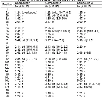
Table 1.1H-NMR data for compounds1-3in CDCl3 Compound1 Compound2 Compound3
Position δ
1.29, brdd, (14.7, 9.2) 1.53, dd (12.0, 9.3)
Merck silica gel 60 (0.063-0.20 mm) was used for column chromatography. Analytical TLC was performed on commercial silica gel 60 F254visualized with vanillin-EtOH-1% H2SO4. All solvents used were reagent grade. A sample of the soft coral Antheliasp. was collected by hand using scuba at Krakatau Island, West Java, Indonesia and was stored in EtOH. The identification of the genus was done by Prof. Junichi Tanaka, Department of Chemistry, Biology, and Marine Science, University of the Ryukyus, Japan.
Instrumentation
Optical rotations were obtained with a JASCO P-1010 digital polarimeter. The IR spectra were taken using a DR 8020 Shimadzu spectrophotometer. The1H and 13C-NMR spectra were recorded on JEOL 500 FTNMR spectrometer. The chemical shifts were expressed in δ (ppm) and coupling constant (J) in Hz. ESIMS data were obtained on a PE QSTAR mass spectrometer and infrared (IR) spectra were recorded on
a DR 8020 Shimadzu spectrophotometer. HPLC was performed on a Hitachi L-6000 pump equipped with a Shodex RI-101 monitor and a Hitachi L-4000 UV detector using a Cosmosil 5 C18AR-II or a Mightysil RP-18 column.
Procedure
Table 2.13C-NMR data for compounds1-3in CDCl3* Compound1 Compound2 Compound3
Position
δC mult. δC mult. δC mult.
1 44.7 C 44.7 C 44.8 C
2 38.7 CH2 37.9 CH2 42.6 CH2
3 27.6 CH2 29.3 CH2 25.2 CH2
4 149.1 C 148.7 C 147.8 C
5 36.7 CH2 31.4 CH2 34.9 CH2
6 26.0 CH2 24.8 CH2 29.3 CH2
7 128.5 CH 57.9 CH 67.2 CH
8 137.1 C 63.6 C 75.9 C
9 27.3 CH2 26.9 CH2 33.8 CH2
10 60.9 CH 56.6 CH 54.5 CH
11 75.5 C 75.9 C 76.9 C
12 49.8 CH 48.4 CH 50.3 CH
13 27.3 CH2 27.3 CH2 27.8 CH2
14 38.0 CH2 36.9 CH2 38.9 CH2
15 24.1 CH3 23.5 CH3 24.2 CH3
16 110.6 CH2 111.3 CH2 113.9 CH2
17 68.3 CH2 65.3 CH2 65.9 CH2
18 74.6 C 74.6 C 75.2 C
19 29.8 CH3 29.7 CH3 29.7 CH3
20 26.4 CH3 26.1 CH3 26.1 CH3
*multiplicity was determined by DEPT and HMQC Spectrum
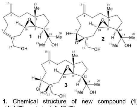
Fig 1. Chemical structure of new compound (1), stolonidiol(2), and clavinflolB(3)
Compound1. Colorless oil; []D 27
-22°(c0.01, CHCl3); IR
(KBr) νmax 3419, 2965, 1683, 1645, 1456, 1377, 1168, 1014, 948, 892 cm-1;1H and13C-NMR see Tables 1 and 2; ESIMS m/z 343.2052 [M+Na]+(calcd for C20H32O3Na 343.2249).
Compound 2. Colorless oil; []D27-37.9°(c 0.29, CHCl3) {[]D -31°(c 1.4, CHCl3) [10]};
1
H and 13C-NMR see Tables 1 and 2.
Compound 3. Colorless oil; []D27 +2.5°(c 0.29, CHCl3) {[]D +8.9°(c 0.36, CH2Cl2) [11]};
1
H and 13C-NMR see Tables 1 and 2.
Cytotoxicity assay
The NBT-T2 cell (BRC-1370) was purchased from Riken and cultured under a standard protocol using
DMEM. NBT-T2 is a cell line derived from chemically induced rat bladder carcinoma cells. The cells were seeded in 1 mL of modified Eagle’s media supplemented with 10% heat-inactivated fetal bovine serum, streptomycin, amphotericin B, and glutamic acid. Cells were exposed to graded concentrations of the new and known compounds as well as their fractions at 37 °C for 72 h and observed under a microscope to evaluate the effects at 48 and 72 h.
RESULT AND DISCUSSION
A sample of the soft coral Anthelia sp. was thoroughly extracted with acetone. After concentration, the residue was partitioned between EtOAc and water. The EtOAc fraction showed a significant toxicity at 1 g/mL against rat bladder tumor cells NBT-T2 was chromatographed on silica gel followed normal phase or reversed-phased HPLC to give a new dolabellane 1 together with known ones2and3(Fig. 1).
The molecular formula of compound 1, C20H32O3 was concluded by its ESIMS and NMR spectrum. It was indicated by five degrees of unsaturation that can be accounted for one trisubtituted epoxide [δH2.63, dd,
Fig 2.Key COSY, HMBC and NOE of new compound1
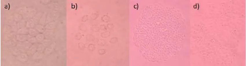
Fig 3.Cytotoxic NBT-T2 Cells assay in a) blank MeOH, 10 μg/mL; b) compound 1, 10 μg/mL; c) compound 2, at
1 μg/mL; d) compound 3, at 0.5 μg/mL
1.21, s, δC 29.8; δH 1.29, s, δC 26.4) and one hydroxymethylene (δH 4.11, s, 3.72, s, δC 68.1) were confirmed by1H and 13C-NMR spectrum. The presence of exo-methylene and hydroxy groups were also supported by IR absorption at 948.1645 cm-1 and 3419 cm-1, respectively. In the1H-1H COSY, it was possible to identify four structural units, which were assembled with the assistance of an HMBC experiment (Fig. 2). The Key HMBC correlations of H3-15 to C-1, C-2, C-11, C-14; H2 -16 to C-3, C-4, C-5; H2-17 to C-7, C-8, C-9; H-10 to C-1, C-8, C-11, C-12 permitted connections of the carbon skeleton. The olefin at C7-C8 was confirmed by HMBC correlations of H-7 and C-5. Furthermore, the isopropyl alcohol group attached at C-12 was confirmed from the simultaneous HMBC correlations of H-20 to C-12, C-19; H-19 to C-12, C-20. On the basis of the above analysis, the planar structure of 1 was established unambiguously. The7,8was assigned asEon the basis of NOE correlation between H-17and H-7.The relative stereochemistry of angular methyl (C-15) was established to be the same as that of 2 by extensive NOE analysis in which H-15 correlated with H-20, H-19, and H-10, suggesting angular methyl (H-15), isopropyl group (H-19, H-20), and H-10 to be the same side of 1 (Fig. 2). As predicted by the similarity of the1H and13 C-NMR data of 1 to stolonidiol (2), the relative stereochemistry of 1 was assigned to be identical with
that of stolonidiol, which stereochemistry was established by X-ray diffraction [10]. Therefore, the relative stereochemistry of1 was 1S*, 10R*, 11R*, and 12S*. Two known compounds (2-3) were verified their structure using 1D NMR as well as their optical rotation that nearly the same (or similar) values as reported in literature [10-11].
All isolated compounds 1-3 were evaluated their toxicity against NBT-T2 rat bladder epithelial cells. The new compound 1 showed moderate toxicity at 10
μg/mL, while known compounds 2-3 showed strong
toxicity at 1, 0.5 μg/mL, respectively. The compounds
1-3 were found as bicyclo [9.3.0]-tetradecane skeleton having thecis-geometry at the ring junction. Compound 1 is featured by the presence of one E double bond instead of one epoxide ring as in 2. In addition, this is the first report that dolabellane diterpenoids are present in the genusAnthelliasp. This finding also implies that the dolabellane-type compound as in1-3may be useful for anti cancer drug development.
CONCLUSION
One new compound (1) along with known ones (2-3) has been isolated and characterized as dolabellane diterpenoid. The new compound1showed
while 2-3 showed strong toxicity at 1 and 0.5 μg/mL, respectively. The compounds show the unique structures containing bicyclo [9.3.0]-tetradecane skeleton.
ACKNOWLEDGEMENT
This work was supported in part by Grant-in-Aids (15406005, 18032061) for Scientific Research from MEXT, the Ministry of Education, Culture, Sports, Science and Technology of Japan.
REFERENCES
1. Uemura, D., Han, C., Hanif, N., Inuzuka, T., Maru, N., and Arimoto, H., 2012, Pure Appl. Chem.,86, 6, 1297–1315.
2. Tanaka, J., Kuniyoshi, M., Tanaka, C., Issa, H.H., Balansa, W., Otsuka, M., Githige, W.P., and Higa, T., 2005,Pure Appl. Chem.,77, 1, 83–89.
3. Blunt, J.W., Copp, B.R., Keyzers, R.A., Munro, M.H.G., and Prinsep, M.R., 2013, Nat. Prod. Rep., 30, 2, 237–323.
4. Bordeleau, M-E., Mori, A., Oberer, M., Lindqvist, L., Chard, L.S., Higa, T., Belsham, G.J., Wagner, G., Tanaka, J., and Pelletier, J., 2006, Nat. Chem. Biol., 2, 4, 213–220.
5. Kashman, Y., and Rudi, A., 2004, Phytochem. Rev., 3, 3, 309–323.
6. Lin, Y-S., Fazary, A.E., Chen, C-H., Kuo, Y-H., and Shen, Y-C., 2011, Helv. Chim. Acta, 94, 2, 273– 281.
7. Mushti, C.S., Kim, J-H., and Corey, E.J., 2006, J. Am. Chem. Soc., 128, 43, 14050–14052.
8. Kim, B.J., Nam, J.H., Kwon, Y.K., So, I., and Kim, S.J., 2013,Basic Clin. Pharmacol. Toxicol., 112, 2, 83–89.
9. Hanif, N., Tanaka, J., Setiawan, A., Trianto, A., de Voogd, N.J., Murni, A., Tanaka, C., and Higa, T., 2007,J. Nat. Prod., 70, 3, 432–435.
10. Mori, K., Iguchi, K., Yamada, N., Yamada, Y., and Inouye, Y., 1987,Tetrahedron Lett., 28, 46, 5673– 5676.