Docking Sulochrin and Its Derivative as α-Glucosidase Inhibitors
of
Saccharomyces cerevisiae
Wening Lestari
1,*, Rizna Triana Dewi
2, Leonardus Broto Sugeng Kardono
2, and Arry Yanuar
3 1Center for Radioisotopes and Radiopharmaceuticals Technology, National Nuclear Energy Agency, Puspiptek, Serpong 15314, Indonesia
2
Research Center for Chemistry, Indonesian Institute of Sciences, Puspiptek, Serpong 15314, Indonesia
3
Faculty of Pharmacy, Universitas Indonesia, Depok 16424, Indonesia
Received April 26, 2016; Accepted August 11, 2016
ABSTRACT
Sulochrin is known to have an activity as inhibitors of the α-glucosidase enzyme. In this report interaction of sulochrin to the active site of the α-glucosidase enzyme from Saccharomyces cerevisiae was studied by docking
method. The crystal structure of α-glucosidase from S. cerevisiae obtained from the homology method using
α-glucosidase from S. cerevisiae (Swiss-Prot code P53341) as a target and crystal structure of isomaltase from
S. cerevisiae(PDB code 3A4A) as a template. These studies show that sulochrin and sulochrin-I could be bound in
the active site of α-glucosidase from S. cerevisiae through the formation of hydrogen bonds with Arg213, Asp215,
Glu277, Asp352. Sulochrin-I has stability and inhibition of the α-glucosidase enzyme better than sulochrin. The iodine atom in the structure of sulochrin can increase the activity as an inhibitor of the α-glucosidase enzyme.
Keywords:sulochrin; sulochrin-I; α-glucosidase inhibitor; S. cerevisiae
ABSTRAK
Sulochrin telah diketahui mempunyai aktivitas sebagai inhibitor enzim α-glukosidase. Pada laporan ini interaksi sulochrin terhadap sisi aktif enzim α-glukosidase dari Saccharomyces cerevisiae telah dipelajari melalui metode
docking. Struktur kristal enzim α-glukosidase dari S. cerevisiae diperoleh melalui metode homologi menggunakan
enzim α-glukosidase dari S. cerevisiae(Swiss-Prot code P53341) sebagai target dan struktur kristal isomaltase dari
S. cerevisiae (kode PDB 3A4A) sebagai templet. Sulochrin dan sulochrin-I dapat terikat di sisi aktif enzim
α-glukosidase dari S. cerevisiae melalui formasi ikatan dengan Arg213, Asp215, Glu277, Asp352. Sulochrin-I
mempunyai stabilitas dan menginhibisi enzim α-glukosidase dari S. cerevisiaelebih baik dari sulochrin. Atom iodium
pada struktur sulochrin dapat meningkatkan aktvitas sebagai inhibitor enzim α-glukosidase.
Kata Kunci:sulochrin; sulochrin-I; inhibitor α-glukosidase; S. cerevisiae
INTRODUCTION
Diabetes mellitus is a disease related to malfunction of insulin. In 2010 people with diabetes reached 220 million and in 2025 it is predicted to reach 300 million people [1]. Most diabetics are type 2 diabetes mellitus. Type 2 diabetes mellitus is characterized by the occurrence of postprandial hyperglycemia [2]. Control of postprandial hyperglycemia is an important step for the treatment of diabetes. It can be done by inhibiting the absorption of glucose through inhibition of carbohydrate
hydrolyzed enzymes such as α-glucosidase and α-amylase [3-4]. The α-glucosidase enzyme converts
carbohydrates into glucose [5-7]. Mechanism of
α-glucosidase inhibitors is reversible inhibition, competitive to the α-amylase enzyme and pancreatic
digestive enzymes in the small intestine such as isomaltase, sucrase, and maltase. These enzymes can inhibit the absorption of glucose and it will be lowering the hyperglycemia after a meal [7-8].
Research on α-glucosidase inhibitor compounds
has been carried out,in silico,in vivo,andin-vitro. Data from in silico study reported that interaction of
α-glucosidase inhibitor through the formation of hydrogen bonds with the α-glucosidase residues.
Interactions between sulfonamide derivatives and
α-glucosidase from Saccharomyces cerevisiae occurs at Asp349, His348, Phe157, Tyr71, Phe177, Arg212 [6]. Salacinol derivatives also showed potential as
α-glucosidase inhibitors, interaction with residues
Asp327, His600, Asp542, Asp203, Arg526 in NtMGAM [9].
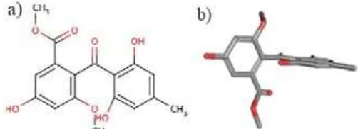
Fig 1.Sulochrin structure a) two dimension and b) three dimension
Methyl 2-(2,6-dihydroxy-4-methylbenzoil)-5-hydroxy
-3-methoxybenzoat (sulochrin) known as α-glucosidase
inhibitors (Fig. 1). It could be isolated from the ethyl acetate extract of A. Terreus. In silico study shown that sulochrin can bind to the binding pocket in the crystal structure of Thermotoga maritima 4-α-glucano
transferase which has some similarities with the crystal structure of S. cerevisiae [10]. Sulochrin also interacts
with the active site of human α-glucosidase at Asp587 with Ki with ΔG values of -6.90 kcal/mol [11]. In vitro
study, reported that sulochrin has IC50 of 8.5 mg/mL when tested against S. cerevisiae. Sulochrin-I, a sulochrin derivative studied with the radioligand binding assay (RBA) method is reported to bind to the
α-glucosidase from S. cerevisiae with Kd value of 26.316 nM [12]. Interaction of sulochrin and its derivative
(sulochrin-I) with α-glucosidase of S. cerevisiae can be determined by docking these molecules (ligand and its receptor). The docking process will obtain the inhibition
constants (Ki) and the free energy (ΔG) to form a stable
conformation at the active site of the enzyme
α-glucosidase. Currently, the crystal structure of the S. cerevisiae α-glucosidase is not yet available then
docking process must be done using a homology structure of S. cerevisiae α-glucosidase. In this study, the homology was performed using S. cerevisiae α-glucosidase MAL12 obtained from Swiss-Prot (code
P53341) as a target and the crystal structure of isomaltase from S. cerevisiae which was obtained from the PDB (PDB code 3A4A, 1.60 Å resolution) as a template [13]. Isomaltase fromS. cerevisiaeis known as oligo-1,6-glucosidase. This selection is based on the similarity of the amino acid sequence between
α-glucosidase and isomaltase from S. cerevisiae.
EXPERIMENTAL METHOD
Materials
The material used in this work was a three-dimensional crystal structure of S. cerevisiae α-glucosidase which obtained from homology method
using the SWISS-MODEL software. The target is the
docked in the three-dimensional structure was sulochrin-I and this structure was prepared using the Chem3D software. The instruments of hardware and software were used. The hardware specifications was a computer equipped with 4 GB of RAM, Intel Xeon E5260 2.4 GHz, Graphic Card nVIDIA GeForce GT-680 (Taiwan), and Microsoft Windows XP operating system (USA). The software of a ChemOffice 2004 (licensed to Wening PRR), Open Babel (The Blueobelisk Group, USA), Autodock (The Scripps Research Institute, USA), PyMOL (DeLano Scientific LLC, Italy), PuTTY (www.PuTTY.org), Cygwin (Red Hat Inc., USA), SWISS-MODEL (http://swissmodel.expasy. org) [14-15], PROCHECK online (www.ebi.ac.uk/ thrnton-srv/databases/pdbsum/generate. html), Clustal W2 (European Bioinformatics Institute, UK), were used.
Homology Modelling
Identification of homologs of α-glucosidase
(Swiss-Prot code P53341) was carried out by performing sequence database searches with a standard tool, UniProt. The coordinate of the crystal structure S. cerevisiae oligo-1,6-glucosidase (PDB ID: 3A4A, 1.6 Å resolution) was used as a template to build the S. cerevisiae structure. Sequence alignment was performed by using the ClustalW2 program. The 3D model of the S. cerevisiae α-glucosidase was built by
SWISS-MODEL software based on the S. cerevisiae
oligo-1,6-glucosidase structure. The final protein model was evaluated by PROCHECK for the evaluation of Ramachandran Pot.
Molecular Docking Simulation
The 3D model of the S. cerevisiae α-glucosidase
was used in docking analysis by using AutoDock. Hydrogen polar were added and Gasteiger charges were calculated. This research was used acarbose as a control for the docking simulation. The structure of sulochrin and sulochrin-I were built by using ChemOffice program. Acarbose, sulochrin and sulochrin-I were docked toS. cerevisiaeα-glucosidase
which used the same parameter for three compounds. Hydrogens polar were added and torsion number were calculated. Docking parameters were grid in 50 Å x 50 Å x 50 Å with X, Y and Z axis -21.727; -0.752; 18.637 respectively. Docking simulation runs with 100 runs using Lamarckian Genetic Algorithm. The free
energy (ΔG) and inhibitions constant (Ki) scoring were
Fig 2.Sequence alignment result between the template protein S. cerevisiaeoligo-1,6-glucosidase (PDB ID 3A4A) and the target protein S. cerevisiae α-glucosidase (Mal12). The asterisks (*) indicate the conserved residues
between the two protein. The red residues form the responsible for the break glycosidic bond
RESULT AND DISCUSSION
In silicostudy was carried out in order to determine the interaction between the ligand to its receptor. In this study, the receptor of a three-dimensional crystal structure of S. cerevisiae α-glucosidase was used.
Unfortunately, its crystal structure is not yet available, therefore the homology modeling must be done in order to obtain a three-dimensional model of the structure. The first step in homology modeling was to determine the target and template. The target used in this study was the amino acid sequence ofS. cerevisiae α-glucosidase MAL12 with 584 amino acids and obtained from Swiss-Prot (code P53341). The template used in this study was the crystal structure of S. cerevisiae oligo-1,6-glucosidase which was obtained from the PDB (PDBID 3A4A) with resolution 1.60 Å and 589 amino acids [16]. The selection of target and template are based on the
sequence similarity their amino acids. Oligo-1,6-glucosidase has amino acid residues which responsible for the break the glycosidic bond. These residues are His112, Asp215, Glu277, His351 and Asp352 [16]. The presence of these residues makes the crystal structure is suitable to be used as template in the homology process.
Crystal structure of S. cerevisiae oligo-1,6-glucosidase (PDB code 3A4A) was aligned with the
S. cerevisiae α-glucosidase MAL12 (Swiss-Prot code
P53341) using the ClustalW2 program. This alignment would be able to identify the similarity sequence of the above-mentioned molecules. The results showed that the alignment similarity of amino acid sequence between the S. cerevisiae oligo-1,6-glucosidase and
S. cerevisiae α-glucosidase MAL12 was 71.92% as
Fig 3.Sulochrin (1), sulochrin-I (2) and acarbose (3) structures
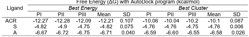
Table 1. Free energy (ΔG) of ligand to S. cerevisiae α-glucosidase (PI: experiments 1, PII: experiments 2, PIII:
experiments 3, ACR: acarbose, S: sulochrin, A: sulochrin-I)
Free Energy (ΔG) with AutoDock program (kcal/mol)
Best Energy Best Cluster
Ligand
PI PII PIII Mean SD PI PII PIII Mean SD
ACR -12.27 -12.28 -12.09 -12.21 0.107 -10.06 -10.04 -10.2 -10.1 0.087 S -4.82 -4.9 -4.75 -4.82 0.075 -4.76 -4.76 -4.75 -4.76 0.006 A -6.67 -6.72 -6.75 -6.71 0.040 -6.59 -6.60 -6.55 -6.58 0.026
oligo-1,6-glucosidase was qualified as a template in the process homology modeling [8]. In a recent publication,
homology modeling of α-glucosidase was performed with Bacillus cereus (O16GB) as a template and the percentage of similarity was found to be 58.4% [8].
Homology modeling of S. cerevisiae α-glucosidase was performed using an SWISS-MODEL software that can be accessed at http://swissmodel.expasy.org. Homology modeling process was done using target
S. cerevisiae α-glucosidase MAL12. The result from this
process was QMEAN Z-score a value that describes the quality of a model by providing an estimate of the ‘degree of nativeness’ of the structural features observed in a model and useful measure quality of theoretical models. Rasouli and Fazeli-Nasab reported that a model with QMEAN Z-score between 0 and 1 was considered to be a good quality model [17-19]. The value of QMEAN Z-Score calculated for S. cerevisiae α-glucosidase MAL12 was off -0.019. This result shows S. cerevisiae α-glucosidase MAL12 was a good quality
of the model because of the estimated reliability of the model was very much close to the value of 0, the value which was considered to be a good quality model.
The evaluation of the obtained models was performed using the PROCHECK program through Ramachandran plot. This evaluation gave the percentage of residual in most favorable regions of 87.7% and 0.2% in disallowed region. The percentage of residues in most favorable regions illustrates the quality of protein models. The above-mentioned results indicated that the model developed was qualified to be used for molecular docking process.
Docking simulation was performed on sulochrin, methyl 2-(2,6-dihydroxy-3-iodo-4-methylbenzoil)-5-hydroxy
-3-metoxybenzoat (sulochrin-I) and acarbose as a control (Fig. 3). Docking was run using AutoDock 4.2 with Lamarckian Genetic Algorithm (Lamarckian GA). A number of the evaluation was chosen the long category with RMSD 0.5 Å. The analysis showed that acarbose
has the lowest of the free energy (ΔG),
-12.21±0.07 kcal/mol. The free energy for sulochrin and sulochrin-I were -4.82±0.07 and -6.71±0.04 kcal/mol, respectively. This results indicating that acarbose has the most stable bond when it interacts with the
S. cerevisiae α-glucosidase compared with sulochrin
and sulochrin-I (Table 1). This analysis also showed that sulochrin-I has more stable bond than the sulochrin when interacted with S. cerevisiae α-glucosidase. This result suggests that the presence
of iodine atoms in the sulochrin molecule improving the stability of sulochrin-S. cerevisiae α-glucosidase.
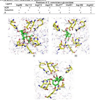
Inhibition constants (Ki) of acarbose, sulochrin, sulocrin-I are shown in Table 2. It can be seen that acarbose has the smallest Ki value compared to that of sulochrin, sulochrin-I. Ki values of acarbose, sulochrin, sulochrin-I were 1.13 10-3±0.2 10-3, 293.69±35.77 and 12.03±0.83 µM respectively. These results suggest that
acarbose has the best ability to inhibit of α-glucosidase.
The presence of iodine atoms in sulochrin was found to decrease Ki values sulochrin from 293.69 to 12.03 µM (sulochrin-I). Lowering Ki value of iodinated sulochrin indicated the increasing of its ability to inhibit the
activity of α-glucosidase. Inhibition of α-glucosidase
occurs through the formation of hydrogen bonds between ligands (i.e. acarbose, sulochrin and sulochrin-I) with the active site of S. cerevisiae α-glucosidase (Table3). Based on results in Table 3, it
Table 2.Inhibition constants (Ki) of ligand toS. cerevisiaeα-glucosidase (PI: experiments 1, PII: experiments 2, PIII:
experiments 3, ACR: acarbose, S: sulochrin, A: sulochrin-I)
Inhibition constant (Ki) with AutoDock program (µM)
Best Energy Best Cluster
Ligand
PI PII PIII Mean SD PI PII PIII Mean SD
ACR 1.01×10⁻³ 1×10⁻³ 1.38×10-31.13×10-3 0.2×10⁻³ 42.58×10⁻³ 43.39×10⁻³ 33.25×10⁻³ 0.039 0.0056 S 294.17 257.68 329.23 293.69 35.77 321.78 329.23 325.32 321.78 3.74 A 12.92 11.91 11.26 12.03 0.830 14.63 15.75 15.027 15.14 0.56
Table 3. Ligand interaction on active site ofS. cerevisiae α-glucosidase (√ indicate interaction with distance ≤ 5Å, - indicate no interaction, ACR: acarbose, A: sulochrin-I)
Residues ofS. cerevisiae α-glucosidase Ligand
Asp69 His112 Arg213 Asp215 Glu277 His351 Asp352 Arg442
ACR √ √ √ √ √ – √ √
Sulochrin – – – – – – – √
A – – √ √ √ – √ √
Fig 4.Interaction of (i) acarbose, (ii) sulochrin, (iii) sulochrin-I with active sites ofS. cerevisiae α-glucosidase
sites onS. cerevisiae α-glucosidase (i.e. Asp69, His112, Arg213, Asp215, Glu277, His351, Asp352 and Arg441) except His351, while sulochrin only interacts with Arg442 and sulochrin-I interacts with residues Arg213, Asp215, Glu277, Asp352, Arg442. The difference in these
interactions causes acarbose have the most stable and good inhibitory ability.
The results of the analysis of docking indicate that the ligands visually occupy the active sites of
The three-dimensional model structure of the
S. cerevisiae α-glucosidase was obtained by homology
using S. cerevisiae α-glucosidase MAL12 (Swiss-Prot code P53341) as a target and the crystal structure of
S. cerevisiae oligo-1,6-glucosidase (PDB code 3A4A, 1.60 Å resolution) as a template. Interaction of sulochrin
to the active site of the α-glucosidase enzyme from S. cerevisiae has been studied by docking method. Model compounds of sulochrin-I were found to form a more stable bond and better inhibition capability towards
α-glucosidase enzymes compared to that of sulochrin.
The presence of iodine atoms on sulochrin was found
enhancing its inhibitory action towards the α-glucosidase
enzymes. Sulochrin-I could be bound in the active site of
α-glucosidase from S. cerevisiae through the formation of hydrogen bonds with Arg213, Asp215, Glu277, Asp352.
REFERENCES
[1] Li, Y., Wen, S., Kota, B.P., Peng, G., Li, G.Q., Yamahara, J., and Roufogalis, B.D., 2005, Punica granatumflower extract, a potent alpha-glucosidase inhibitor, improves postprandial hyperglycemia in Zucker diabetic fatty rats, J. Ethnopharmacol., 99 (2), 239–244.
[2] Kim, Y.M., Jeong, Y.K., Wang, M.H., Lee, W.Y., and Rhee, H.I., 2005, Inhibitory effect of pine extract on
α-glucosidase activity and postprandial
hyperglycemia,Nutrition, 21 (6), 756–761.
[3] van de Laar, F.A., Lucassen, P.L., Akkermans, R.P., van de Lisdonk, E.H., Rutten, G.E., and van
Weel, C., 2005, α-glucosidase inhibitors for patients
with type 2 diabetes results from a Cochrane systematic review and meta-analysis, Diabetes Care, 28 (11), 154–163.
[4] Zeng, Y.F., Lu, Z.R., Yan, L., Oh, S., Yang, J.M., Lee, J., and Ye, Z.M., 2012, Towards alpha-glucosidase folding induced by trifluoroethanol: Kinetics and computational prediction, Process Biochem., 47 (12), 2284–2290.
[5] Gao, H., Huang, Y.N., Gao, B., Li, P., Inagaki, C., and Kawabata, J., 2008, Inhibitory effect on
α-glucosidase by Adhatoda vasica Nees, Food Chem., 108 (3), 965–972.
[6] Bharatham, K., Bharatham, N., Park, K.H., and Lee, K.W., 2008, Binding mode analyses and pharmacophore model development for sulfonamide
chalcone derivatives, a new class of α-glucosidase
inhibitors,J. Mol. Graphics Modell., 26, 1202–1212.
[8] Park, H., Hwang, K.Y., Kim, Y.H., Oh, K.H., Lee, J.Y., and Kim, K., 2008,Discovery of novel alpha-glucosidase inhibitors based on the virtual screening with the homology-modeled protein structure,Bioorg. Med. Chem., 16 (1), 284–292. [9] Nakamura, S., Takahira, K., Tanabe, G.,
Morikawa, T., Sakano, M., Ninomiya, K., Yoshikawa, M., Muraoka, O., and Nakanishi, I., 2010, Docking and SAR studies of salacinol
derivatives as α-glucosidase inhibitors, Bioorg. Med. Chem. Lett., 20 (15), 4420–4423.
[10] Dewi, R.T., Anita, Y., Istyastono, E.P., Darmawan, A., and Hanafi, M., 2009, The applicability of the crystal structure of Termotoga maritima 4- -glucanotransferase as the template for sulochrin as -glucosidase inhibitors, Indones. J. Chem., 9 (3), 487–490.
[11] Farkhani, A., 2012, Molecular Dynamic Analysis of
Docking Product of Complex α-Glucosidase with
Sulochrin, Undergraduate Thesis, Universitas Indonesia, 35-38.
[12] Lestari, W., Susilo, V.Y., Setiyowati, S., Triningsih, Ariyanto, A., Widayati, P., Kardono, L.B.S., and Yannuar, A., 2014, Synthesis of sulochrin-125I and
its binding affinity as α-glucosidase inhibitor using
Radioligand Binding Assay (RBA) method, Atom Indonesia, 40 (1), 22–26.
[13] Lestari, W., 2013, Synthesis and Binding Study Sulochrin-125I as α-glucosidase Inhibitor using Radioligand Binding Assay (RBA) and Molecular Docking Methods, Thesis, Universitas Indonesia, 42.
[14] Arnold, K., Bordoli, L., Kopp, J., and Schwede, T., 2006, The SWISS-MODEL workspace: A web-based environment for protein structure homology modeling,Bioinformatics, 22 (2), 195–201.
[15] Kiefer, F., Arnold, K., Künzli, M., Bordoli, L., and Schwede, T., 2009, The SWISS-MODEL Repository and associated resources, Nucleic Acids Res., 37, D387–D392.
[16] Yamamoto, K., Miyake, H., Kusunoki, M., and Osaki, S., 2010, Crystal structures of isomaltase from Saccharomyces cerevisiae and in complex with its competitive inhibitor maltose, FEBS J., 277 (20), 4205–4214.
[17] Benkert, P., Biasini, M., and Schwede, T., 2010, Toward the estimation of the absolute quality of individual protein structure models,Bioinformatics, 27 (3), 343–350.
protein in bread wheat,American-Eurasian J. Agric. Environ. Sci., 14 (10), 1044–1048.
[19] Rahman, M.A., Chaturvedi, N., Sinha, S., Pandey, P.A., Gupta, D.K., Sundaram, S., and Tripathi, A.,
2013, Computational protein structure modeling and analysis of UV-B stress protein in