Perkembangan Plasenta dan Pertumbuhan Janin
pada Tikus Hamil yang Diinfeksi
Porphyromonas Gingivalis
Placental Development and Fetal Growth in
Porphyromonas Gingivalis-Infected
Pregnant Rats
Banun Kusumawardani1, Yuliana MD Arina2, Azham
Purwandhono3
1Department of Biomedical Sciences, Faculty of Dentistry, Universitas Jember, East Java-Indonesia, 2Department of Periodontology, Faculty of Dentistry, Universitas Jember, East Java-Indonesia, 3Department of Pathology, Faculty of Medicine, Universitas Jember, East Java-Indonesia
Corresponding : kusumawardani_banun@yahoo.co.id
Abstract
Maternal Porphyromonasgingivalis infection on periodontal tissue can result in Porphyromonasgingivalis
dissemination to umbillical cord. Porphyromonas gingivalis presumably gain access to the systemic circulation via local tissue inflammation, and may affect the placental development and the fetus itself. This study aimed to analize the effect of periodontal infection with Porphyromonas gingivalis on placental development, and to determine its effect on fetal growth in a pregnant rat model. Female rats were infected with live-Porphyromonas gingivalis at concentration of 2x10⁹ cells/ml into subgingival sulcus of the maxillary first molar before and/or
during pregnancy. They were sacrified on gestational day (GD) 20. Fetuses were evaluated for weight and length. All placentas were fixed in 10% buffered formalin, processed for paraffin embedding, and stained with hematoxylin and eosin.The histopathological analysis of placentas on GD 20 showed that trophoblast cells in labyrinth and junctional zone had a greater density in control group than Porphyromonas gingivalis-infected periodontal maternal group. The nucleated-erythrocytes were found more abundant in the fetal blood vessels of
Porphyromonas gingivalis-infected periodontal maternal group than in the fetal blood vessels of control group. In conclusion, the impaired placental morphology influenced the normal function of placenta to maintain the growth and development of fetus. The decreased placental weightresulted in the decreased of fetal weight andlength.
Key words: Porphyromonasgingivalis, periodontitis, pregnancy, placental development, fetal growth
Abstrak
Infeksi Porphyromonas gingivalis pada jaringan periodontal maternal dapat mengakibatkan penyebaran
Porphyromonas gingivalis ke tali pusat janin. Porphyromonas gingivalis mungkin mendapatkan akses ke sirkulasi sistemik melalui peradangan jaringan lokal dan mempengaruhi perkembangan plasenta dan janin itu sendiri. Penelitian ini bertujuan untuk menganalisis pengaruh infeksi periodontal oleh Porphyromonas gingivalis
pada perkembangan plasenta, dan menentukan pengaruhnya terhadap pertumbuhan janin pada model tikus hamil. Tikus betina diinfeksi dengan live-Porphyromonas gingivalis konsentrasi 2x10⁹ sel/ml pada sulkus
Porphyromonas gingivalis. Eritrosit-berinti ditemukan lebih banyak di dalam pembuluh darah janin dari kelompok periodontal maternal yang diinfeksi Porphyromonas gingivalis daripada di dalam pembuluh darah janin dari kelompok kontrol. Kesimpulannya, gangguan morfologi plasenta mempengaruhi fungsi normal plasenta untuk mempertahankan pertumbuhan dan perkembangan janin. Penurunan berat plasenta mengakibatkan penurunan berat badan janin dan panjang janin.
Kata kunci: Porphyromonasgingivalis; periodontitis; kehamilan; perkembangan plasenta; pertumbuhan janin
Introduction
Porphyromonas gingivalis is a Gram-negative, black-pigmented anaerobe asso-ciated with several periodontal diseases1. This bacteria has most frequently been detected in deep periodontal pockets and has exhibited a low prevalence in healthy perio-dontal tissue without destructive inflamma-tion2. Porphyromonas gingivalis has been found to adhere to various oral surfaces in periodontitis patients3 and has also been detected within gingival tissues in vivo4. It has also been shown that this organism invades oral epithelial cells in vitro5. The abilities of Porphyromonas gingivalis to adhere and invade have been strongly implicated in the periodontal pathogenicity of this organism. Our previous study showed that Porphyromonas gingivalis infection in maternal periodontal tissue can result in Porphyromonas gingivalis dissemination to umbillical cord and induction of fetal growth restriction, but Porphyromonas gingivalis was not always detected in the umbillical cord from abnormal pregnancies6.
The growth and survival of fetus is dependent on placenta, which forms the interface of feto-maternal circulation, facilitates metabolism, gas exchange, and waste disposal of the fetus. In addition, the placenta produces hormones altered maternal physiology during pregnancy, and forms a barrier against the maternal immune system7. Murine placenta consists of decidual zone,
juntional zone and labyrinth zone. Labyrinth zone consists of branched villi designed for the efficient exchange of nutrients8. Maternal and fetal blood flows are adversely in the labyrinth to maximize the transport of nutrients9. If the labyrinth does not obtain the proper pattern, branching and dilatation of vascularization, then perfused placenta will be disrupted so that the diffusion of oxygen and nutrients to be disturbed10.
Therefore, we hypothesized that Porphyromonas gingivalis and its lipopoly-saccharide from periodontal tissue can spread into the uterus through the circulatory system, then induces placental inflammatory response resulting in fetal growth restriction. The aims of this study were to analize the effect of periodontal infection with Porphyromonas gingivalis on placental development, and to determine its effect on fetal growth in a pregnant rat model.
Materials And Methods
gingivalis before and during pregnancy (Pg-BD). Each group consisted of five pregnant rats.
Induction of experimental periodontitis was performed by injection of 0.05 ml live-Porphyromonas gingivalis ATCC 33277 with a concentration of 2x10⁹cells/ml into the distopalatal and distobuccal gingival sulcus area of maxillary first molar. Injection was repeated every 3 days for 30 days. For infection after pregnancy, it was also performed by a repeated injection every 3 days for 19 days. Control group rats were injected with saline 0.05 ml as the treatment schedule of the treatment group rats. Then,
The pregnant rats were sacrificed on GD 20. Fetuses were removed post-mortem from the uterus and surrounding membranes. Each fetus was removed from chorioamniotic sac. Placental weight, fetal weight and fetal length were weighed to the nearest microgram. The resorption site and viable fetuses were counted and recorded for each rat. The viability of each fetus was assessed visibly. Fetuses were evaluated for weight and crown-tail length. Blood of umbillical cord was collected from each fetus and pooled per dam.
All placentas were fixed in 10% buffered formalin, dehydrated, processed for paraffin embedding, serial section at 5 μm and stained with hematoxylin and eosin. Samples were analyzed using descriptive histology. The density of trophoblast cells in labyrinth zone, junctional zone and decidual zone, and erythrocytes in the fetal blood
vessels were examined by light microscopy. Descriptive histological anal y sis was carried out by a trained ex aminer who was blind to the groups.
Numerical variables consisted of placental weight, fetal weight and fetal length. These variables were performed by statistical analyzes to determine effect of Porphyromonas gingivalis infection in pregnant rats to fetal growth. Independent samples T-test was performed to compare the placental weight, fetal weight and fetal length of maternal periodontal infection. Linear regression analysis was to analyze the linear corelation between numerical varia-bles. Value of significance was determined as P<0.05. Numerical data were presented in mean ± standard deviation.
Results
Placental development and fetal growth was observed on GD 20. This study only observed morphological defects, because abnormalities of the function such as central nervous system disorders could not be placental weight affected (P<0.05) fetal weight and fetal length (Table 2). Each addition of 1 gram of placental weight, it will increase fetal weight at 7.014 grams. In addition, it will also increase fetal length at 58.773 mm.
cells in labyrinth and junctional zone had a nucleated-erythrocytes were found more abundant in the fetal blood vessels of Porphyromonas gingivalis-infected perio-dontal maternal group than in the fetal blood vessels of control group.
Discussion
Pregnancy is a natural condition that must be followed to be able to accept and tolerate the fetus as well in order to obtain good results. This study showed that Porphyromonas gingivalis exposure had disrupted this balance. During the experiment, pregnant rats infected with Porphyromonas gingivalis has decreased motion activity, but no pregnant rats died spontaneously.
During periodontal infection, the amount of periodontal pathogens may increase dramatically, causing transient bacteremia11, which resulted in a selective bacterial colonization in other body parts12. Our previous studies showed that Porphyromonas gingivalis from maternal periodontal tissue can spread through the blood circulation until colonizes in placenta6.
Trophoblast cells in labyrinth and junctional zone of the Porphyromonas gingivalis-infected periodontal maternal group had less density than control group. It is thought to be due to Porphyromonas gingivalis exposure that causes an increase in apoptosis of trophoblast cells in the labyrinth and junctional zone. Furthermore, the
nucleated-erythrocytes were found more abundant in the fetal blood vessels of Porphyromonas gingivalis-infected perio-dontal maternal group than in the fetal blood vessels of control group. On GD 20, erythrocytes in the fetal blood vessels should not be nucleated.
nucleated erythrocytes were found in premature infants from pregnancies complicated by intrauterine infection without acidosis or hypoxemia18. So it can be postulated that the increased number of nucleated erythrocytes may be due to the inflammatory response of the fetus in the placenta.
Impaired fetal growth of the Porphyromonas gingivalis-infected perio-dontal maternal grouphas impaired placental morphology which is characterized by decreased density of trophoblast cells. Impaired placental morphology greatly affects the normal function of the placenta to maintain the growth and development of the fetus. Placental functions include adequate trophoblast invasion, increased uteropla-cental blood flow during pregnancy, transport of nutrients such as glucose and amino acids from mother to fetus, as well as the production and transfer of regulating growth hormones19.
The establishment of functional fetal and placental circulation is the earliest events during the development of the embryo or placenta. Increased transplacental exchange that supports the exponential increase in fetal growth during the last half of pregnancy, especially depending on the dramatic growth of placental blood vessels and the resultant increase in umbilical and uterine blood flow20. Placental vascular development and function will have an impact on fetal growth and development.
This study showed that placental weight was closely correlated with fetal weight and
fetal length. Decreased placental weight will result in decreased fetal weight and fetal length. Strong correlation between placental weight to fetal weight and fetal length may be influenced by differences in morpho-logical structure of placenta, characterized by the density of trophoblast cells in labyrinth zone. This will probably lead to an increase in the volume fraction of labyrinth zone, and a decrease in the volume fraction of junctional zone. In addition, this study also have found decreased fetal blood vessels and space interhaemal labyrinth of placenta from the Porphyromonas gingivalis-infected periodontal maternal group, that will lead to lower diffusion ability of placenta. It can reduce oxygen levels, nutrient absorption, and fetal placental blood flow.
Conclusion
The impaired placental morphology influenced the normal function of placenta to maintain the growth and development of fetus. The decreased placental weight resulted in the decreased fetal weight and fetal length.
Acknowledgements
Table 1. Maternal periodontal infection correlated with placental weight, fetal weight and fetal length on GD 20
Variable
Maternal periodontal infection
Pg-BD
P
Control
Placental weight, g, n(x±SD) 30(0,62±0,09) 43(0,22±0,03) 0
Fetal weight, g, n(x±SD) 30(4,08±0,43) 43(0,56±0,17) 0
Fetal length, mm, n(x±SD) 30(48,97±1,75) 43(18,39±1,61) 0
Pg-BD : Porphyromonas gingivalis-infected periodontal maternal group
Table 2. Effect of placental weight to fetal weight and fetal length on GD 20
Variable
Placental weight (gram)
N R2 B P
Fetal weight, gram 141 0,656 7,014 0,000
Fetal length, mm 141 0,609 58,773 0,000
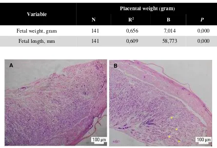
A B
*
Figure 1. The placenta of control group (A) and Porphyromonas gingivalis-infected periodontal maternal group (B) on GD 20. The placenta of the control group (A) had a greater density of fetal blood vessels and trophoblast cells in the labyrinth zone and junctional zone than Porphyromonas gingivalis-infected periodontal maternal group. Placentas from Porphyromonas gingivalis-infected periodontal maternal
group had a lot of spaces between the trophoblast cells (*), resulting in trophoblast cells became less density (B). 40x Magnification. Labyrinth zone of Porphyromonas gingivalis-infected perio-dontal maternal group group (D) had more nucleated erythrocytes in the fetal blood vessels than the control group (C). 400x Magnification.
References
1. Darveau, RP., Tanner, A., Page, RC. 2000 1997. The microbial challenge in periodontitis. Periodontol, 14: 12-32. 2. Griffen, Al., Becker, MR., Lyons, SR.,
Moechberger, ML., Leys EJ. 1998. Prevalence of Porphyromonas gingivalis and periodontal health status. J Clin Mi-crobiol 36: 3239-42.
3. Christersson, LA., Fransson, CL., Dun-ford, RG., Zambon, JJ., 1992. Subgingi-val distribution of pathogenic microor-ganism in adult periodontitis. J Perio-dontol, 63:418-25.
4. Papapanou PN., Sandros ,J., Lindberg K, Duncun, MJ., Niederman, R., Nannmark, U., 1994. Porphyromonas gingivalis may multiply and advance within stratified human junctional
epi-thelium in vitro. J Periodontal Res, 29: 374-5.
5. Lamont, RJ., Chan, A., Belton, CM., Izutsu, KT., Vasel, D., Weinberg, A., 1995. Porphyromonas gingivalis inva-sion of gingival epithelial cells. Infect Immun, 63: 3878-85.
6. Kusumawardani,B., Soesatyo, M., Dasuki, D., Asmara, W., 2011. Fetal growth restriction in Porphyromonas gingivalis-infected pregnant rats. Denti-ka Dent J, 16: 26-30.
7. Cross, JC., 2003. Simmons, DG., Wat-son, ED., 2003. Chorioallantoic mor-phogenesis and formation of the placen-tal villous tree. Ann NY Acad Sc, 995:84-93.
8. Rossant, J., Cross, JC., 2001. Placental development: lessons from mouse mu-tants. Nat Rev Genet, 2: 538-48.
9. Adamson, SL., Lu, Y., Whiteley, KJ., Holmyard D, Hemberger M, Pfarrer C, Cross, JC., 2002. Interaction between trophoblast cells and the maternal and fetal circulation in the mouse placenta. Dev Biol, 250:358-73.
10.Pardi, G., Manconi, AM., Cetin, I., 2002. Placental-fetal interrelationship in IUGR fetuses-a review. Placenta, 23(Suppl A): S136-S141.
11.Daly, CG., Mitchell, DH., Highfield, JE., Grossberg, DE., Stewart, D., Bacte-remia due to periodontal probing: a clin-ical and microbiologclin-ical inves-tigation. J Periodontol, 72: 210-4.
12.Haraszthy , VI., Zambon, JJ., Trevisan, M., Zeid, M., Genco, RJ., 2002. Identi-fication of periodontal pathogens in ath-eromatous plaques. J Periodontol, 71:1554-60.
13.Dame, Ch., Juul, SE., 2000. The switch from fetal to adult erythropoiesis. Clin Perinatol, 27:507–24.
14.Mandel, D., Littner, Y., Mimouni, FB., Dollberg, S., 2003. Nucleated red blood cells in polycythemic infants. AmJ Obsted Gynecol, 188:193–5.
15.Baschat, AA., Gembrouch, U., Reiss, I., Gortner, L., Harman, CR., Weiner, CP., 1999. Neonatal nucleated redblood cell counts in growth-restricted fetuses: relationship in arterial and venous Doppler studies. Am J Obstet Gynecol, 181:190–5.
16.Maier, RF., Gunther, A., Vogel, M., Dudenhausen, JW., Obladen, M., 1994. Umbilical venous erythrpoietin andumbilical arterial pH in relation to morphologic placental abnormalities. Obstet Ginecol, 84:81–7.
17.Leikin, E., Garry, D., Visintainer, P., Verma, U., 1997. Correlation of nucle-ated redblood cell counts in preterm in-fants with histologic chorioamnionitis. Am J Obstet Gynecol, 177: 27–33. 18.Korst, LM., Phelan, JP., Ahn, MO.,
Martin, GI., 1996. Nucleated red blood cells: an update on the markerfor fetal asphyxia. Am J Obsted Gynecol, 175: 843–6.
19.Murphy, VE., Smith, R., Giles, WB., Clifton, VL., 2006. Endocrine regulation of human fetal growth: the role of the mother, placenta, and fetus. Endocrine Rev, 27: 141-69.