CHAPTER II
LITERATURE REVIEW
II. 1. Anatomy and physiology of retina
The retina is a thin, multilayered sheet of neural tissue that lines
the inner aspect of the posterior wall of the eye globe. This layer is vital
for the vision process since it contains neurons that are capable to sense,
amplify, integrate and transmit the visual signals to the brain. The
photoreceptors respond to the light particles and convert them into
electrical impulses. Subsequently, these impulses travel down the optic
nerve to the visual cortex for further processing and visual perception.
The retina is a highly organized structure that comprises two
distinct layers: the outer pigmented layer called the retinal pigment
epithelium (RPE) and the adjacent inner layer of nerve tissue called the
neurosensory retina (14;15).
II. 1. 1. The retinal pigment epithelium
The RPE is a monolayer of hexagonal cells that is located between
the light-sensitive outer segments of the photoreceptors and the choroid.
This layer plays an essential role in the retinal physiology by forming the
outer high-resistance basolateral blood-retinal barrier and supporting the
function of the photoreceptors (17).
II. 1. 2. The neurosensory retina
The complex of neurosensory retina formed by the neural
extension of the brain is responsible for the detection and transmission of
the signals from the photoreceptors to the brain cortex. This inner layer
of retina comprises of six major types of cells: the amacrine cells, bipolar
cells, ganglion cells, horizontal cells, Müller cells and the photoreceptor
cells. The neurosensory part of retina is divided into three distinct layers.
The outermost layer is formed by the photoreceptor cells which convert
the light signal into an electrical impulse. These impulses are then
transferred to the second layer consisting of nuclei of interneurons
(bipolar, horizontal and amacrine cells). The third layer comprises of
ganglion cells, which collect the signals of from the bipolar cells, and
their axons will subsequently transmit this information to the visual
I1. 1. 3. The photoreceptor cells
Photoreceptors are photosensitive neurons in the outer part of
the retina. Morphologically, two types can be distinguished: rod and cone
cells. The retina contains approximately 120 million rods and 6 million
cones (21;22).
Rods are particularly sensitive to light, being able to detect even
a single photon (23). This photoreceptor type allows vision in dim light.
In contrast, cones are less (30-100 fold) sensitive to the light, playing an
important role in mediating bright-light and color vision (16,24). Rods
are predominantly located in the peripheral area of retina whereas cones
are predominantly located in the center of retina, called the fovea (14).
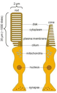
Figure 2. The photoreceptor cells: rod and cone
All photoreceptors consist of four main components an outer
segment, which contains the visual pigments, an inner segment, a
perikaryal region containing the cell nucleus, and an axon. The molecular
components in the outer segment are needed for light absorption and
conversion to electrical signals, whereas the inner segment harbors the
machinery for protein synthesis and production of energy. The electrical
light response is then transmitted to the horizontal and bipolar cells
(24-26).
The blood supply of retina provided by two main sources: the
choriocapillaris immediately outside Bruch's membrane, which supplies
the outer third part of the retina, including the outer plexiform and outer
nuclear layers, the photoreceptors, and the retinal pigment epithelium;
and branches of the central retinal artery, which supply the inner
two-thirds. The fovea is supplied entirely by the choriocapillaris (14).
II. 2. Visual process
Two main processes are essential for the mechanism of vision in
the retina, phototransduction - the process in which absorbed photons are
converted into electrical responses - and visual cycle. Studies of genes
linked to human hereditary blindness have been crucial for improving the
II. 2. 1. Phototransduction
Phototransduction is the process of absorbing light stimulus and
creating a response by photoreceptor cells. This biochemical cascade
takes place in the outer segment of a photoreceptor (27). The
membranous discs of outer segment are loaded with photosensitive
pigments, rhodopsin in rods and cone opsins in cones. These pigments
covalently linked to 11-cis-retinaldehyde (11-cis-RAL) by a Schiff-base
linkage. Upon absorption of a light photon, the opsin activates by
11-cis-RAL isomerization into all-trans-retinal. The activated form of the
rhodopsin photopigment, metarhodopsin II, triggers the activation of a
trimeric G-protein called transducin by allowing the change of the bound
GDP nucleotide to GTP. The alpha subunit of transducin then activates
the phosphodiesterase (PDE) alpha and beta subunits which subsequently -GMP. The reducing of the cytoplasmic cGMP
concentration modifies the conductance of the cGMP-gated channels
leading to their closure and thereby blocking the influx of sodium cations
towards the cells. As a result, positively charged potassium flows out of
cell more rapidly than sodium and calcium is transported to the cell. The
cell becomes depolarized and suppresses its release of glutamate
neurotransmitter enabling the signal to travel down the optic nerve
neurotransmitter (26-28). This cascade provides enormous signal
500 transducin molecules, leading to hydrolyzation hydrolysis of about
105 cGMP (29;31;32).
In the termination phase of this cascade, rhodopsin and other
molecules are rapidly deactivated and recycled. The activated rhodopsin
will be phosphorylated by rhodopsin kinase which then allows the
protein arrestin to bind to rhodopsin. The binding of arrestin blocks the
activity. Finally, rhodopsin will be recycled back to a form that can
absorb light (33-35).
II. 2. 2. The retinoid cycle
The retinoid cycle is a complex recycling system that supplies
the 11-cis-retinal chromophore of rod and cone visual pigments after
isomerized to all-trans-retinal by light. This cycle takes place in retinal
rod and cone photoreceptor outer segments and the retinal pigment
epithelium. After inactivation of metarhodopsin II, all-trans-RAL will dissociate from the opsin protein and transported into the bilayer of outer
segment disc membranes. ATP-binding cassette transporter called ABCR
has been implicated to facilitate reduction of all-trans-RAL by transport
it from disc membranes to the cytoplasmic space to become
all-trans-retinol (all-trans-ROL). Retinal dehydrogenase will subsequently
catalyze all-trans-RAL to all-trans-ROL. All-trans-ROL will be released
Lecithin retinol acyl transferase (LRAT) is the major retinyl ester
synthase in RPE cells, which catalyzes the transfer of a fatty acyl group
from phosphatidylcholine to all-trans-ROL. The isomerases in RPE cells
will catalyze the conversion from all-trans-retinoid to the sterically
constrained 11-cis-ROL. Eventually, 11-cis-ROL will be oxidated to
formed 11-cis-RAL(33-34).
II. 3. Retinitis pigmentosa
II. 3. 1. Definition and clinical manifestations
RP is a clinically and genetically diverse group of hereditary
retinal dystrophies affecting primarily rods, with subsequent cone
photoreceptor cell degeneration. The onset and progression vary between
individuals with RP (2;4;36). The first symptom of RP is night blindness
at an early stage, followed by visual field constriction and central severe
visual impairment due to photoreceptor degeneration as the disease
progresses (37). These clinical symptoms correlate with a predominantly
affected rod photoreceptor cells followed by the cone photoreceptors
degeneration causing complete blindness at the final stage (38). This
disease can be subdivided into non syndromic retinitis pigmentosa, in
which there is no systemic abnormalities, and syndromic retinitis pigmentosa where the retinal degeneration is associated with other
disorders. The most frequent syndromic form of RP is Usher Syndrome,
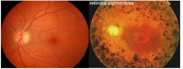
RP patients displays a classic triad of RP in the fundus:
bone-spicule pigmentation, arteriole attenuation and pale, waxy of the disc
optic (Figure 3) (39;40).
Figure 3. Fundus appearance of normal eye and RP (a) Normal fundus appearance; (b) Fundus appearance of RP patients which shows bone-spicule like pigmentation, attenuation of arterioles and pallor of the optic disc (16).
II. 3. 2. Pathophysiology of retinitis pigmentosa
RP is a group of retinal dystrophies which primarily affect
rod photoreceptor cells followed by cone degenerations. The
degeneration of photoreceptor cells is subsequently followed by
retinal outer layers degenerations, whereas the inner layer of retina is
still well preserved until the later stage of the disease. In the previous
study in mice model implicated that this degeneration are due to the
disability to form rod outer segment (OS) and consecutively lead to
rod photoreceptor cells loss. Cone degenerations which triggered by
toxic byproducts of rod cells degeneration or due to the loss of trophic
factors that are normally produced by rod, will subsequently occur and
lead to a successive loss of outer retinal layers. The loss of
integrity. In the normal retina, photoreceptor layer is located between
RPE and retinal vessels. The loss of photoreceptor layer will result in
approximation of RPE to retinal vessels. This contact stimulates the
detachments of RPE cells from B
along the adjacent retinal vessels to perivascular sites in the retina.
The nuclei of RPE cells are translucent, whereas the cell body is dark
due to melanin. The RPE cells will form three-dimensional pigmented
cell clusters around retinal vessels which are known as bone-spicule
pigmentations.
Attenuation of retinal blood vessels is a funduscopic hallmark
of RP which caused by the occlusion of vessels lumina by
extracellular matrix deposits. Thickening of the blood vessels wall and
the occlusion of lumina correlates with the retinal vasculature atrophy
and sclerosis. RPE cells will also reestablished the blood-retina
barrier by forming tight junctions thereby reducing leakage from
vascular endothelial fenestrations (39).
Another typical appearance of RP fundus is disc optic atrophy. It
is suspected that the ganglion cells axon are compressed by the retinal
blood vessel and lead to the loss of ganglion cells. At the later stage,
more ganglion cells will be lost and cause the pale, waxy, atrophic optic
Figure 4. RPE cell migration along retinal vessels at late stages.
(a) Light micrograph of a retinal vessel in the inner retina surrounded by apparently upward migrating RPE cells. (b; c) Cross sections of a retinal vessel (indicated by an arrowhead) in the inner retina that are sheathed by the RPE cells. (d) Retinal capillary (arrowhead), illustrating fenestrations of the vessel (arrows). (e) detail of figure d, arrow indicates the endothelial fenestrations; ECM is indicated by an asterisk (39).
II. 3. 3. Histopathology appearance of retina with RP
The rod photoreceptor cells outer segments (OS) shortening is
one of the first histopathology finding in the retina at the early stage. At
the later stage, RPE cells will be migrate upwards along the retinal
vessels (39). The pigmented cells will cluster around retinal vessels and
forms bone spicule pigmentation (Figure 4). Extracellular matrix (ECM)
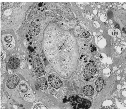
Figure 5. Electron micrograph of bone spicule pigment in retina
An occlusion of retinal blood vessel by accumulated extracellular matrix (*) associated with the encircling layer of RPE cells (R). IPL: inner plexiform layer; GC: ganglion cells (40).
II. 3. 4. Molecular genetics of retinitis pigmentosa
RP can be inherited in an autosomal dominant (adRP, 30 - 40%),
autosomal recessive (arRP, 50 - 60%) X-linked (XLRP, 5 - 20%) and
digenic fashion (1;3;5). A
person in the family. Hence, both autosomal recessive and dominant
inheritance, arising from de novo alterations as well as X-linked inheritance in isolated male cases, are possible. About 50 genes are
currently known to be causative for RP, several displaying allelic
heterogeneity; some have been reported to cause both dominant and
recessive modes of inheritance (1;2). Although many genes have been
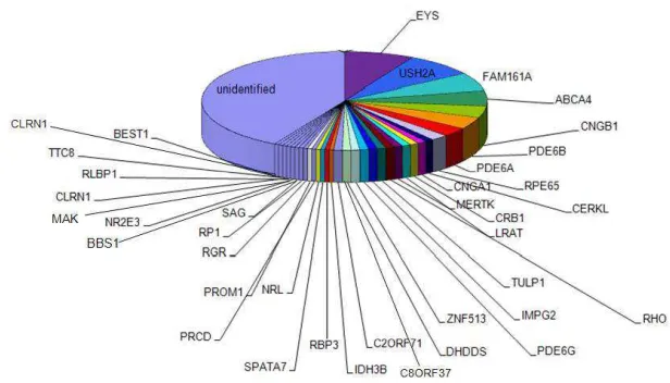
described as causative for RP, 50% of the genetic defects remain
mutations and new genes that are responsible for the remaining cases
(Figure 6).
Figure 6. All identified RP genes
Currently, 39 genes has been reported responsible for arRP (courtesy of R.W.J Collin)
Based on the molecular pathway and the retinal structure,
RP-associated genes can be classified as involved in: the rod
phototransduction cascade, the retinoid cycle, and ciliary transport
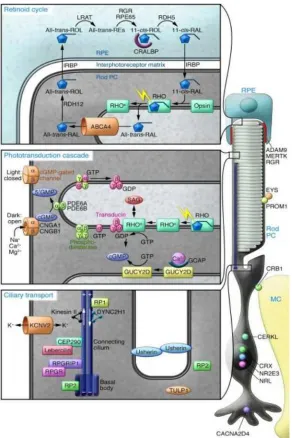
(illustrated in Figure 7). Thus, knowledge about molecular mechanisms
of vision is important to understand the involvement of the genes in the
Figure 7. Three major processes in human rod photoreceptor cells
There are three major processes that are essential in human rod photoreceptor cells and the RPE: retinoid cycle, phototransduction cascade and cilliary transport (6)
II. 4. Molecular genetic analysis
II. 4. 1. Homozygosity mapping
Homozygosity mapping has been proven to be an effective
arRP genes (41-45). A homozygous mutation is likely to reside within
homozygous region detectable with a high resolution genome-wide
single nucleotide polymorphism (SNP) genotyping array. The application
of homozygosity mapping leads to the identification of the genetic
lesions both in consanguineous and non-consanguineous cases, as shown
in patients with autosomal recessive kidney diseases (44). This technique
has been proven to be a powerful method for detection of new mutations
in known genes implicated in retinal dystrophies and mutations in new
disease genes.
In autosomal recessive mode of inheritance, alterations on both
alleles are required for the disease manifestation. Homozygosity mapping
is a method that utilizes homozygous regions shared by the affected
individuals of the same family. In consanguineous families, affected
individuals are more likely to inherit two copies of the same disease
allele from their common ancestor. Therefore, the region surrounding
disease causing mutations will be homozygous. A closer relative
marriage will present a larger homozygous region (46-48).
In non-consanguineous families, homozygous regions can be
found as well, due to a common ancestor who cannot be traced from the
available family history. The size of homozygous region is related to the
number of generations between the parents of the patient and their
common ancestor. The closer the generation of the founder, the bigger
II. 4. 2. Pathogenecity prediction of missense mutations
In a missense mutations, in silico web based prediction
programs, including Polymorphism Phenotype (PolyPhen), Sorting Intolerant from Tolerant (SIFT) and Align GVGD were used to predict the pathogenecity of missense mutation. These programs are able to
calculate the potential functional and structural impacts based on
Grantham scores (comparison of the differences in physical properties of
the amino acids side chains) and PhyloP scores (evolutionary
conservation of the nucleotide). Project hope will be also used to
II.4.2.1 SIFT
This web based program uses sequence homology to predict
whether an amino acid substitution effect to the protein function and
contribute to a disease phenotype. The idea of this approach is that
important amino acids tend to be highly conserved across species. SIFT,
which assigns scores from 0 to 1, predicts substitutions with scores less
than 0.05 as deleterious, whereas those greater than or equal to 0.05 are
considered to be tolerated.
II.4.2.2 Polyphen
PolyPhen includes the evolutionary conservation of the amino
the wild-type and mutated amino acid residue and the consequence of the
amino acid change for the structural properties of the protein to predict
the effect of mutation.
II.4.2.3 Align GVGD
Align GVGD combine Grantham variation (GV) and Grantham
deviation (GD) to predict the pathogeneicity of mutations. GV measures
the degree of biochemical variation among amino acids found at a given
position in the multiple sequence alignment, whereas GD reflects the
'biochemical distance' of the mutant amino acid from the observed amino
acid at a particular position. Align-GVGD can be used to predict the
transactivation activity of each missense substitution. A score of GV=0
consider as a residue that is invariant in the alignment, a value of GV of
60 65 is the upper limit of conservative variation across species, and a
value of GV>100 is indicative of positions that are under little functional
constraint. A value of GD=0 corresponds to a missense substitution that
is within the cross-species range of variation at its position in the protein;
at invariant positions (GV=0); GD=60 65 is the upper limit of a
conservative missense substitution.
II.4.2.4 Project hope
This web-based program was used to analyze the effect of a
structural information from a several of sources, including calculations
on the 3D protein structure, sequence annotations and protein predictions
from other prediction tools. This informations were combined to analyze
the effect of a certain mutation on the protein crystal structure. Hence, we
can predict and visualized the missense mutation effect on the mutant
protein structure and function.
II. 4. 3 Novel mutations confirmation
Mutation analysis of candidate genes residing within the
homozygous regions may revealed either known mutations or novel
mutations. Pathogeneicity of the known mutations can be determined
from the available mutations database, such as Human Genome Mutation Database (HGMD). This website provide informations and link of other databases which have a complete informations regarding the mutations.
The available informations are including the pathogenecity prediction,
molecular mechanism prediction, the protein expression, knock-out
murine phenotype and human phenotype with this specific mutation.
Conversely, in a novel mutation, those data are not available
since the functional studies has not been performed for these novel
mutations. Therefore, supporting data to predict the pathogenecity of
mutations are needed. Several in silico web based programs such as
SIFT, align GVGD and Polyphen are useful for the mutations
effect of the mutations but cannot confirm whether this gene defects are
really the disease-causing mutations for probands or not. Thus,
segregation analysis and frequency analysis in unaffected ethnically
matched controls are needed. Segregation analysis is performed using
In
the segregation analysis, mutations should exclusively found in the
affected individual, whereas in the unaffected individual, mutations will
be either absence or found in a heterozygous state. By performing this
method, the mode of inheritance can also be determined and risk of the
next generation can be calculated.
Frequency analysis in the unaffected ethnically matched controls
is performed to prove that the variants found is a mutation not a common
variants. RFLP PCR and ARMS PCR are the effective techniques to
perform frequency analysis. A mutation should be absent in all of the
control samples. If the same amino acid changes found in many of the
control samples, it means that this is a common variant, and not a
mutation. The technique of RFLP PCR and ARMS PCR explained
below.
II.4.3.1 RFLP PCR
Restriction Fragment Length Polymorphism (RFLP) is is used to
identify a base pair change in the DNA sequence that occurs at a site
bacteria that recognize specific short sequences of DNA and cut the DNA
at those sites. The restriction enzyme is added to the DNA being
analyzed and incubated for several hours, the restriction enzyme will cut
at its recognition sites. By running the DNA in the gel electrophoresis,
the fragments of DNA will separate according to size. DNA fragments
can be visualized and assesed whether or not the DNA was cut by the
enzyme by comparing the band size using a certain marker.
In this study, RFLP was used to confirm whether the base pair
changes in the DNA are mutations or a common variant by performing
this method in the ethnically matched control samples. This technique
was also performed in the screening of all identified mutations in
Indonesian population that were found from the previous study.
Volume, incubation temperature and time of the restriction
enzymes are different for each enzyme. This informations provided by
the enzyme manufacturer which can be found in the enzyme manual kit.
In some mutations, RFLP PCR can not be performed due to
unavailability of the restriction enzymes that cut in the mutation sites.
Therefore, ARMS PCR was used as the next approach in this situation.
II.4.3.2 ARMS PCR
ARMS PCR method is a technique for point mutation or small
one containing an ARMS primer specific for the normal DNA sequence
and cannot amplify mutant DNA at a given locus and the other one
containing a mutant-specific primer and does not amplify normal DNA.
Figure 8. An ilustration of RFLP and ARMS PCR.
(a) RFLP PCR, a change in the DNA sequence can create or abolish recognition site of the enzyme, thus affecting the quantities and lengthe of the DNA fragments resulting from enzyme digestion which can be visualized by gel electrophoresis.
(b) ARMS PCR, the figure above display an interpretation of ARMS PCR, Lane 1 shows the DNA size ladder. Lane 2 presents the results from a normal individual using the ARMS primer with the normal or wild-type sequence, resulting in the target DNA product. Lane 3 also presents the results from a normal individual, but the ARMS primer now corresponds to the mutant sequence; thus, PCR amplification only occurs with the control reaction. Lanes 4 and 5 reveal the results from a patient with the mutation on both alleles (homozygote). Lane 4 represents the results with an ARMS primer that corresponds to wild-type sequence, with PCR amplification therefore only occurring in the control reaction; lane 5 uses an ARMS primer that corresponds to the mutant sequence, with amplification of both target and control DNA. Lanes 6 and 7 reveals the results with a heterozygous individual. In lane 6, the ARMS primer corresponds to wild-type sequence with PCR amplification of target DNA (from one allele) plus the control DNA, and in lane 7, the ARMS primer corresponds to mutant sequence with PCR amplification of target DNA (from the other allele) plus the control DNA (52)
The genotype of an individual can be determined by analysis of
the amplified products: for homozygote individual PCR, products were
primer for probands with homozygous mutations) and for a heterozygote
genotype PCR products were obtained in both reactions. ARMS PCR
will be used in this study to confirm whether the amino acid changes are
mutations or a common variant by performing this method in the
ethnically matched control samples. This technique will be also
performed in the screening of all identified mutations in Indonesian
population that were found from the previous study.
II. 5. The important role of molecular diagnostics
Accurate molecular genetic diagnosis has been proven to be
essential to determine the prognostic and therapeutic approach for
individuals with inherited eye disorders. Knowledge of the underlying
molecular mechanism of the disease is critical in providing information
about its nature, course, and prognosis. The development of gene therapy
for retinal dystrophies is making molecular diagnosis increasingly
important.
Some syndromic diseases involve retinal dystrophy as the first
sign preceding the other organ involvement, as in Senior Loken
syndrome. This syndrome involves retinal dystrophy followed by
nephrolithiasis at the later stage (49). Molecular diagnosis at an early
the involvement of other organs and
slow the progression of the disease.
Genetic diagnosis is also important to provide the patient and
the family with genetic counseling about the inheritance manner, the
recurrence risk of the diseases and the possibility to perform an early
intervention to slow the disease progression.
II. 6 Early intervention for RP
Gene therapy is a very promising approach to treat RP patients.
Nonetheless, this approach still not applicable for the patients. Several
studies need to be done to prove the safety and efficacy of this therapy.
Thus far, therapy for RP patients are very limited. Physicians should
emphasize the therapies that are available to help patients. The aim of the
patient management is to slow the disease progression and help patients
retain their vision to maintain the normal daily function.
Vitamin A (beta-carotene) is an antioxidants that has been
implicated to slow RP progression. A recent comprehensive
epidemiologic study concluded that very high daily doses of vitamin A
palmitate (15,000 U/d) slow the progress of RP by about 2% per year.
The effects are modest and there is a risk of hepatotoxicity and
teratogenicity caused by this regiment. Liver enzymes and vitamin A
level tests are need to be performed annually for the patients whose
Docosahexaenoic acid (DHA) is an omega-3 polyunsaturated
fatty acid and antioxidant. Studies have shown a correlation of ERG
amplitudes with patients' erythrocyte-DHA concentration. Others studies
reported trends of less ERG change in patients with higher levels of
DHA. However, a recent study compared DHA plus vitamin A to vitamin
A alone in patients with RP over 4 years. In this study, the benefit of
DHA was not seen. Further clinical trials must be done to determine
DHA benefit (50).
Ascorbic acid consumption (1000 mg/d) has been
recommended, but there are no evidence exists that ascorbic acid is
helpful. Although several agents has been suggested to slow the disease
progression, there were still no strong evidence that prove the effectivity
of these therapy (50). Therefore,
these agents has to be consumed wisely with also considering the side
effects to the other organ and the effectivity of the agents. Meanwhile,
avoidance to oxidants agents may slow the disease progression, such as
UV light avoidance, stop smoking and alcohol consumption.
II. 7. Genetic Counseling
Genetic counseling of the affected individual with RP or their
relatives will improve their understanding about the disease. The aim of
genetic counseling is to inform patients about the hereditary nature of
genotype and also the risk of the next generations. Counseling modalities
consist of genetic counseling, psychological counseling, and low vision
rehabilitation counseling. The risk of passing the disease to the next
generations can be calculated based on the mode of inheritance.
Informations gained from counseling will help the affected individuals in
the decision making regarding the future strategy, such as pregnancy,
vocational choices and medical interventions. Counseling about
prognosis should be included with providing informations regarding the
great variation among and within inheritance groups, families, and
individuals with respect to age of onset and natural history of the
disorder. Because no treatment is currently available for most
RP patients, genetic counseling and supportive follow-up should be
viewed as an essential service for this common group of genetic
disorders, and co-operation with the ophthalmologists should be actively
sought. The availability of support groups are very useful for the RP
patients. Patients can shared their experience, knowledge and latest
information about RP. Furthermore, patients will also get a psychological
II.9. Conceptual Framework
Non Syndromic Retinitis Pigmentosa Autosomal recessive
(homozygous/ compound heterozygous)
Autosomal Dominnant X-Linked
Inherited Gene Mutation