.
VETERINARSKI ARHIV 81 (4), 535-543, 2011
Subcutaneous xanthoma in a cockatiel (
Subcutaneous xanthoma in a cockatiel (
Nymphicus hollandicus
Nymphicus hollandicus
))
- a case report
- a case report
Marija LiparMarija Lipar11*, Danijela Horvatek*, Danijela Horvatek22, Estella Prukner-Radov, Estella Prukner-Radovččiićć22, Andrea , Andrea Gudan Kurilj
Gudan Kurilj33, Berislav Radiši, Berislav Radišićć11, Dražen Vnuk, Dražen Vnuk11, Nikica Prvanovi, Nikica Prvanovićć44, , Branimir Škrlin
Branimir Škrlin55, and Dražen Mati, and Dražen Matiččiićć1 1 1
1ClinicClinic forfor Surgery, Orthopaedics and Ophthalmology, Faculty of Veterinary Medicine, University of Zagreb, Surgery, Orthopaedics and Ophthalmology, Faculty of Veterinary Medicine, University of Zagreb,
Croatia Croatia
2
2Department of Avian Diseases, Faculty of Veterinary Medicine, University of Zagreb, Croatia Department of Avian Diseases, Faculty of Veterinary Medicine, University of Zagreb, Croatia 3
3Department of General Pathology and Pathological Morphology, Faculty of Veterinary Medicine, University Department of General Pathology and Pathological Morphology, Faculty of Veterinary Medicine, University
of Zagreb, Croatia of Zagreb, Croatia
4
4Clinic for Obstetrics and Reproduction, Faculty of Veterinary Medicine, University of Zagreb, CroatiaClinic for Obstetrics and Reproduction, Faculty of Veterinary Medicine, University of Zagreb, Croatia 5
5Department of Radiology, Ultrasonography and Physical Therapy, Faculty of Veterinary Medicine, Department of Radiology, Ultrasonography and Physical Therapy, Faculty of Veterinary Medicine,
University of Zagreb, Croatia University of Zagreb, Croatia
LIPAR, M., D. HORVATEK, E. PRUKNER-RADOVČIĆ, A. GUDAN KURILJ, B.
LIPAR, M., D. HORVATEK, E. PRUKNER-RADOVČIĆ, A. GUDAN KURILJ, B.
RADIŠIĆ, D. VNUK, N. PRVANOVIĆ, B. ŠKRLIN, D. MATIČIĆ
RADIŠIĆ, D. VNUK, N. PRVANOVIĆ, B. ŠKRLIN, D. MATIČIĆ: Subcutaneous Subcutaneous xanthoma in a cockatiel (
xanthoma in a cockatiel (Nymphicus hollandicusNymphicus hollandicus) - a case report) - a case report.Vet. arhiv 81,
535-543, 2011. ABSTRACT
Subcutaneous tumor in a 17-year-old female cockatiel is described. The yellowish, elliptic tumor measuring Subcutaneous tumor in a 17-year-old female cockatiel is described. The yellowish, elliptic tumor measuring 4×3 cm was noticed by the owner of this bird, on the right side of the cloaca. The skin was damaged and the wound 4×3 cm was noticed by the owner of this bird, on the right side of the cloaca. The skin was damaged and the wound was bleeding. After the clinical examination, surgical removal of the tumor was suggested. Histopathological was bleeding. After the clinical examination, surgical removal of the tumor was suggested. Histopathological examination revealed the presence of a xanthoma. In a period of one year, neither the reoccurrence of the skin examination revealed the presence of a xanthoma. In a period of one year, neither the reoccurrence of the skin xanthoma nor any health problems were observed.
xanthoma nor any health problems were observed. Key words:
Key words: xanthoma, cockatiel, xanthoma, cockatiel, Nymphicus hollandicusNymphicus hollandicus, hyperlipidemia, metabolic disorders, , hyperlipidemia, metabolic disorders,
subcutaneous tumors subcutaneous tumors
Introduction Introduction
The most common benign tumors in pet birds are lipoma and xanthoma. They are The most common benign tumors in pet birds are lipoma and xanthoma. They are typically granulomatous lesions that occur in the skin, subcutaneous tissues, tendons typically granulomatous lesions that occur in the skin, subcutaneous tissues, tendons and internal organs of humans (
and internal organs of humans (KRUTH, 1985KRUTH, 1985), dogs (), dogs (ROMANUCCI et al., 2008ROMANUCCI et al., 2008), cats ), cats *Corresponding author:
Marija Lipar, PhD, DVM, Clinic for Surgery, Orthopaedics and Ophthalmology, Faculty of Veterinary Medicine, University of Marija Lipar, PhD, DVM, Clinic for Surgery, Orthopaedics and Ophthalmology, Faculty of Veterinary Medicine, University of Zagreb, Heinzelova 55, 10000 Zagreb, Croatia, Phone: +385 1 2390 128; Fax: +385 1 2390 380; E-mail: [email protected] Zagreb, Heinzelova 55, 10000 Zagreb, Croatia, Phone: +385 1 2390 128; Fax: +385 1 2390 380; E-mail: [email protected]
((VOGELNEST, 2001; CHANUT et al., 2005VOGELNEST, 2001; CHANUT et al., 2005), geckos (), geckos (GARNER et al., 1999GARNER et al., 1999), and birds ), and birds ((GRUNDY, 1999; SCOTT et al., 2001; REAVILL, 2004GRUNDY, 1999; SCOTT et al., 2001; REAVILL, 2004). Occasionally in birds, they appear ). Occasionally in birds, they appear as a massively swollen thickened area of yellow skin (
as a massively swollen thickened area of yellow skin (HARCOURT-BROWN, 2000HARCOURT-BROWN, 2000). ). Xanthomas are caused by a hyperlipidemia that develops due to disturbances in lipid Xanthomas are caused by a hyperlipidemia that develops due to disturbances in lipid synthesis, lipid metabolism and lipid transport (
synthesis, lipid metabolism and lipid transport (GRUNDY, 1999; LATIMER 1994; BARRIE GRUNDY, 1999; LATIMER 1994; BARRIE and WATSON, 1955
and WATSON, 1955). Cholesterol is an important precursor molecule for the synthesis ). Cholesterol is an important precursor molecule for the synthesis of vitamin D and steroid hormones, including the adrenal gland hormones cortisol and of vitamin D and steroid hormones, including the adrenal gland hormones cortisol and aldosteron, and steroids. Increased triglyceride and cholesterol levels in the plasma is aldosteron, and steroids. Increased triglyceride and cholesterol levels in the plasma is defi ned as hyperlipidemia. The causes of hyperlipidemia can be familial (inherited) as defi ned as hyperlipidemia. The causes of hyperlipidemia can be familial (inherited) as described in humans (
described in humans (FIRTH and MARAIS, 2008FIRTH and MARAIS, 2008), dogs and cats (), dogs and cats (GRUNDY, 1999; BARRIE GRUNDY, 1999; BARRIE and WATSON, 1955
and WATSON, 1955); it may occur after the treatment of metabolic diseases. Estrogens, ); it may occur after the treatment of metabolic diseases. Estrogens, progesterone, corticosteroids and retinoids can cause hyperlipidemia. Response to injury progesterone, corticosteroids and retinoids can cause hyperlipidemia. Response to injury could be a trigger to lipid aggregation and the appearance of xanthoma.
could be a trigger to lipid aggregation and the appearance of xanthoma. Materials and methods
Materials and methods
History.
History. A 17-year-old female cockatiel was presented for feather plucking and a A 17-year-old female cockatiel was presented for feather plucking and a wound at the right side of the cloaca. The edema was observed by owner who had been wound at the right side of the cloaca. The edema was observed by owner who had been monitoring the animal for two months and had not noticed any behavioral changes. A monitoring the animal for two months and had not noticed any behavioral changes. A veterinarian aspirated the edema, which became temporarily smaller. However, in a few veterinarian aspirated the edema, which became temporarily smaller. However, in a few days the swelling became larger and the bird could no longer fl y. Historical data on tumor days the swelling became larger and the bird could no longer fl y. Historical data on tumor content were not available.
content were not available. Clinical fi ndings.
Clinical fi ndings. The tumor measured 4×3 cm on the right side of the cloaca. The The tumor measured 4×3 cm on the right side of the cloaca. The skin was damaged and the wound was bleeding. X-ray examination was performed. There skin was damaged and the wound was bleeding. X-ray examination was performed. There were no bone defects or herniation of the abdominal organs, the tumor was demarked, were no bone defects or herniation of the abdominal organs, the tumor was demarked, and contrast was not applied to the gastrointestinal system (Fig. 1. and Fig. 2.). Surgical and contrast was not applied to the gastrointestinal system (Fig. 1. and Fig. 2.). Surgical removal of the tumor was suggested to the owner.
removal of the tumor was suggested to the owner. Preanesthetic preparation and anesthesia.
Preanesthetic preparation and anesthesia. The bird had food withheld for one hour The bird had food withheld for one hour before the surgery. Ketamine (Narketan
before the surgery. Ketamine (Narketan® ® 10, Vetoquinol, Switzerland) in doses of 15 10, Vetoquinol, Switzerland) in doses of 15 mg/kg was injected into the pectoral muscle (
mg/kg was injected into the pectoral muscle (HALL and CLARKE, 1991HALL and CLARKE, 1991). The bird was ). The bird was confi ned in a warm, darkened box until the anesthetic started to work.
confi ned in a warm, darkened box until the anesthetic started to work. Anesthesia was maintained by 1.5% sevofl urane (SevoFlo
Anesthesia was maintained by 1.5% sevofl urane (SevoFlo®®, Abbott, UK) applied , Abbott, UK) applied through a face mask.
through a face mask.
The bird was placed on a heating pad during surgery and recovery. The bird was placed on a heating pad during surgery and recovery. Surgical procedure.
Surgical procedure. The surgical The surgical fifi eld was disinfected by 10% iodine solution eld was disinfected by 10% iodine solution (Betadin
(Betadin®®, Alkaloid, FYR of Macedonia) and margined by a sterile drape. The aspiration , Alkaloid, FYR of Macedonia) and margined by a sterile drape. The aspiration of 15 mL of liquid contents was performed prior to incision. An elliptic incision was made of 15 mL of liquid contents was performed prior to incision. An elliptic incision was made into the skin and subcutis. The capsule of the tumor was blunt dissected and extracted. The into the skin and subcutis. The capsule of the tumor was blunt dissected and extracted. The
Fig. 1. On lateral (LL) x-ray image (arrow marks xanthoma)
Fig. 2. Dorso-ventral (DV) x-ray image (arrow marks xanthoma)
abdominal wall muscles were not damaged. Blood vessel ligation was performed using a abdominal wall muscles were not damaged. Blood vessel ligation was performed using a Vicryl 2-0 simple interrupted suture. The subcutis was closed with a Vicryl 2-0 running Vicryl 2-0 simple interrupted suture. The subcutis was closed with a Vicryl 2-0 running suture. The skin was closed with a simple interrupted Nylon 4-0 suture. The ulcerated suture. The skin was closed with a simple interrupted Nylon 4-0 suture. The ulcerated skin, the entire tumor and surrounding necrotic soft tissue were surgically removed. The skin, the entire tumor and surrounding necrotic soft tissue were surgically removed. The duration of the surgery was 15 minutes.
Pain management was maintained with Butarphanol (Butomidor
Pain management was maintained with Butarphanol (Butomidor®®, Richterfarma, , Richterfarma, Germany) in doses of 1-2 mg/kg BID for 3 days (
Germany) in doses of 1-2 mg/kg BID for 3 days (CARPENTER, 2005CARPENTER, 2005). ). Post-surgical nursing
Post-surgical nursing: Ringer’s lactate solution (5 mL) was administered orally by a : Ringer’s lactate solution (5 mL) was administered orally by a crop cannula as fl uid replacement. The temperature of the Ringer’s lactate solution was crop cannula as fl uid replacement. The temperature of the Ringer’s lactate solution was adjusted to normal bird body temperature.
adjusted to normal bird body temperature.
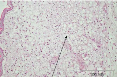
Fig. 3. Xanthoma histology. Aggregation of cholesterol in vacuoles.
According to our instructions, the owner mixed the antibiotic enrofl oxacin (30 mg/kg According to our instructions, the owner mixed the antibiotic enrofl oxacin (30 mg/kg PO q12h; Vetofl ok 10 %, Pliva, Zagreb, Croatia) in the bird’s drinking water (the patient PO q12h; Vetofl ok 10 %, Pliva, Zagreb, Croatia) in the bird’s drinking water (the patient was managed as an outpatient) for 5 days.
was managed as an outpatient) for 5 days. Histopathology.
Histopathology. For histological examination, the tissue sample was For histological examination, the tissue sample was fifi xed in 7% xed in 7% neutral buffered formalin, processed routinely using standard Hematoxilin-Eosin and neutral buffered formalin, processed routinely using standard Hematoxilin-Eosin and Ziehl-Nielsen staining.
Ziehl-Nielsen staining. Follow up.
Follow up. No postoperative complications, such as bleeding or automutilation, No postoperative complications, such as bleeding or automutilation, occurred. Skin sutures were removed 10 days postoperatively. The owner started giving occurred. Skin sutures were removed 10 days postoperatively. The owner started giving a low fat diet. In a period of one year, neither a reoccurrence of the skin xanthoma nor a low fat diet. In a period of one year, neither a reoccurrence of the skin xanthoma nor health disturbances were observed.
health disturbances were observed. Results
Results
Aspirated liquid content was examined. While there was no presence of bacteria, Aspirated liquid content was examined. While there was no presence of bacteria, some blood cells and many macrophages and heterophyles were found. The concentration some blood cells and many macrophages and heterophyles were found. The concentration of triglyceride (25.0 mmol/L) and cholesterol (10.0 mmol/L) was very high. Serum of triglyceride (25.0 mmol/L) and cholesterol (10.0 mmol/L) was very high. Serum cholesterol (10.3 mmol/L) and triglyceride (10.5 mmol/L) levels were also elevated. cholesterol (10.3 mmol/L) and triglyceride (10.5 mmol/L) levels were also elevated.
Histopathological examination:
Histopathological examination:
Numerous large macrophages with abundant foamy
cytoplasm (occasionally vacuolated) were diffused throughout the dermis (Fig. 4). Occasionally they were associated with cholesterol cleft formation (Fig. 3). A mass of heterophiles were scattered throughout. Some lakes of fi ne, amorphous, slightly acellular eosinophilic material were found between the cells. The Ziehl-Nielsen staining for acid-fast bacteria was negative.Discussion
Xanthomas (Greek xanthos, meaning “yellow”) are yellow colored tumors, deposition of cholesterol rich material in various body parts. In this bird, on the right side of the cloaca the yellowish, elliptic tumor measured 4×3 cm, the skin was damaged and the wound was bleeding. The development of lipomas or xanthomas is associated with obesity, hyperlipidemia, hypothyroidism, genetic factors, and systemic diseases such as diabetes mellitus. In addition, some medicines can cause disturbances in lipid metabolism. Serum cholesterol is sometimes elevated due to impossibility to metabolize all cholesterol. Skin, the largest organ, is susceptible to a wide variety of tumors as the body desperately tries to rid itself of foreign matter. Xanthomas are situated in the skin or in subcutis in the majority of cases, like in this one. A lipoma is benign tumor composed of fatty tissue, they are the most common form of soft tissue tumor.
Although the etiology of xanthoma is not identifi ed, there are other theories (except a fatt rich diet) such as the ingestion of toxic fat-soluble substances that might initiate
infl ammation and trauma (LATIMER, 1994). In humans, xanthomas frequently appear in tendons probably after tendon trauma or other factors. Tendons, arteries and corneas have a dense connective tissue matrix composed of collagen and proteoglycan. Proteins of this connective tissue matrix or the response to injury predisposes to cholesterol deposition. Xanthogranulomatous infl ammation is an uncommon but well recognizeddisease process in the human kidneys and gallbladder (SPINELLI et al., 2006), characterizedby aggregations of lipid-laden foamy macrophages and parenchymadestruction. It has also been reported less often at many othersites in the human body, including an ovary, fallopian tube, endometrium,lymph node, bone, lung, appendix (COZZUTO and CARBONE, 1988), colon (LO et al., 1996), stomach (KUBOSAWA et al., 2007) andparotid gland (COCCO et al., 2005).
FIRTH and MARAIS (2008) described inherited hypercholesterolemia in humans. Familial hypercholesterolemia (FH) is an autosomal dominantly inherited disorder characterized by elevated plasma low-density lipoprotein (LDL) cholesterol levels, tendon xanthoma (80% patients suffering from FH) and premature ischaemic heart disease that are controlled by medications. 55% of all patients were white and only 0.5% black. In this theory, a possible cause of xanthoma is a locally prevalent defective gene mutation.
Like any rapidly growing mass, they can ulcerate when bumped or when they outgrow their blood supply and cells die off. Xanthomas within the skin are frequently associated with infl ammation of underlying tissues (LATIMER, 1994)andif bones are involved, the affected bone may require amputation.
A xanthoma may develop in the body cavity of bird, near the trachea and compress it (MONKS et al., 2006) causing respiratory disorders.
The diagnosis of cutaneus xanthomas in this bird was based primarily on the characteristic histological appearance of foamy machrophages, giant cells, free cholesterol and lipid (MONKS et al., 2006).
Atypical multiple, papilliform, xanthomatous, cutaneus neoplasia in a goose (Anser anser) was treated by chemotherapeutic cisplatin with poor results. Serum lipid and lipoprotein analysis revealed a persistent hypercholesterolemia and hypertriglyceridemia without biochemical evidence of an underlying metabolic disease (JAENSCH et al., 2002).
Lipids in avian blood are similar in quantity and quality to those of mammals. Age, heredity, nutrition and various diseases affect cholesterol levels in avian blood. The normal serum cholesterol range for most birds is 2.7-5.5 mmol/L. Hypercholesterolemia has been associated with starvation, high levels of dietary fat, hypothyroidism and hepatic disease. In this case, hepatic and thyroid disorders were not observed by clinical and x-ray examination.
CHANUT et al. (2005) reported systemic xanthogranulomatosis in a 4-month-old domestic cat. Necropsy revealed multiple nodular lesions on most abdominal organs (liver, spleen, kidney, adrenal glands, mesentery, and colon). The cause was probably gene dysfunction because multiple nodules had appeared in the kitten.
GARNER et al. (1999) reported xanthomas in the brains of geckos that suffered from hydrocephalus. Tumor masses in the brain had caused disturbances and obstruction in cerebrospinal fl uid fl ow and production. Damage was mechanical. In this case, a functional problem was noted.
Multifocal cutaneous xanthoma lesions were reported in a goose (JAENSCH et al., 2002), just as our patient had suffered from hypercholesterolemia and hypertriglyceridemia. Due to normal serum lipid profi les in humans with xanthoma, it is unlikely that the pathogeneses of that syndrome seen in birds is the same. Previous injury also can be a trigger for cholesterol and triglyceride aggregation due to neovascularisation during the wound healing process. In this case, the female cockatiel was housed with a male thus small injury was possible. Both birds were fed the same diet, but were not the same age.
Conclusions
Although xanthomas are locally invasive tumors, they are not real neoplasia, it needs additional factors to develop. Such factors are hypercholesterolemia, hypertriglyceridemia, hormonal disorders and genetic. Prognosis of a solitary xanthoma is favorable; however, it depends on the affected organ and surgical approach. Chemotherapeutics are not helpful in xanthoma therapy. The surgical treatment of subcutaneous xanthoma has a favorable outcome. Recovery is rapid after volatile anesthesia. This type of tumor was diagnosed by the histopathological analysis of tissue.
References
BARRIE, J., T. D. WATSON (1955): Hyperlipidemia. In: Bonagura JD, editor. Kirk’s Current Veterinary Therapy XII. Saunders, Philadelphia. pp. 430-434.
CARPENTER, J. W. (2005): Exotic Animal Formulary, 3rd ed., Elsevier Saunders. Missouri. pp.
135-343.
CHANUT, F., M. A. COLLE, J. Y. DESCHAMPS, O. ALBARIC, M. WYERS (2005): Systemic xanthogranulomatosis associated with hyperchylomicronemia in a cat. J. Vet. Med. A. Physiol. Clin. Med. 52, 272-274.
COCCO, A. E., G. T. MacLENNAN, P. LAUVERTU, J. K. WASSMAN (2005): Xanthogranulamatous sialadenitis: a case report and literature review. Ear Nose Throat J. 84, 369-374.
COZZUTTO, C., A. CARBONE (1988): The xanthogranulomatous process. Xanthogranulomatous infl ammation. Pathol. Res. Pract. 183, 395-402.
FIRTH, J. C., A. D. MARAIS (2008): Familial hypercholesterolaemia: The Cape Town experience. S. Afr. Med. J. 98, 99-104.
GARNER, M. M., N. P. LUNG, S. MURRAY (1999): Xanthomatosis in geckos: fi ve case. J. Zoo Wildl. Med. 30, 443-447.
GRUNDY, S. M. (1999): Xanthomatoses and lipoprotein disorders. In: Fitzpatrick’s Dermatology in General Medicine. (Freedberg, I. M., A. Z. Eisen, K. Wolff, Eds.). McGraw-Hill. New York. pp. 1804-1811.
HALL, L. W., C. V. CLARKE (1991): Anaesthesia of birds. In: Veterinary Anaesthesia. 9th edition.
Saunders Philadelphia. pp. 345-347.
HARCOURT-BROWN, N. H. (2000): Psittacine birds. In: Avian Medicine. (Tully, T. N., M. P. C. Lawton, G. M. Dorrestein, Eds.). Butterworth Heinemann Oxford, Auckland, Boston, Johannesburg, Melbourne, New Delhi. pp. 112-142.
JAENSCH, S. M., R. BUTLER, A O’HARA, S. R. RAIDAL, K. WYATT (2002): Atypical multiple, papilliform, xanthomatous, cutaneous neoplasia in a goose (Anser anser). Aust. Vet. J. 80, 277-280. KRUTH, S. H. (1985): Lipid deposition in human tendon xanthoma. Am. J. Pathol. 121, 311-315. KUBOSAWA, H., K. YANO, K. ODA, M. SHIOBARA, K. ANDO, M. NUNOMURA, H.
SARASHINA (2007): Xanthogranulamatous gastritis with pseudosarcomatous changes. Pathol. Int. 57, 291-295.
LATIMER, K. S. (1994): Oncology. In: Avian Medicine: Principles and Application. (Ritchie, B. W., G. J. Harrison, L. R. Harrison, Eds.). Wingers. Florida. pp. 642-668.
LO, C. Y., T. G. LORENTZ, C. S. P. POON (1996): Xanthogranulomatous infl ammation of the sigmoid colon. Aust. N. Z. J. Surg. 66, 643-644.
MONKS, D. J., H. P. ZSIVANOVITS, J. E. COOPER, N. A. FORBES (2006): Successful treatment of tracheal xanthogranulomatosis in a red-tailed hawk (Buteo jamaicensis) by tracheal resection and anastomosis. J. Vet. Med. Surg. 20, 247-252
REAVILL, D (2004): Tumors of pet birds. Vet. Clin. North. Am. Exot. Anim. Pract. 7, 537-560. ROMANUCCI, M., D. MALATESTA, P. GUARDIANI, P. FRESCURA, L. DELLA SALDA
(2008): Xanthogranulomatous infl ammation of the small bowel in a dog. Vet. Pathol. 42, 207-211.
SCOTT, D. W., W. H. MILLER, C. E. GRIFFIN (2001): Muller and Kirk’s Small Animal Dermatology. 6th edition. Saunders, Philadelphia. pp. 873-874, 1148-1153.
SPINELLI, A., G. SCHUMACHER, A. PASCHER, E. LOPEZ-HANNINEN, H. AL-ABADI, C. BENCKERT, I. M. SAUER, J. PRATSCHKE, U. P. NEUMANN, S. JOANS, J. M. LANGREHR, P. NEUHAUS (2006): Extended surgical resection for xanthogranulomatous cholecystitis mimicking advanced gallbladder carcinoma. World J. Gastroenterol. 12, 2293-2296.
VOGELNEST, L. J. (2001): Cutaneous xanthomas with concurrent demodicosis and dermatophytosis in a cat. Aust. Vet. J. 79, 470-475.
Received: 27 April 2010 Accepted: 17 March 2011
LIPAR, M., D. HORVATEK, E. PRUKNER-RADOVČIĆ, A. GUDAN KURILJ,
LIPAR, M., D. HORVATEK, E. PRUKNER-RADOVČIĆ, A. GUDAN KURILJ,
B. RADIŠIĆ, D. VNUK, N. PRVANOVIĆ, B. ŠKRLIN, D. MATIČIĆ
B. RADIŠIĆ, D. VNUK, N. PRVANOVIĆ, B. ŠKRLIN, D. MATIČIĆ: Supkutani
ksantom u nimfe (Nymphicus hollandicus) - prikaz slučaja.Vet. arhiv 81, 535-543,
2011. SAŽETAK
U radu je opisan potkožni tumor u nimfe stare 17 godina. S lijeve strane kloake vlasnik je uočio žućkastu, eliptičnu tvorevinu veličine 4×3 cm. Iznad otekline koža je bila oštećena te je krvarila. Nakon kliničkog pregleda predloženo je kirurško odstranjenje tumora. Histopatološki je ustanovljen ksantom. U razdoblju od jedne godine ksantom se nije ponovo pojavio, a nisu zabilježene ni druge zdravstvene poteškoće.
Kjučne riječi: ksantom, nimfa, Nymphicus hollandicus, hiperlipidemija, metabolički poremećaji, potkožni