* Corresponding author. Tel/Fax : +62-411-586498 Email address : [email protected]
EFFECT OF HIRA PROTEIN ON TRANSCRIPTIONAL REGULATIONS OF GENES IN
VERTEBRATE CELLS
Ahyar Ahmad
Biochemistry and Biotechnology Lab., Department of Chemistry, Faculty of Natural Sciences, Hasanuddin University, Makassar 90245, Indonesia
Received 2 June 2008; Accepted 2 July 2008
ABSTRACT
For better understanding of DNA replicating-coupled chromatin assembly and transcription regulation in eukaryotes, we studied biochemical and genetic analysis of nuclear-related proteins from chicken DT40 cell lines. The genetic analysis of some nuclear proteins, such as HIRA and CAF-1, indicated that these proteins could play overlapping roles in chromatin dynamics and is consistent with the finding that HIRA protein exhibited binding ability to histones and ASF-1, as also ASF-1 bound directly with CAF-1p60. In this study, revealed not only that the N-terminal and C-N-terminal halves of HIRA mediate individually transcription repressions but also that even one of the seven WD dipeptide motifs and the LXXLL motif of HIRA are required for these mediations in vivo. Finally, we found that HIRA-mediated repression is sensitive to tricostatin TSA and it co-represses transcription together with HDAC-2. We believe our findings will contribute to a major break-through in future studies on the specific, individual roles of HIRA involved in numerous DNA-utilizing processes, through the formation and/or maintenance of the chromatin structure in vertebrate cells.
Keywords: histone regulator, transcriptional repression, tricostatin, chromatin
INTRODUCTION
Mammalian HIRA has been named for its homology to Hir1p and Hir2p (Hir is an acronym for histone regulator), two Saccharomyces cerevisiae transcriptional corepressors that strongly repress the transcription of one copy each of H2A and H2B histone genes located at the HTA1-HTB1 locus throughout most of the cell cycle, and transiently recruit a SWI/SNF chromatin remodelling complex, thereby leading to transcription activation at this locus at the G1/S transition [1, 2]. HIRA proteins with seven WD repeat domains exhibited binding ability as to histones [3] and ASF-1 [4], and then ASF-1 interacted directly with CAF-1p60 [5, 6]. The HIRA protein family was predicted to form a -propeller structure, which may be involved in protein-protein interactions, in its N-terminal half most similar to Hir1p and CAF-1p60 [1, 7-9]. The C-terminal half of HIRA similar to Hir2p is responsible for its binding ability as to homeodomains, Hir3p, Pax3 and core histones [3, 10]. Alterations in chromatin structure through the acetylation of core histones have been thought to be of fundamental importance in numerous DNA-utilizing processes [11-16]. The acetylation and deacetylation of core histones are catalyzed by HATs and HDACs, respectively. The members of the HAT and HDAC families play individual particular roles, and then participate in combination with one another in the processes of acetylation and/or deacetylation of core histones for the regulation of numerous cell functions. On the other hand, CAF-1p48 with seven WD repeat motifs, together with the p46 polypeptide, was found to
be associated with transcription repressor complexes of HDAC-1 and 2, and mSin3, Rb, or Mi2 plus MecP2.
Recently, we revealed not only that all seven WD dipeptide motifs of both HIRA and CAF-1p48 are necessary for their in vitro interaction but also that both the typical LXXLL motif of HIRA and a C-terminal region of HDAC-2 are necessary for their in vitro as well as in vivo interactions [17]. In this study, we revealed not only that the N-terminal and C-terminal halves of HIRA mediate individually transcription repressions but also that even one of the seven WD dipeptide motifs and the LXXLL motif of HIRA are required for these mediations in vivo. Finally, we also found that HIRA-mediated repression is sensitive to TSA and HIRA co-represses transcription together with HDAC-2.
MATERIALS AND METHODS
Material
The pM, pG5probasinLuc, pRL-CMV plasmids and Dual-LuciferaseR assay system were purchased from Promega. FuGENE 6 transfection reagent were purchased from Roche Molecular Biochemicals. DMEM medium and fetal bovine serum were purchased from GIBCO, Invitrogene Life Technologies.
Plasmids construction
Indo. J. Chem., 2008, 8 (3), 454 - 458
455
followed by treatment with T4 polymerase. Resultant blunt-ended plasmids, the NdeI/XhoI blunt-ended fragments containing the full-length HIRA and its derivative cDNAs were excised and subcloned into the SmaI site of the pM vector to constructed pMHIRA, pMHIRA-(1-443), pMHIRA-(444-1019), pMHIRA-(WT7AA), pMHIRA-(L993A), pMHIRA-(WT7AA, L993A), pMHIRA-(1-443, WT7AA) and pMHIRA-(444-1019, L993A). Each end point of the deletions and point mutation plasmids was determined by sequence analysis (ABI 373A Sequencer DNA from Applied Biosystems Inc.) by the dye terminator method.
Cell culture
Adherent cell cultures of HeLa were maintained in DMEM supplemented with 10% fetal bovine serum (GIBCO, Invitrogene Life Technologies) and grown at 37oC in 5% CO2 in air in a humidified incubator.
Luciferase assays
HeLa cells (2 X 10⁴) were cultured in 24-well dishes for 24 hr until 60-70% confluence. HeLa cells (60-70% confluence, 24-well plate) were cotransfected with 1.6 g of pM or pMHIRA and its derivates constructs, 0.4 g of pG5probasinLuc, and 10 ng of pRL-CMV as internal control in 500 l of DMEM containing 10% fetal bovine serum by lipofection with FuGENE 6 transfection reagent (Roche Molecular Biochemicals). After 24 hr, the medium was replaced, and cells were harvested and assayed for luciferase activity 36-48 hr after transfection. Luciferase assays were performed with the Promega Dual-LuciferaseR Assay System according to the manufacturers instructions. The reaction tube was discarded and procceded to the next samples assay. Each transfection was performed in duplicate and repeated at least three times. The relative luciferase activity was normalized by dividing the measurement for the firefly luciferase activity with that for the renilla luciferase activity.
RESULT AND DISCUSSION
We previously reported that all the WD dipeptide motifs in the N-terminal half and the LXXLL motif in the C-terminal half of HIRA were essential for its in vitro and in vivo interactions with CAF-1p48 and HDAC-2, LXXLL motif are responsible for the transcription repression in vivo, HIRA and its derivates were fused to the DNA-binding domain of GAL4, and then repressor
activity of the fusion proteins was assayed in HeLa cells. A reporter plasmid, containing 5 copies of the GAL4 binding site (5X GAL4 probasinLuc), the cloned upstream of the SV40 promoter and the direction of expression of luciferase, was used to monitor the activity of GAL4-HIRA fusion proteins. Fold repression activity was determined as a luciferase activity relative to the basal transcription activity of the GAL4 (BD) reporter alone as a control.
HIRA, its N-terminal and C-terminal halves, and their single and/or double missense mutant derivats as to WD dipeptide and/or LXXLL motifs were fused to GAL4, and transiently expressed in HeLa cells, and then repressor activities were assayed as by luciferase activity. Figure 1 showed not only that GAL4-HIRA could repress the promoter activity to about 40% that of the control BD protein (lanes 2 and 1) but also that GAL4-HIRA-(1-443) and GAL4-HIRA-(444-1019) exhibited certainly repression activity, i.e. their repression activities were about 50% and 75%, respectively (lanes 3 and 4). To address whether or not the WD dipeptide motifs in N-terminal half and the LXXLL motif in C-terminal half of HIRA were responsible for these repressions, we co-transfected the high basal reporter plasmid 5X GAL4 probasinLuc along with GAL4-HIRA point mutations in the WD dipeptide and LXXLL motifs. The repression activity of the double point mutation (WT7AA) of the WD repeat motif was about 75% (lane 5), similar to that of GAL4-HIRA-(444-1019), indicating that this activity was due to the C-terminal half of HIRA.
Figure 1. Necessity of WD dipeptide and LXXLL motifs in N-terminal and C-terminal halves of HIRA for the mediation of transcription repression in vivo.
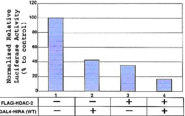
Figure 2. HIRA protein requires the HDAC-2 enzyme activity for transcriptional repression.
Mi2 plus MecP2 [20]. Further studies are necessary to resolve this repression mechanism.
Figure 1 showed not only that the N-terminal and C-terminal halves of HIRA mediate individually transcription repressions but also that even one of the seven WD dipeptide motifs and the LXXLL motif of HIRA are required for these mediations in vivo. On the other hand, HDAC activity has been shown to repress transcription [21]. Since HIRA associates with histone deacetylase activity and binds with HDAC-2 via its LXXLL motif in vivo [19], we evaluated whether HIRA
Indo. J. Chem., 2008, 8 (3), 454 - 458
457
synergistic repressive effect on transcription (Figure 2, lane 4).
We next asked whether the repression mediated by GAL4-HIRA could be relieved by treatment with the specific HDAC inhibitor TSA. HeLa cells were transfected as in Figure 2. Cells were treated (lanes 2 and 4) or not (lanes 1 and 3) with the HDAC inhibitor TSA (0.4 M) harvested after 16 hr, and then assayed
for luciferase activity. The basal activity of the reporter is normalized to a value of 100%. As depicted in Figure 3, the repressive effect observed with GAL4-HIRA on reporter activity was relieved by the addition of TSA. Collectively, these results indicate that HIRA contains a transcriptional repressor domain, encompassing the LXXLL motif, and requires the HDAC-2 activity to exert this repressive effect.
Figure 3. HIRA-mediated transcriptional repression is sensitive to histone deacetylases inhibitor TSA.
As illustrated in Figure 4, if the C-terminal of HIRA bound to HDAC caused deacetylation of chromatin induces on close conformation that allows the down regulation of transcription and expression of gene activity. Oppositely, if N-terminal region of HIRA bound to p48 subunit caused acetylation of chromatin induces an open conformation that allows the transcription machinery acces to promoter and up-regulation of transcription and expression of gene activity. Thus, these HIRA-participating interactions with CAF-1p48 and HDAC-2 (and probably other HIRA-carrying complexes) should be involved directly or indirectly in chromatin assembly or maintenance and alterations of chromatin structure, involving gene transcription. Our preliminary analysis of the HIRA-deficient DT40 mutant obtained showed not only that the HIRA-deficiency caused delayed cell growth, but also that the N-terminal and C-terminal halves of HIRA were oppositely involved in transcription regulation of various cell cycle-related genes [22]. Analysis of the HIRA-deficient and AFS1 conditional DT40 mutant [23], together with the CAF-1-deficient conditional mutant cells [24], will be of powerful tools for understanding the role of HIRA and the other terminal half of HIRA mediate transcription repression in vivo, but also that HIRA-mediated repression is sensitive to TSA and it co-represses transcription together with HDAC-2. We believe our findings will contribute to a major break-through in future studies on the specific, individual roles of HIRA, as well as CAF-1 involved in numerous DNA-utilizing processes, through the formation and/or maintenance of the chromatin structure in higher eukaryotes, including vertebrate cells.
ACKNOWLEDGEMENT
The author wishes to express sincere gratitude to Dr. Tatsuo Nakayama and Dr. Yasunari Takami for excellent support, advice, and inspiration for this research. This work was supported in part by a Grant-in-Aid from the Ministry of Education, Sciences, Sports and Culture of Japan.
REFERENCES
1. Spector, M.S., Raff, A., DeSilva, H., Lee, K., and Osley, M.A., 1997, Mol. Cell. Biol., 17, 545-552 2. Dimova, D., Nackerdien, Z., Furgeson, S., Eguchi,
S., and Osley, M.A., 1999, Mol. Cell, 4, 75-83
3. Lorain, S., Quivy, J.P., Monier-Gavelle, F., Scamps, C., Lecluse, Y., Almouzni, G., and Lipinski, M., 1998, Mol. Cell. Biol., 18, 5546-5556.
4. Sharp, J.A., Fouts, E.T., Krawitz, D.C., and Kaufman, P.D., 2001,Curr Biol., 11, 463-473 5. Tyler, J.K., Collins, K.A., Prasad-Sinha, J., Amiott.
7. Roberts, C., Daw, S.C., Halford, S., and Scambler, P.J., 1997, Hum. Mol. Genet., 6, 237-245 and Scambler, P.J., 1998, Nat. Genet., 20, 74-77. 11. Wolffe, A.P., and Pruss, D., 1996, Cell, 84,
817-819
12. Pazin, M.J., and Kadonaga, J.T., 1997, Cell, 89, 325-328
13. Pennisi, E., 1997, Science, 275, 155-157
14. Wolffe, A. P., Wong, J. and Pruss, D. 1997, Genes Kao, G.D., Ye T.J., Harper, J.W., and Adams, P.D., 2002, Mol. Cen, ll. Biol., 22, 7459-7472.
23. Sanematsu, F., Takami, Y., Barman, H.K., Ono, T., Fukagawa, T., Shibaraha, K.I., and Nakayama, T., 2006, J. Biol. Chem., 281, 13817-13827.