Molecular detection and differentiation of canine hemoplasma
infections using RFLP-PCR in dogs in southern Iran
Mohammad A. Hasiri*, Hassan Sharifiyazdi, and Tayebeh MoradiDepartment of Clinical studies, school of Veterinary Medicine, shiraz University, shiraz, iran ________________________________________________________________________________________ HASiri, M. A., H. SHArifiyAzdi, T. MorAdi: Molecular detection and differentiation of canine hemoplasma infections using RFLP-PCR in dogs in southern Iran. Vet. arhiv 86, 529-540, 2016.
ABSTrACT
Two hemoplasma species are known in dogs: Mycoplasma haemocanis (Mhc) and Candidatus Mycoplasma haematoparvum (CMhp). The aim of the present study was to develop a novel restriction fragment length polymorphism (RFLP)-PCR method based on the 16S rDNA gene, using endonuclease hind iii, for detection and differentiation of canine hemoplasmas. Also, analysis of risk factors, clinical features and hematologic changes of positive cases was performed in dogs living in the Shiraz area of Iran. Blood samples were collected
from anemic (packed cell volume (PCV) ≤35; n = 26) and control dogs (PCV >35; n = 27) and were examined
for the presence of canine hemoplasmas, using RFLP-PCR and 16S rDNA Sanger sequencing. The presence
of Mhc (4 out of 53 cases; 7.5%) and CMhp (3 out of 53 cases; 5.7%) was confirmed by RFLP analysis of the amplified 16S rDNA and sequencing. No association was found between hemoplasma infection and
anemia, health status, age, breed, gender, type of housing or the presence of other dogs in this study. Only the platelet number in Mhc infected dogs was statistically higher compared to CMhp positive and hemoplasma negative dogs. The present report documents the occurrence of Mhc and CMhp in southern Iran, and these
hemotropic Mycoplasma infections must be expected even in the absence of clinical symptoms or hematologic abnormalities in dogs. For the first time, it has been indicated that RFLP-PCR assay is able to successfully
distinguish hemotropic Mycoplasma in dogs.
Key words: hemoplasma, Mycoplasma haemocanis (Mhc), Candidatus Mycoplasma haematoparvum (CMhp), dog, Iran
________________________________________________________________________________________
Introduction
Hemotropic Mycoplasma (also called hemoplasma) species are facultative intracellular, cell wall-less, epierythrocytic parasites, comprising the group of uncultivable Mycoplasma species (MeSSICk, 2004).
*Corresponding author:
M. Abbaszadeh Hasiri, Assistant Professor of Small Animal Internal Medicine, Department of Clinical Studies, School of Veterinary Medicine, Shiraz University, P.O. Box 1731, 71345, Shiraz, Iran, Phone: +98 71 3228 8660, +98 91 7707 0881, Fax: +98 71 3228 6950, E-mail: abbaszadeh@shirazu.ac.ir; vetmail@gmail.com
Two species of hemotropic mycoplasma that infect dogs are known so far: Mycoplasma hemocanis (Mhc) and Candidatus Mycoplasma hematoparvum (CMhp)
(BARKER et al., 2009; GREENE, 2012). Transmission through blood sucking arthropods,
such as the tick rhipicephalus sanguineus or by ingestion (or transfusion) of infected blood have been proven (DANTAS-TORReS, 2008; WENGI et al., 2008). These extracellular
parasites attach to the surface of canine erythrocytes, causing hemolytic anemia, mostly
through extravascular destruction of erythrocytes by the mononuclear phagocyte system
(GREENE, 2012). Infection with these hemoplasmas generally only induces clinically
significant anemia in splenectomized or immunocompromised dogs, although latent
infections may cause subclinical anemia (BRINSON and MESSICK, 2001; GREENE,
2012). Most non-splenectomized dogs infected with hemoplasma do not develop clinical
evidence of disease and do not have sufficient numbers of organisms present in the blood to be recognized during routine blood film examinations. Therefore, molecular techniques
that are simpler, faster and usually more sensitive have been developed for hemoplasma species detection (CRIADO-FORNELIO et al., 2003; GREENE, 2012).
The objective of this study was the molecular characterization of hemotropic Mycoplasma species and differentiation of Mhc and CMhp in a population of 26 anemic
and 27 non-anemic (PCV ≥ 35) dogs, in the south of Iran by RFLP-PCR. In addition,
complete blood count (CBC) parameters and epidemiological factors were evaluated to determine if there was an association between them and Mycoplasma infection status.
Materials and methods
Sample collection: In total, 26 anemic dogs (PCV ≤35) and 27 control dogs (PCV >35) were selected from cases referred to the School of Veterinary Medicine, Shiraz
University, Fars province, Iran during a one-year period (October 2013 to September 2014). Complete histories, including: age, breed, gender, type of housing (indoor, outdoor), presence of another dog in the household and whether they were symptomatic
or not, were recorded. A physical examination, which included TPR (temperature, pulse
and respiratory rate), was performed. A blood sample (4 mL) was obtained via cephalic vein puncture, and placed into tubes containing eDTA.
PCr assay. Amplification of the 16S rDNA sequences for both Mhc and CMhp was performed using the following primers: 5’- GGCCCATATTCCT(AG)CGGGAAG -3’
and 5’- AC(AG)GGATTACTAGTGATTCCA-3’ (HOeLzLe et al., 2011). These primers
amplified a ~1000 bp PCR product encompassing the polymorphic site for the Hind III restriction enzyme. The PCR reaction (25 μL) was performed in 10 mM Tris-HCl, pH 8.4, 50 mM KCl, 2 mM MgCl2, 100 μM of each dNTP, 20 pmol of each primer (Cinnagen Inc., Tehran, Iran), and 2 U Taq DNA polymerase (Cinnagen Inc., Tehran, Iran) using 2 μL of DNA extracted from the blood as a template. The target gene was amplified using a DNA
followed by 45 amplification cycles (94 °C for 45 s, 55 °C for 45 s, 72 °C for 45 s) and a final extension cycle (72 °C for 5 min). Finally, the PCR products from positive cases
were sequenced using the Sanger method (ABI 3730 Capillary DNA analyzer, Applied
Biosystems, Foster City, CA, USA) to find possible existing patterns of polymorphism in the 16S rDNA gene. Sequences from the hemoplasma species identified in this study
were used in the phylogenetic analyses, along with other related sequences of hemotropic Mycoplasma, with the Clustal W and neighbor-joining methods using MEGA4 software
(TAMURA et al., 2007).
Finally, on the basis of the obtained pattern of polymorphism in this gene in Iran
and data already existing in GenBank, an appropriate PCR-RFLP method was designed.
Two canine hemoplasma sequence analysis showed a polymorphism for restriction
enzyme (Hind III; 5’-A/AGCTT- 3’). RFLP analysis predicted a unique fragment without
digestion for Mhc, so this species could be clearly distinguished from CMhp, which produced two fragments of 873 bp and 123 bp. Accordingly, PCR products were digested
with endonucleases Hind III (Jena Bioscience, Germany) to differentiate two canine hemoplasma agents. Enzyme digestion was performed in a 20 μL mixture containing 12 μL of the 16S rDNA PCR product, 0.5 μL (2 U) of enzyme, 2 μL buffer and 5.5 μL of
distillated water at 37 °C for at least 5 hrs. After digestion with Hind III, the presence of
PCR products was determined by electrophoresis of 10 μL of each reaction product in 1.2% (w/v) agarose gel with trisborate EDTA electrophoresis buffer, and visualized under
UV light.
statistical analysis. The data were analyzed using SPSS software (version 15) by
means of chi-square, Fischer exact test and Mann-Whitney test. Differences between means were considered statistically significant if the P value was less than 0.05.
Results
The PCR yielded an amplicon of approximate length of 1001 bp and 975 bp for CMhp and Mhc, respectively. All positive PCR products (n = 7) were purified and sequenced
using both forward and reverse primers. Partial 16S rDNA sequence analysis revealed
that 4 (7.5%) and 3 (5.7%) samples were positive for Mhc and CMhp, respectively. All samples positive by the PCR assay were subjected to RFLP analysis of the amplification
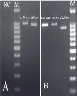
product. There was a complete correlation between the results obtained with the sequencing method and RFLP typing design. Fig. 1 shows the different RFLP patterns for two species of Mhc and CMhp after digestion with Hind III restriction enzyme. No atypical or ambiguous RFLP patterns occurred, and all positive samples could be correctly assigned to a species according to the RFLP pattern. The 16S rDNA sequences obtained
in this study were submitted to the GenBank, with accession numbers kU886262 to
Fig. 1. (A) Gel electrophoresis of PCR products amplified from positive cases for Candidatus Mycoplasma haematoparvum (CMhp) and Mycoplasma haemocanis (Mhc) compared to molecular marker (M) and negative control (NC). (B) RFLP patterns after digestion of PCR product using Hind III for Mhc and CMhp compared to uncut PCR product and molecular marker
(M).
Sequencing results showed that all the isolates for Mhc (n = 4) shared similar sequences in amplified regions of the 16S rDNA gene (KU886264), whereas a point
mutation of T to C in position 902 (based on our accession numbers) was detected in Iranian CMhp, and two genotypes A(kU886262) and B (kU886263) of these organisms were characterized in Iran. Furthermore, multiple alignments of other isolates of CMhp
reported in GenBank showed additional polymorphisms base d on 16S rDNA analysis
(Table 1).
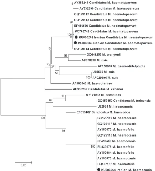
Phylogenetic analysis of the sequence alignment using a neighbor-joining algorithm is shown in Fig. 2.
Out of 53 blood samples (27 non-anemic dogs and 26 anemic dogs), 7 (13.2%) were
positive for one Mycoplasma species. The results showed that only 3 (11.53%) anemic dogs were detected as positive cases. Unexpectedly, 4 (14.81%) of the non-anemic dogs were also positive by PCR assay. The lowest hematocrit levels (10.5% and 19.3%) were
of examined dogs based on PCR results are presented in Tables 2 and 3. Factors such
as age, breed, gender and the presence of another dog in the same household, showed
no significant differences between infected or non-infected dogs (P>0.05). Although six (85.7%) of the infected dogs were kept outdoors (in a yard or garden), the type of housing (indoor, outdoor) was not statistically significant either (P = 0.40).
Table 1. Intraspecies variation in the IranianCMhp 16S rDNA sequences (genotype A: kU886262
and genotype B: KU886263) and other existing CMhp sequences in the GenBank database
Nucleotid
Positions Main Base
Nuleotide polymorphisms in 16S rDNA for Candydatus Mycoplasma hematoparvum CMhp k C762746 AY383241 GQ1291 13 GQ1291 12 GQ1291 14
eF416569 AY532390 kU886262 kU886263
314 G G G G G A G G G G 379 T T T T T T T G T T 493 T T T T T T T G T T 503 T T T T T T T G T T 745 G G G G G G G T G G 746 T T T T T T T G T T 751 T T T T T T T G T T 902 T C T T T T T T C T
Table 2. Comparison of hematological variables (Mean ± Se) in dogs with haemotropic
Mycoplasma (either Mhc or CMhp) infection and PCR negative dogs
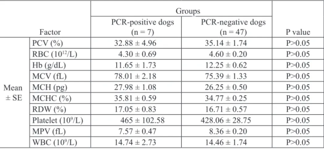
Factor Groups P value PCR-positive dogs (n = 7) PCR-negative dogs (n = 47) Mean ± Se PCV (%) 32.88 ± 4.96 35.14 ± 1.74 P>0.05 RBC (1012/L) 4.30 ± 0.69 4.60 ± 0.20 P>0.05 Hb (g/dL) 11.65 ± 1.73 12.25 ± 0.62 P>0.05 MCV (fL) 78.01 ± 2.18 75.39 ± 1.33 P>0.05 MCH (pg) 27.98 ± 1.08 26.25 ± 0.50 P>0.05 MCHC (%) 35.81 ± 0.59 34.77 ± 0.25 P>0.05 RDW (%) 17.05 ± 0.83 16.71 ± 0.57 P>0.05 Platelet (109/L) 465 ± 102.58 428.06 ± 28.75 P>0.05 MPV (fL) 7.57 ± 0.47 8.36 ± 0.20 P>0.05 WBC (109/L) 14.74 ± 2.73 14.46 ± 1.74 P>0.05
Fig. 2. Phylogenetic tree based on 16S rDNA sequences, constructed according to the Neighbor-Joining method, showing the position of Iranian Mhc and CMhp compared to other hemotropic
Mycoplasma species. Bootstrap values, calculated from 1,000 repetitions, are placed at each branch point.
The number of platelets were significantly increased in Mhc infected dogs compared to the CMhp positive dogs (P = 0.01) (Table 3).
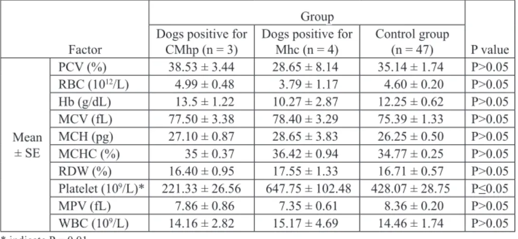
Table 3. Comparison of hematological variables (Mean ± Se) in dogs with Mhc, CMhp and negative cases
Factor
Group
P value Dogs positive for
CMhp (n = 3) Dogs positive for Mhc (n = 4) Control group(n = 47)
Mean ± Se PCV (%) 38.53 ± 3.44 28.65 ± 8.14 35.14 ± 1.74 P>0.05 RBC (1012/L) 4.99 ± 0.48 3.79 ± 1.17 4.60 ± 0.20 P>0.05 Hb (g/dL) 13.5 ± 1.22 10.27 ± 2.87 12.25 ± 0.62 P>0.05 MCV (fL) 77.50 ± 3.38 78.40 ± 3.29 75.39 ± 1.33 P>0.05 MCH (pg) 27.10 ± 0.87 28.65 ± 3.83 26.25 ± 0.50 P>0.05 MCHC (%) 35 ± 0.37 36.42 ± 0.94 34.77 ± 0.25 P>0.05 RDW (%) 16.40 ± 0.95 17.55 ± 1.33 16.71 ± 0.57 P>0.05 Platelet (109/L)* 221.33 ± 26.56 647.75 ± 102.48 428.07 ± 28.75 P≤0.05 MPV (fL) 7.86 ± 0.86 7.35 ± 0.61 8.36 ± 0.20 P>0.05 WBC (109/L) 14.16 ± 2.82 15.17 ± 4.69 14.46 ± 1.74 P>0.05 * indicate P = 0.01 Discussion
Hemoplasmas are obligate epierythrocytic organisms that attach to the erythrocytes of dogs. In some cases, hemoplasma infection is associated with hemolytic anemia of variable severity, ranging from nonclinical hemolysis to severe anemia (WILLI et al.,
2007).
In this study, we applied a novel RFLP-PCR for detection and differentiation of two distinct haemotropic mycoplasma in dogs from Shiraz, Iran.
The results showed that the discriminatory power of RFLP analysis of 16S rDNA was perfect and this method allows the accurate typing of canine hemotropic mycoplasma
species. To our knowledge, this is the first discrimination of the hemotropic mycoplasma
in a dog population by a RFLP approach.
Despite the identification of canine hemoplasma agents in dogs in Iran (TORkAN et
al., 2014), 16S rDNA gene sequencing and phylogenetic analysis of both causative agents
had not been identified in this area. Based on this, a decision was made to check the
phylogenetic position of both canine hemotropic mycoplasmas based on the 16S rDNA gene. Sequencing results and the RFLP PCR pattern of the samples obtained in the present study, with those previously reported for different hemotropic mycoplasma, revealed that
This study represents an expansion of the phylogenetic data available for
mycoplasmas, including hemoplasmas, based on 16S rDNAsequence data. A previous
phylogenetic study revealed that hamoplasmas had been classified into Haemominutum
and Haemofelis clusters based on 16S rDNA analyses (keNNY et al. 2004). Similarly, our phylogenetic tree based on this molecular marker showed that Iranian canine hemotropic Mycoplasma species were divided into two major groups. Further studies will be required, including phylogeny with non-16S rRNA gene sequences, to describe more fully the relationship between the canine and feline isolates.
In addition, the comparison between the sequence alignments of the Iranian 16S rDNA for the CMhp undertaken in this study, showed the occurrence of genetic polymorphisms and heterogeneity in this ribosomal locus, in which two different genotypes of CMhp (A and B) were characterized in Iran. Despite the occurrence of this genetic variation in
Iranian CMhp, 16S rDNA sequences in Mhc, were completely conserved (100% identity)
in the present study.
In the current study, 13.2% of samples were positive for hemoplasma. It was
hypothesized that this high prevalence may be associated with the presence of rhipicephalus sanguineus, a proposed vector for canine hemoplasmas. CMhp and Mhc
infections were diagnosed at 5.7% and 7.5%, respectively. Similarly, TORkAN et al. (2014) found the prevalence of CMhp and Mhc infections among dogs in Isfahan (central
Iran) was 10% and 13%, respectively (TORkAN et al., 2014). A survey conducted in
Brazil to compare infection rates for Mhc between urban and rural areas showed that
twenty (11.3%) out of 176 dogs living in rural areas were positive, whereas 6 of 104
(5.8%) dogs from urban areas harbored the organism (SANTOS, 2008). In France, blood
samples from dogs (n = 460) were analyzed by PCR to evaluate hemoplasma infection status. Seventy-one dogs (15.4%) were positive. Of these, 44 (9.6%) were infected with an organism closely related to CMhp; 15 (3.3%) were infected with Mhc, and 12 dogs
(2.6%) were infected with both organisms (keNNY et al., 2004). Although both canine
hemoplasma species were detected in the population examined in the present study, they
were not detected in the same dog concurrently. The present results are in agreement with previous studies in Sudan and France, where Mhc was more prevalent than CMhp
(INOKUMA et al., 2006; KENNY et al., 2004). In another study conducted in europe, canine
hemoplasmas were detected in 82 (9.6%) out of 850 blood samples (NOVACCO et al.,
2010). The hemoplasma prevalence was significantly higher in Portugal (40%) than in
Italy (9.5%) and Spain (2.5%) (NOVACCO et al., 2010).
In a recent investigation performed in the United States, hemoplasma prevalence was
1.3% (7 out of 506), with Mhc and CMhp prevalence of 0.6% and 0.8%, respectively
(COMPTON et al., 2012). Difficulties exist in comparing results from different studies
types of animals are sampled in different studies (e.g., variable percentages of healthy and
sick animals, demographics). Vector distribution could also influence results (
DANTAS-TORReS, 2008).
In the present research, there was no association between canine hemotropic mycoplasma infection with the presence of another dog in the household, fever, respiratory signs such as sneezing or coughing, and gastrointestinal signs such as vomiting or diarrhea.
Some research indicates that kennel-kept dogs were significantly more frequently infected
with hemoplasmas than dogs living in private homes (NOVACCO et al., 2010). Although in
the present study six (85.7%) of the infected dogs were kept outdoors (in a yard or garden), type of housing (indoor, outdoor) was not statistically significant either. However a larger sample size is required for accurate statistical analysis to evaluate this finding.
Five infected dogs were male and 5 positive cases were older than 2 years old, but
the gender or age factors had no significant effect on infection occurrence. In contrast to
our results, NOVACCO et al. (2010) showed that PCR-positive dogs for canine hemotropic
mycoplasma infections were significantly younger than PCR negative dogs (NOVACCO
et al., 2010). In the present study, although breed type or general signs (depression and/
or lethargy) showed no statistical differences between infected or non-infected dogs,
infection prevalence was higher in mixed breed dogs (than pure breeds) or those that had
depression and/or lethargy. NOVACCO et al. (2010) found that crossbred dogs are more
likely to be PCR-positive than purebred dogs. Similar to our study, some research showed that the prevalence of canine hemoplasma infections did not differ between clinically healthy and sick dogs (COMPTON et al., 2012; NOVACCO et al., 2010) and there was no association between anemia and hemoplasma infection (NOVACCO et al., 2010; ROURA et
al., 2010). Some of these disagreements between studies may also be related to different sample sizes and the various stages of infection. As the stages of infection in the PCR-positive dogs were unknown, we could not determine whether the dogs represented chronic carriers that may have recovered from acute illness and thus lacked clinical signs, or acutely infected animals.
Although the anemia and infection had no statistical relation in our results, the fewest
hematocrits (10.5% and 19.3%) were detected in dogs with Mhc infection. This could
be attributed to the more severe hemolytic anemia induced by Mhc compared to CMhp
during the acute phase of infection. Interestingly, statistical analysis showed a significant
increase in platelet count in Mhc infected dogs (647.75×10⁹/L) compared to CMhp
positive dogs (221.33×10⁹/L) (P = 0.01). Platelet elevation may be related to secondary
Conclusion
We conclude from the present study that both forms of canine hemoplasma infection must be expected in the south of Iran. The hemoplasma infected dogs may not exhibit clinical signs clearly attributable to a specific canine haemoplasma agent. Accordingly,
the RFLP-PCR presented in this study provides a rapid and easy-to-use method in a wide variety of laboratories, to detect and discriminate between two species of haemoplasma in dogs.
_______
Acknowledgements
The researchers would like to thank the Research Council of Shiraz University and the School of Veterinary
Medicine, Shiraz University, for their financial support of this study (Grant No. 87-GR-VT-47).
References
BARkeR, e. N., S. TASkeR, M. DAY, S. M. WARMAN, K. WOOLLEY, R. BIRTLES, K. C. GEORGES, C. D. EZEOKOLI, A. NEWAJ-FYZUL, M. D. CAMPBELL, O. A. E. SPARAGANO, S. CLEAVELAND, C. R. HELPS (2009): Development and use of real-time
PCR to detect and quantify Mycoplasma hemocanis and ‘‘Candidatus Mycoplasma hemato-parvum’’ in dogs. Vet. Microbiol. 140, 167-170.
BRINSON, J. J., J. B. MeSSICk (2001): Use of a polymerase chain reaction assay for detection of
hemobartonella canis in a dog. J. Am. Vet. Med. Assoc. 218, 1943-1945.
BUSS, D. H., A. W. CASHELL, M. L. O’CONNOR, F. RICHARDS II, L. D. CASE (1994): Occurence, etiology, and clinical significance of extreme thrombocytosis: a study of 280 cases.
Am. J. Med. 96, 247-253.
COMPTON, S. M., R. G. MAGGI, E. B. BREITSCHWERDT (2012): Candidatus Mycoplasma
haematoparvum and Mycoplasma haemocanis infections in dogs from the United States. Comp. Immunol. Microbiol. Infect. Dis. 35, 557-562.
CRIADO-FORNELIO, A., A. MARTINEZ-MARCOS, A. BULING-SARANA, J. C.
BARBA-CARReTeRO (2003): Presence of Mycoplasma hemofelis, Mycoplasma hemominutum and piroplasmids in cats from southern europe: A molecular study. Vet. Microbiol. 93, 307-317. DANTAS-TORReS, F. (2008): The brown dog tick, rhipicephalus sanguineus (Latreille, 1806)
(Acari: Ixodidae): from taxonomy to control. Vet. Parasitol. 152, 173-185.
GREENE, C. E. (2012): Infectious diseases of the dog and cat. 3rd ed. Philadelphia, USA: Saunders,
310-319.
HOELZLE, K., M. WINKLER, M. M. KRAMER, M. M. WITTENBRINK, S. M. DIECKMANN,
L. e. HOeLzLe (2011): Detection of Candidatus Mycoplasma haemobos in cattle with anaemia. Vet. J. 187, 408-410.
HULMe-MOIR, k. L., e. N. BARkeR, A. STONeLAke, C. R. HeLPS, S. TASkeR (2010): Use of real-time quantitative polymerase chain reaction to monitor antibiotic therapy in a dog with
INOkUMA, H., M. OYAMADA, B. DAVOUST, M. BONI, J. DeReURe, B. BUCHeTON,
A. HAMMAD, M. WATANABE, K. ITAMOTO, M. OKUDA, P. BROUQUI (2006):
epidemiological survey of ehrlichia canis and related species infection in dogs in eastern Sudan. Ann. NY. Acad. Sci. 1078, 461-463.
KENNY, M. J., S. E. SHAW, F. BEUGNET, S. TASKER (2004): Demonstration of two distinct
hemotropic mycoplasmas in French dogs. J. Clin. Microbiol. 42, 5397-5399.
MeSSICk, J. B. (2004): Hemotrophic mycoplasmas (hemoplasmas): a review and new insights into pathogenic potential. Vet. Clin. Pathol. 33, 2-13.
NOVACCO, M., M. L. MELLI., F. GENTILINI, F. MARSILIO, C. CECI, M. G. PENNISI, G. LOMBARDO, A. LLORET, L. SANTOS, T. CARRAPICO, B. WILLI, G. WOLF, H. LUTZ,
R. HOFMANN-LeHMANN (2010): Prevalence and geographical distribution of canine hemotropic mycoplasma infections in Mediterranean countries and analysis of risk factors for infection. Vet. Microbiol. 142, 276-284.
ROURA, X., I. R. PeTeRS, L. ALTeT, M. D. TABAR, e. N. BARkeR, M. PLANeLLAS, C.
R. HELPS, O. FRANCINO, S. E. SHAW, S. TASKER (2010): Prevalence of hemotropic
mycoplasmas in healthy and unhealthy cats and dogs in Spain. J. Vet. Diagn. Invest. 22, 270-274.
SANTOS, A. P. (2008): Infecção por hemoplasmas em felinos domésticos da região de Porto
Alegre, Rio Grande do Sul, Brasil. Porto Alegre, f. Dissertação (Doutorado)-Universidade Federal do Rio Grande do Sul, p. 162.
SYkeS, J. e. (2014): Canine and Feline Infectious Diseases. 1st ed., elsevier, pp 391-392. TAMURA, K., J. DUDLEY, M. NEI, S. KUMAR (2007): MEGA 4: Molecular Evolutionary
Genetics Analysis (MEGA) software version 4.0. Mol. Biol. Evol. 24, 1596-1599.
TORkAN, S., S. J. ALDAVOOD, A. SekHAVATMANDI, S. MOSHkeLANI (2014): Detection of haemotropic Mycoplasma (haemobartonella) using multiplex PCR and its relationship with
epidemiological factors in dogs. Comp. Clin. Pathol. 23, 669-672.
WENGI, N., B. WILLI, F. S. BORETTI, V. CATTORI, B. RIOND, M. L. MELI, C. E. RUSCH, H.
LUTz, R. HOFMANN-LeHMANN (2008): Real-time PCR-based prevalence study, infection follow-up and molecular characterization of canine hemotropic mycoplasmas. Vet. Microbiol. 126, 132-141.
WILLI, B., F. S. BORETTI, S. TASKER, M. L. MELI, N. WENGI, C. E. REUSCH, H. LUTZ,
R. HOFMANN-LeHMANN (2007): From haemobartonella to haemoplasma: molecular methods provide new insights. Vet. Microbiol.125, 197-209.
Received: 10 May 2015 Accepted: 22 March 2016
________________________________________________________________________________________ HASiri, M. A., H. SHArifiyAzdi, T. MorAdi: Molekularni dokaz infekcija uzrokovanih hemoplazmama u pasa u južnom iranu i njihovo razlikovanje na osnovi polimorfizma dužine restrikcijskog fragmenta. Vet. arhiv 86, 529-540, 2016.
SAžeTAK
U pasa su poznate dvije vrste hemoplazama: Mycoplasma haemocanis (Mhc) i Candidatus Mycoplasma haematoparvum (CMhp). Cilj je ovog istraživanja bio razviti novu metodu za dokaz i razlikovanje pasjih hemoplazama temeljenu na određivanju polimorfizma dužine restrikcijskog fragmenta (PDRF) gena 16S rDNA
uporabom endonukleaze hind iii. Analizirani su i rizični čimbenici, kliničke osobitosti i hematološke promjene u inficiranih pasa na području Shiraza u Iranu. Uzorci krvi bili su prikupljeni od anemičnih (hematokrit ≤35; n = 26) i kontrolnih pasa (hematokrit >35; n = 27) te pretraženi na prisutnost pasjih hemoplazama metodom RFLP-PCR i Sangerovom metodom sekvenciranja 16S rDNA. Prisutnost Mhc (4 od 53 slučaja; 7,5%) i CMhp (3 od 53 slučaja; 5,7%) bila je potvrđena analizom polimorfizma restrikcijskog fragmenta 16S rDNA i sekvenciranjem. Nije ustanovljena veza između infekcije hemoplazmama i anemije te zdravstvenog stanja, dobi, pasmine, spola, načina držanja i prisutnosti drugih pasa. Jedino je broj trombocita u pasa inficiranih vrstom Mycoplasma haemocanis bio statistički značajno veći od onih inficiranih Candidatus Mycoplasma haematoparvum i pasa
negativnih na hemoplazme. Ovo izvješće potkrepljuje prisutnost Mhc i CMhp u južnom Iranu. Infekcije hemotropnim mikoplazmama mogu se očekivati i u pasa bez kliničkih znakova ili hematoloških poremećaja. Prvi put je pokazano da se metodom RFLP-PCR mogu uspješno razlikovati hemotropne mikoplazme u pasa.
Ključne riječi: hemoplazma, Mycoplasma haemocanis (Mhc), Candidatus Mycoplasma haematoparvum (CMhp), pas, Iran