www.elsevier.com/locate/jinsphys
Typical ventilatory pattern of the intact locust is produced by the
isolated CNS
H.P. Bustami
*, R. Hustert
Institut fu¨r Zoologie und Anthropologie der Universita¨t Go¨ttingen, Berliner Str. 28, 37073 Go¨ttingen, Germany
Received 18 January 2000; received in revised form 27 January 2000; accepted 27 January 2000
Abstract
Ventilatory rhythms of locusts are generated in the central nervous system (CNS). The primary oscillator or central pattern generator (CPG) is located in the metathoracic ganglion. We studied the different patterns of ventilation by recording long-term efferent discharges from the isolated metathoracic ganglion.
Two different basic patterns occur: continuous ventilation and discontinuous ventilation. These patterns can be found in the isolated nerve cord as well as in intact animals. In intact animals sensory feedback usually elicits high frequency continuous ventilation as is the case in most physiological experiments. Many studies of ventilation-associated interneurones were performed under what we call stressed conditions i.e. with strong sensory feedback. Under these conditions many interneurones may be recruited which probably do not belong to the basic CPG. In isolated nerve cords of locusts we recognised the two basic types of ventilation. This provides an experimental approach to the origin of rhythmogenesis in ventilation. We can now examine single interneurones under less stressed or even discontinuous ventilatory conditions in the isolated CNS.
We suggest the dominance of intrinsic rhythmogenesis of ventilation in the metathoracic ganglion of locusts. 2000 Elsevier Science Ltd. All rights reserved.
Keywords:Ventilation; Locusts; CPG; Isolated nerve cord
1. Introduction
Ventilation in insects is one of the basic behaviours of rhythmical functions of the body providing a suf-ficient supply of the tissues with oxygen. Depending on the insect body size ventilatory mechanisms range from passive gas exchange by diffusion to active convection in the tracheal system, mainly mediated by co-ordinated motor activity (Chapman, 1998). This motor activity depends on patterned neural control, which in locusts is generated by a central pattern generator (CPG) that is supposed to be located in the metathoracic ganglion (review: Burrows, 1996). The neural patterns from this ventilatory CPG exhibit a wide range of bursting fre-quencies. They depend mainly on behavioural or meta-bolic conditions influencing the animal. A stressed locust
* Corresponding author. Tel.: +49-551-395-436; fax: + 49-551-395-427.
E-mail address:[email protected] (H.P. Bustami).
0022-1910/00/$ - see front matter2000 Elsevier Science Ltd. All rights reserved. PII: S 0 0 2 2 - 1 9 1 0 ( 0 0 ) 0 0 0 5 0 - 0
will show strong and high frequency abdominal venti-lation movements. By contrast a locust at rest shows ventilation patterns of slow and discontinuous abdominal pumping movements. Between these extremes tran-sitional patterns exist. They originate from the underly-ing CPG. It is a network — of partly known interneu-rones (Ramirez and Pearson, 1989) — that provides the rhythms to the motor neurones which supply the venti-latory muscles.
Backed by the basic rhythmicity of this CPG, meta-bolic, hormonal, sensory, neural and behavioural influ-ences can modify the rhythmic output and many of these aspects have been studied.
In locusts, Ramirez and Pearson (1989) analysed pat-tern generation in rapid ventilation studying CPG-related interneurones with rhythm resetting properties (i.e. they depolarised in phase with extracellular ventilatory activity and current injection led to a reset of the rhythm). These interneurones are located in the metatho-racic ganglion.
consumption) were studied for locust respiration and tra-cheal ventilation during flight activity (Weis-Fogh, 1967), for the flight metabolism in reference to oxygen consumption of the dragonfly Erythemis simplicicollis
(Harrison and Lighton, 1998), and for running in differ-ent cockroaches species (Herreid and Full, 1984).
The influence of hypoxia and hypercapnia on venti-latory movements has been studied in the aquatic insect
Corydalus cornutus (Kinnamon et al., 1984). The relation of internal physiological parameters as pH, pCO2and tracheal gas levels has been studied in relation
to the ventilation rate in the grasshopperMelanoplus dif-ferentialis and the locust Schistocerca americana
(Krolikowski and Harrison, 1996). It could be shown that pCO2 influences ventilation rates in locusts.
Tem-perature changes cause transitions from continuous to discontinuous CO2 release in honeybees (Lighton and
Lovegrove, 1990). The relationship of respiratory pat-terns to water loss in Drosophila melanogaster have been characterised (Williams et al., 1997; Williams and Bradley, 1998), including genetic differences in venti-lation patterns developed by popuventi-lations of Drosophila bred under varying humidity conditions.
One typical ventilatory pattern of resting or quiescent insects has been termed discontinuous ventilation (DV) which often can be found in insects as, for example, in desert ants (Lighton, 1990; Lighton and Berrigan, 1994), grasshoppers (Harrison et al., 1995), honeybees (Lighton and Lovegrove, 1990) and other insect species (Kestler, 1984). The endogenous activity of the CPG undisturbed by afferent feedback has been never been studied in greater depth (but see: Case, 1961; Komatsu, 1982).
In most of the previous studies on the ventilatory net-work the distortion of the abdomen caused by dissection has provoked unnatural afferent feedback to the basic rhythm generator neurones, mainly resulting in stressed ventilation. Therefore we have tried a new approach along with the “traditional” method of searching interne-urones of the CPG intracellularly: ventilation patterns from an isolated nerve cord might be comparable to pat-terns found in intact resting animals. In that case the motor output from an isolated CNS should nearly rep-resent the pure endogenous activity of the ventilatory CPG. Long-term experiments with the isolated locust CNS are able to characterise the wide range of “unstressed motor patterns”, which were also shown for intact animals in earlier studies (Hustert, 1975). Thus a major goal of our recent examinations was to establish a basic preparation for further experiments on the intra-cellular level of the network (CPG) that produces endogenous ventilatory patterns, to describe those pat-terns and to compare them with the results of earlier studies that have been done on ventilation in locusts and in various species of the hexapods.
2. Material and methods
2.1. Animals
Three species of locusts, Schistocerca gregaria,
Locusta migratoriaandTaeniopoda equeswere used in this study. They are all inhabitants of dry or desert areas.
Taeniopoda eques was used for better comparison with respiration studies on this grasshopper (Harrison et al., 1995). Locusta migratoria and Schistocerca gregaria
were bred in our colony (gregarious form) in cages with a food supply of wheat. Taeniopoda eques were reared similarly from egg pods kindly provided by Dr. Jon Har-rison, Arizona State University.
2.2. Recording efferent discharges of ventilatory nerves in an isolated nerve cord
2.2.1. Preparation
Before isolating the nerve cord the animals were nar-cotised by cooling them down to 0°C. The head was cut off as were the rear segments of the abdomen. The guts were removed and the dorsal part of the animal was cut away with lateral incisions. The ventral part containing the nerve cord was fixed with insect needles in a small petri dish on Sylgard (Dow Corning) and flooded with saline. Then the nerve cord was isolated by detaching the peripheral nerves from the prothoracic to behind the first unfused abdominal ganglion (i.e. actually the fourth abdominal ganglion since the first three ones are fused with the metathoracic ganglion). This part of the central nervous system (CNS) was transferred to another small petri dish and superfused in oxygenated locust saline (according to Clements and May (1974), pH 6.8, with sucrose). Ventilatory activity from isolated nerve cords was monitored at the relevant efferent nerve stumps. This persisted reliably for at least 3 h, extending often to 4 or 5 h. A tracheal supply was maintained in some experiments (Fig. 4) with the two major ventral tracheal trunks remaining intact at the isolated ganglion and the more distant cut ends of which were positioned to open into the atmosphere above the saline’s surface (Fig. 1).
2.2.2. Recording efferent activity on tape
Rhythmic activity was recorded with suction elec-trodes from the stumps of the ventral or the median nerves of the third abdominal ganglion which is fused with the metathorathic ganglion.
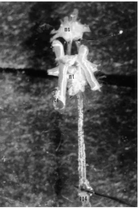
Fig. 1. Photograph of an isolated locust CNS (Taeniopoda eques) with its ventral tracheal supply. The isolated nerve cord contains the mesothoracic (ms), metathoracic (mt) and the first unfused abdominal ganglion (ag4). The nerve cord is covered with a low level of saline and one of the cut ends open to the surface (ce). This provides a good gas exchange by diffusion. The photograph was taken during collabor-ation with Jon Harrison, Arizona State University, Tempe, 1999.
cut to a diameter that was suitable for sucking in nerves with a tight fit in the tip. Bursting of ventilatory motone-urones was recorded from the proximal stumps of ventral abdominal nerves with mainly expiratory activity and of the median nerves with mainly antagonistic inspiratory activity. During in situ preparations the abdominal pumping movements correspond to the observed output, i.e. ventilatory bursts.
External conditions during the recordings (temperature, saline, pH, oxygen supply) were kept stable in a narrow range. Temperature was measured regularly with a thermometer. The initial level and con-centration of the saline was kept stable by replacing care-fully evaporated H2O with distilled water when working
without bath perfusion. We used a buffered saline (Clements and May, 1974) with the pH set at 6.8. By superfusing the nerve cord with oxygenated saline the accumulation e.g. of CO2 could be excluded (in the
experiments without maintained tracheal supply) and the supply of oxygen was kept stable. Saline levels of less than 1 mm above the ganglion further insured sufficient oxygen supply and CO2 release.
For comparison, efferent patterns were recorded in some experiments (not illustrated here) under varied conditions (saline, pH, temperature, saline with sugar or without) for at least 30 min up to 4 h. The data were amplified 1000×and stored on magnetic tape (TASCAM portastudio 424 tapedeck).
2.2.3. Analysis of the data
The stored data of efferent ventilatory activities could be printed continuously on an 8 channel printer (UNISCRIPT digital, PICKER International Ltd).
To obtain average records of ventilatory activity, the number of ventilatory bursts were counted in subsequent 1 min intervals. Only clear bursting activity (above sometimes prevailing tonic discharge) was counted as breathing activity. Whenever at least one interval elapsed without breathing patterns, this inactivity was taken as discontinuous ventilation (DV) according to earlier stud-ies (Lighton, 1990; Harrison et al., 1995).
The results of this analysis were visualised as sequen-tial histograms showing the neuronal ventilatory activity against the time recorded.
For analysing the data as described above we defined some important parameters as follows:
Burst: A high frequency discharge of spikes from ventilatory motorneurones lasting about 0.5–1.5 s.
Bursting period: Period of time in which repetitive bursting activity occurs, separated from adjacent per-iods by at least 1 min without any bursting activity.
Pauses: Period of at least 1 min duration without any bursting activity.
Counting Interval: For our sequential histograms we used time intervals of 1 min. Within these intervals the number of bursts were counted.
Continuous ventilation: Long lasting bursting periods with a wide range of frequencies (Fig. 1A).
Discontinuous ventilation: Period of time in which bursting periods are separated by silent or just tonic discharge intervals of at least 1 min (Fig. 1B).
To describe more detailed the observed patterns we made pause duration — histograms based on the sequen-tial histograms (see Section 3.2).
3. Results
3.1.1. Ventilation patterns in Taeniopoda eques
The spiking discharge patterns of ventilatory motor units observed in nerves of the isolated CNS show a wide range between total inactivity to continuous rhyth-micity (Fig. 3C and D and 4). In order to classify within this range two basic types of ventilation are defined. The results show the two types with transitions between them. Discontinuous ventilation pattern occurs quite often.
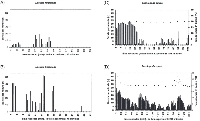
In Fig. 3C ventilation begins with high frequencies ranging over 30 bursts/min. After 30 min discontinuous patterns develop with pauses of max. 16 min between two burst periods.
Fig. 3D shows the results of an experiment in which the nerve cord was isolated but not removed from the abdomen. As in the other examples the connectives of the nerve cord were cut in front of the prothoracic gang-lion and behind the first abdominal ganggang-lion. The con-nections to the tracheae were maintained supplying the ganglia with oxygen. Abdominal muscle movements of the posterior segments agitated the saline around the ganglia. During the first 113 min the ganglia performed continuous ventilation patterns, but with extreme fre-quency variations. A pause of several minutes initiated a period of discontinuous ventilation which lasted until the end of the recording. This long term recording (215 min) shows patterns of strong continuous ventilation rhythms as well as DV and transition between both extremes. Similar patterns were found in several other experiments. At 175 min the temperature of the saline was increased to see possible reaction in the patterns. A rise in burst per min developed — as the temperature increased — and decreased with falling temperature in the saline. The maximum ventilation rate was 30 bursts per min.
3.1.2. Ventilation patterns in Locusta migratoria
The ventilation patterns in the isolated nerve cord of
Locusta migratoria show a wide range between total inactivity and continuous rhythmicity including discon-tinuous ventilation patterns. Discondiscon-tinuous ventilation is visible in Fig. 3A. In this recording the maximum venti-lation rate was 19 bursts per min. A different sequence of patterns from another isolated nerve cord of Locusta migratoriais shown in Fig. 3B with discontinuous venti-lation and irregular oscilventi-lations occurring during a burst period. Similar patterns of Locusta migratoria at rest could be observed by making movement video rec-ordings from undisturbed animals. The maximum venti-lation rate was 55 bursts per min.
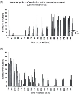
As a reference a long term recording that lasted more than 400 min was made (Fig. 4). The tracheal supply of the isolated nerve cord was maintained. A clear and stable DV-rhythm was visible. At the end of the rec-ording the pauses between the bursting periods showed a regular duration of 15–20 min. Also the length of the
bursting periods was nearly regular at 5–6 min. The fre-quency of 10–15 bursts per min was equal for the last bursting periods. Similar rates were found by Miller (1960) at temperatures between 18–20°C. The tempera-ture in saline was 18–19°C constantly.
3.2. Pause duration histograms
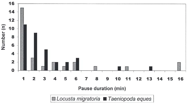
The typical range of pause duration between bursts (only for experiments with trachea removed) of venti-latory patterns in recordings of Taeniopoda eques (Fig. 5) lay between 1 or 2 min. In Locusta migratoria(Fig. 5) the majority of the pauses lasted about 1 min. There was a preference for short pauses similar to the distri-bution of pause length in Taeniopoda eques.
Miller (1960) made continuous recordings from prep-arations of Schistocerca gregaria. The bursting periods were interrupted by 1 min pauses, as for Locusta migratoria.
4. Discussion
The results of this study show that — just as in intact animals — in an isolated nerve cord a wide range of neural ventilatory patterns are generated. We can tinguish two basic types: continuous ventilation and dis-continuous ventilation. These definitions describe the opposite ends of a scale with many transitional forms of ventilatory patterns in between, occurring both in intact insects and in the isolated CNS.
All the patterns of fictive ventilation found inLocusta migratoria (Figs. 3A and B and 4) are comparable to those observed in Taeniopoda eques (Fig. 3C and D), suggesting dominant intrinsic rhythm generation (basic CPG) in the fused metathoracic ganglion of locusts in general. We can record from elements of the CPG intra-cellularly and thereby study integral parts of the venti-latory system unchanged by sensory influence and com-pare it with the neural network of high frequency ventilatory pattern generation (Ramirez and Pearson, 1989) and with results from studies in intact locusts and even other insects.
Recent studies of ventilation patterns in a variety of insect groups focussed on specific aspects of insect ven-tilation. Oxygen consumption and ventilation during rest and locomotion in the cockroach Blaberus giganteus
(Bartholomew and Lighton, 1985) follow ventilatory patterns similar to those of our study. Discontinuous ventilation patterns of emitted CO2-volume could be
recorded in intact Taeniopoda eques (Harrison et al., 1995). In the dragonflyErythemis simplicicollisHarrison and Lighton (1998) found different patterns of CO2
Fig. 2. Efferent ventilatory activity recorded extracellularly from the stump of efferent nerves (metathoracic median nerve) in an isolated locust CNS (Locusta migratoria). (A) long term recording with variable inspiratory bursting patterns which is also demonstrated as sequential histogram in Fig. 4. The data from (A) clearly indicate discontinuous ventilation pattern. (B) Recording during high frequency bursting.
“gulping ventilation” and “chewing ventilation”. These types were confirmed by the study of Hughes and Mill (1966), although Hughes and Mill (1966) pointed out that between the three types a wide range occurs. These patterns of ventilation show similarities to the patterns that we have found in the isolated CNS of locusts.
The studies of Williams et al. (1997) in Drosophila, Lighton (1990) in ants, Lighton and Lovegrove (1990) (and other authors) in honeybees focussed on different aspects of ventilation and were performed under com-pletely different conditions. However, they found in
Fig. 3. Efferent ventilatory activity in long-term recordings from efferent nerves (metathoracic median nerve) in the isolated locust CNS ofLocusta migratoria(A,B) andTaeniopoda eques(C,D). Sequential histograms of bursts per min. Graphs (A) and (B) show discontinuous ventilation. In (C) and (D) temperature is indicated by diamond symbols and scale at the right side. The initial continuous ventilatory periods are followed by discontinuous ventilation patterns. The continuous parts in (D) show a characteristic irregular oscillation from high to low and again to high frequencies. In (D) a rise in frequency corresponding to a rise in saline temperature is visible. In (D) the nerve cord was not removed from the semiintact animal — in contrast to (A–C) where the nerve cord without tracheae was removed from the animal. In (D) the nerves were deafferented (connections to tracheae were maintained). The experiments were carried out at room temperature.
common basic ventilatory or gas exchange patterns com-parable to those found in this study. These can be div-ided in the two basic patterns as defined in this study for fictive ventilatory rhythms. Irregular rhythms of ven-tilation seem to be a typical structure in insect gas exchange or ventilatory movements comparable to our analysis (ventilatory activity against time recorded).
Ventilation rates in the isolated Taeniopoda equesCNS without tracheal supply (Fig. 2C) are similar to those of alert animals, but pause lengths are much lower than in
Taeniopoda eques in rest in which pause lengths extend from 20 min to beyond 60 min (Harrison, personal communication). In long term recordings forTaeniopoda equeswith maintained tracheal supply we found similar pause durations as in intact animals (Bustami et al., 1999). It is probable that internal stimuli like partial hypoxia influence ventilatory rhythms in ganglia with tracheae removed.
The specific patterns of DV or irregular oscillations within a bursting period have been previously called “group ventilation” (Hustert, 1974). These patterns can also be found in other insects. For example, Lighton (1990) has been able to show transitions from pure DV to fast continuous ventilation caused by activity in the desert ant Camponotus detritus. Transitional forms occur, corresponding to the patterns we found in our
Fig. 4. Efferent ventilatory activity in a long-term recording from the median nerve of the third abdominal neuromere of the metathoracic ganglion in an isolated CNS ofLocusta migratoria. The tracheal supply was maintained Continuous sequential histograms of bursts per minute. In (A) and (B) in this recording a pronounced returning DV-rhythm is visible. In (A) most of the bursting periods last 5 to 6 min. In (B) pauses between bursting periods extend to 15–20 min. Saline at 18–19°C.
study (e.g. Fig. 3C inTaeniopoda eques). Earlier studies on efferent ventilation pattern from in situ nerve cords in insects have presented only regular neural output (Case, 1961; Miller, 1960; Komatsu, 1982) in short term rec-ordings without discontinuous ventilation. A closer look at the data of Miller (1960) in resting desert locusts (Schistocerca gregaria) indicates the presence of discon-tinuous ventilation in his records of abdominal pumping movements. Recent studies even in slices of an insect ganglion (Schistocerca gregaria) have shown the per-sistence of ventilatory rhythms in isolated preparations (Ramirez et al., 1999).
The increase of ventilation rate in the isolated nerve cord due to a rise in saline temperature (Fig. 3D) resembles a switch in intact honeybee ventilation rate and pattern caused by an increase in temperature (Lighton and Lovegrove, 1990), comparable to the increase in ventilatory activity in the grasshopper
Tmethis pulchripennis with increasing temperature (Prange, 1990). Apparently, temperature changes affect ventilation patterns of an isolated insect nerve cord as it does in intact animals. Further studies are required for a reliable data basis on this subject.
Strong continuous ventilation patterns occur in differ-ent insects — as well as in a locust isolated nerve cord. For intact animals that we had forced to jump for 2 min (according to Krolikowski and Harrison, 1996) we found patterns similar to those high frequency ventilation pat-terns we found (Fig. 3C and D). The vigorous move-ments of jumping apparently increases the metabolic rate and therefore also the abdominal pumping.
Lighton (1988) found in Psammodes striatus (Tok-Tok beetle) discontinuous gas exchange and a corre-lation between oxygen uptake and contractions of the abdomen. The discontinuous gas exchange is based upon DV movements generated in the CNS. These patterns of oxygen uptake in Psammodes striatus show similarities to the DV fictive ventilation as shown very clearly in Fig. 5. The comparison between the results from differ-ent investigations in insects and our data lead to the question of the underlying principles of rhythmogenesis of ventilation — besides many external factors described and studied by the cited authors.
Krolikowski and Harrison (1996) suggested partial pressure of CO2 and O2 in the tracheae as the important
parameters regulating ventilation in different kinds of
Fig. 5. Pause duration histogram of long-term recordings from ventilatory efferent nerves (median or ventral metathoracic nerves) in isolated nerve cords fromTaeniopoda eques(7 experiments) andLocusta migratoria(7 experiments) without tracheal supply. Pause lengths between two bursting periods were counted. Apparently pause duration of 1 or 2 min dominate in the preparations. Longer pauses — as occurring in the experiments with maintained tracheal supply (Fig. 4) — are rare.
insects. The change of intratracheal partial pressures due to gas exchange or retention is supposed to have a trigger function for the performance of ventilation according to the momentary needs, e.g. in situations of discontinuous ventilation. In the isolated nerve cord no change of tra-cheal partial pressures exist since just “efferent venti-lation” patterns are generated. So this parameter cannot be the main factors for regulation of ventilatory rhythmogenesis in the isolated nerve cord. The present results suggest intrinsic regulatory or trigger mech-anisms on the level of the neural net of the central pat-tern generator, largely independent of sensory influ-ences, on the level of cell metabolism of the CNS or within ventilatory interneurones.
The results seen in the long term recording shown in Fig. 4 is the clearest evidence that mechanisms within the CNS underlie the basic rhythms of ventilation. Earl-ier studies (Harrison, 1997) suggested a PCO2-influenced
rhythmogenesis in locusts. In this model, if a certain trig-ger level of CO2 within the tracheal system is reached,
the ventilatory network begins to operate in order to release the CO2. Possible central chemoreceptors
sens-ible for O2 and CO2 inside the ganglion or inside its
ganglionic tracheae have been suggested by earlier work and also by more recent studies (Miller, 1960; Harrison, 1997; Bustami et al., 1999). These could influence the CPG-output in case of misbalance (i.e. hypoxia/hyperoxia or hypercapnia) of one of those gases in the surrounding saline or in the tissue Fig. 5.
5. Conclusion
discontinuous ventilation to continuous, stressed venti-lation. Investigations in intact insects (Hustert, 1974; Herreid and Full, 1984; Bartholomew and Lighton, 1985; Lighton, 1990; Lighton and Lovegrove, 1990; Slama and Coquillaud, 1992; Lighton and Berrigan, 1994; Krolikowski and Harrison, 1996; Williams et al., 1997) show similarities to the patterns in the isolated locust nerve cord.
The results suggest that the ventilatory patterns are of neural or physiological origin. Furthermore we can postulate a “general principle” of ventilatory patterns which seem to have developed in different families of insects (e.g.: ants (Lighton, 1990), dragonflies (Hughes and Mill, 1966), locusts (Harrison, 1997), cockroaches (Herreid and Full, 1984; Bartholomew and Lighton, 1985) and beetles (Chown and Holter, 2000). So we find continuous as well as discontinuous ventilation pat-terns — with transitional pattern — throughout the dif-ferent insect groups. Genetic differences as in ventilation patterns of Drosophila due to selection by imposed des-iccation (Williams and Bradley, 1998) may also occur in other insects and may serve the long term adaptation of insect populations to altering environments.
The cellular mechanism of rhythmogenesis in venti-lation, may be based on ionic changes in context with the bicarbonate system. This questions requires further studies.
Acknowledgements
This study has been supported by: Graduiertenkolleg “Organisation und Dynamik neuronaler Netze”, Go¨tting-en.
References
Bartholomew, G.A., Lighton, J.R.B., 1985. Short communication — Ventilation and oxygen consumption during rest and locomotion in a tropical cockroach,Blaberus giganteus. Journal of Experimental Biology 118, 449–454.
Burrows, M., 1996. The Neurobiology of an Insect Brain. Oxford Uni-versity Press, Oxford, New York, Tokyo, 682 pp.
Bustami, H.P., Harrison, J., Hustert R., 1999. Hints to an oxygen recep-tor in the CNS of insects which influences ventilation; Talk and abstract at: Reporting seminar at Reinhausen of the “Graduiertenk-olleg: Organisation and Dynamics of Neuronal Networks” at the University of Go¨ttingen, Germany.
Case, J., 1961. Organisation of the cockroach respiratory centre and effects of acids on an isolated insect respiratory centre. Biological Bulletin 121, 385.
Chapman, R.F., 1998. The Insects — Structure and function, 4th ed. Cambridge University Press, Cambridge, 770 pp.
Chown, S.L., Holter, P., 2000. Discontinuous gas exchange cycles in
Aphodius fossor(Scarabaeidae): a test of hypotheses concerning origins and mechanisms. Journal of Experimental Biology 203 (2), 397–403.
Clements, A.N., May, T.E., 1974. Studies on locust neuromuscular
physiology in relation to glutamic acid. Journal of Experimental Biology 60, 673–705.
Harrison, J.F., 1997. Ventilatory mechanism and control in grass-hoppers. American Zoologist 37, 73–81.
Harrison, J.F., Hadley, N.F., Quinlan, M.C., 1995. Acid-base status and spiracular control during discontinuous ventilation in grasshoppers. Journal of Experimental Biology 198, 1755–1763.
Harrison, J.F., Lighton, J.R.B., 1998. Oxygen-sensitive flight metab-olism in the dragonflyErythemis simplicicollis. Journal of Experi-mental Biology 201, 1739–1744.
Herreid, C.F., Full, R.J., 1984. Cockroaches on a treadmill: aerobic running. Journal of Insect Physiology 30 (5), 395–403.
Hughes, G.M., Mill, P.J., 1966. Patterns of ventilation in dragonfly larvae. Journal of Experimental Biology 52, 167–175.
Hustert, R., 1974. Morphologie und Atmungsbewegungen des 5. Abdominalsegments von Locusta migratoria migratorioides. Zoologische Jahrbu¨cher der Physiologie 78, 157–174.
Hustert, R., 1975. Neuromuscular Co-ordination and Propioceptive Control of Rhythmical Abdominal Ventilation in intact Locusta migratoria migratorioides. Journal of Comparative Physiology (A) 97, 159–179.
Kestler, P., 1984. Respiration and respiratory water loss. Chapter 6. In: Hoffmann, K.H. (Ed.), Environmental Physiology and Bio-chemistry of Insects, 1st ed. Springer Verlag, Berlin, Heidelberg, pp. 169–183.
Kinnamon, S.C., Kammer, A.E., Kiorpes, A.L., 1984. Control of venti-latory movements in the aquatic insectCorydalus cornutus: central effect of hypoxia. Physiological Entomology 9, 19–28.
Komatsu, A., 1982. Respiratory nervous activity in the isolated nerve cord of the larval dragonfly, and location of the respiratory oscil-lator. Physiological Entomology 7, 183–191.
Krolikowski, K., Harrison, J.F., 1996. Haemolymph acid-base status, tracheal gas levels and the control of postexcercise ventilation rate in grasshoppers. Journal of Experimental Biology 199, 391–399. Lighton, J.R.B., 1988. Simultaneous measurement of oxygen uptake
and carbon dioxide emission during discontinuous ventilation in the tok-tok beetlePsammodes striatus. Journal of Insect Physiology 34, 361–367.
Lighton, J.R.B., 1990. Slow discontinuous ventilation in the namib dune-sea ant Camponotus detritus (Hymenoptera Formicidae). Journal of Experimental Biology 151, 71–82.
Lighton, J.R.B., Lovegrove, B.G., 1990. A temperature-induced switch from diffusive to connective ventilation in the honeybee. Journal of Experimental Biology 154, 509–516.
Lighton, J.R.B., Berrigan, D., 1994. Questioning paradigms: caste-spe-cific ventilation in harvester ants, Messor pergandeiand M. jul-ianus (Hymenoptera: Formicidae). Journal of Experimental Biology 198, 521–530.
Miller, P.L., 1960. Respiration in the desert locust; I. The control of ventilation. Journal of Experimental Biology 37, 224–236. Prange, H.D., 1990. Temperature regulation by respiratory evaporation
in grasshoppers. Journal of Experimental Biology 154, 463–474. Ramirez, J.M., Pearson, K.G., 1989. Distribution of intersegmental
interneurones that can reset the respiratory rhythm of the locust. Journal of Experimental Biology 141, 151–176.
Ramirez, J.M., Elsen, F.P., Robertson, R.M., 1999. Long-term effects of prior heat on neuronal potassium currents recorded in a novel insect ganglion slice preparation. Journal of Neurophysiology 81, 795–802.
Slama, K., Coquillaud, M.-S., 1992. Homeostatic control of respiratory metabolism in beetles. Journal of Insect Physiology 38 (10), 783–791.
Tonner, F., 1936. Mechanik und Koordination der Atem-Schwimmbewegung bei Libellenlarven. Zeitschrift fu¨r wissen-schaftliche Zoologie 147, 433–454.
and other flying insects. Journal of Experimental Biology 47, 561–587.
Williams, A.E., Rose, M.R., Bradley, T.J., 1997. C02 release patterns inDrosophila melanogaster: the effect of selection for desiccation resistance. Journal of Experimental Biology 200, 615–624. Williams, A.E., Bradley, T.J., 1998. The effect of respiratory pattern
on water loss in desiccation-resistant Drosophila melanogaster. Journal of Experimental Biology 201, 2953–2959.