MOLECULAR STUDY OF ISOLATES Edwardsiella tarda FROM
SEVERAL ISLANDS IN INDONESIA
SITI NARWIYANI*, KURNIASIH**, ASMARANI K.**
*Balai Besar Karantina Ikan Hasanuddin Makassar
**Fakultas Kedokteran Hewan -UGM,Yogyakarta
ABSTRACT
Edwardsiella tarda is an enteric pathogen that causes diarrhea, wound
infection, and death due to septicemia. This species is capable of invading fish
and human epithelial cell lines, and E. tarda escapes from the endocytic vacuole
within minutes of entry and then replicates within the cytoplasm moving directly
from cell to cell. Edwardsiella tarda was much more frequently isolated from lake
fish. The aim of research was to find out the strain variation of several isolates of
E. tarda from different islands in Indonesia. Four isolates of E.tarda from catfish
(Java and Sumatra), Cyprinus carpio (Kalimantan), Brazilian turtle (imported),
and ATCC from Singapura were identified by morphology. All isolates were
DNA extracted and DNA amplified by PCR. Products of amplification were
digested with AluI and HaeIII enzymes (RFLP) for 1 hour. They were visualized
with electrophoresis in 8% polyacrylamid gel and stained with silver. The
restriction endonuclease result showed that 3 isolates from fish had the same
bands. Two isolates from imported turtle had the same band as ATCC isolates.
Digestion with HaeIII enzyme gave more different bands than with AluI enzyme.
It was thought that isolates of E. tarda from turtle and from ATCC were
originated from human.
INTRODUCTION
Edwardsiella tarda is an enteric pathogen that causes diarrhea, wound
infection, and death due to septicemia. This species is capable of invading fish
and human epithelial cell lines, and E. tarda escapes from the endocytic vacuole
within minutes of entry and then replicates within the cytoplasm moving directly
from cell to cell (Strauss et al., 1997). Edwardsiella tarda was much more
frequently isolated from lake fish (Van Damme & Vandepitte., 1980). Eye
tumefaction, inflammation, haemorrhages, ascites and the presence of a purulent
fluid were the main macroscopic lesions observed (Padros et al., 2006). Internal
organs were congested, and livers showed patchy discoloration and petechiae.
Histologically, the liver, kidney, and spleen had severe multifocal necrotizing
inflammation. No bacteria were isolated from infected fish after 6 d Post infection
(Darwish & Plumb, 1989; Sahoo et al, 2000). Clinical signs and internal gross
lesions declined by 8 days post infection and were absent thereafter.
MATERIALS AND METHODS
Five isolates of Edwardsiella tarda were identified conventionally in the
Microbiology laboratory, Faculty of Veterinary medicine, Gadjah Mada
University. Each isolate (2mlx108) of Edwardsiella tarda was DNA exctracted
with Qiagen kit, Germany. They were amplified with specific primers of E. tarda
from fish. Eta 2-351 F (5’-TAG GGA GGA AGG TGT GAA-3’). Edwsp-780 R
(5’-CTC TAG CTT GCC AGT CTT-3’). And from human Eta 1-363 F(5’ – GTG
TCC GTG TTA ATA GCA - 3’).
The PCR programme was denaturation in 940C for 2 minutes, following
by 26 cycles of programme in 940C for 30 seconds, 500C for for 30 seconds, 720C
for 1 minute, and final extension in 720C for 5 minutes.
The PCR products were then digested with AluI and HaeIII enzymes for
one hour. All digestion products were visualized with electrophoresis in 8%
RESULT AND DISCUSSIONS
The isolation and identification of 5 different isolates showed the same
result (Table 1)
Table 1. The result of biochemistry characteristic of Edwardsiella tarda
Media A
TCC
Kalima
ntan
Sum
atra
Java Brazi
lia
Gram test - - - - -
Oxidase + + + + +
Catalase + + + + +
Motility + + + + +
Indole + + + + +
H2S in TSIA + + + + +
Voges-proskover
+ + + + +
Urea hydrolysis - - - - -
Lysine decarboxylase +/ - +/- +/- +/- +/- Ornithine decarboxylase - - - - - Gelatine hydrolysis + + + + +
Gas of glucose + + + + +
Acid of:
Glucose + + + + +
Lactose +/
-
+/- +/- +/- +/-
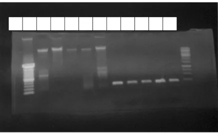
The result of DNA extraction had the different contents of DNA, and the
DNA amplification product showedt he bright band in 216 bp.(Figure 1). It is the
same as reported by Baird et al. (2003). Isolate of Edwardsiella tarda from
imported turtle could only be amplified with human primer.
Figure 1. The result of DNA extraction of E. tarda (lane 1-5), and the PCR
product of E. tarda (lane 6-10). Lane 1, 2, 3, 4, and 5 were isolate from ATCC,
Kalimantan, Sumatra,Brazil, and Java. The sequence of lane 6-10 was the same as
lane 1-5.
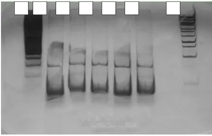
The endonuclease restriction using AluI enzyme had no different among 5
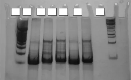
isolates of Edwardsiella tarda (Figure 2). However, using HaeIII enzyme showed
2 different groups of endonuclease restriction sites. Isolate of E. Tarda from fish
in Kalimantan, Java and Sumatra had the same bands restriction. Isolate of E.
Tarda from imported turtle showed the same band as isolate of E. Tarda from
Figure 2. The RFLP result of E. tarda (lane 1-5) used AluI enzyme. The sequens isolate in lane 1,
Figure 3. The RFLP result of E. tarda (lane 1-5) used HaeIII enzyme. The sequence Isolates in
lane 1, 2, 3, 4, and 5 were isolate from ATCC, Kalimantan,Sumatra,Brazil, and Java.
The phenotype characterization and numerical analysis of E. Tarda in wild
Asian Swamp eel in Malaysia were share similar numerical taxonomy and the
same strain of E. Tarda (Najiah & Lee, 2006). The prevalence of site-specific
genotypes with PCR-RFLP of 16s rDNA was found to be specific to detect habitat
specific isolates, or all the fish isolates were found only in fish, not in water or
sediment (Acharya et al., 2007).
CONCLUSION
Four isolate of E. Tarda from Indonesia had 2 strain variation compared
with ATCC. Isolate of E. Tarda from fish in Kalimantan, Java and Sumatra were
the same strain/ group. Isolate of E. Tarda from imported turtle was the same as
REFERENCES
Acharya, M., Maiti, N.K., Mohanty, S., Mishra, P., And Samanta, M. Genotyping
Of Edwarsiella Tarda Isolated From Freshwater Fish Culture System. J.
Comparative Immunology,.Microbiology & Infections. 30 :33-40. 2007.
Baird, K.D., Chikarmane, H.M., Smolowitz, R., And Uhlinger, K.R. J. Biological
Laboratory. 205 : 235-236. 2003.
Damme, I.R.V., And Vandepitte, J. Frequent Isolation Of Edwarsiella Tarda And
Plesiomonas Shigelloides From Healthy Zairese Freshwater Fish: A
Possible Source Of Sporadic Diarrhea In The Tropics. J. Applied And
Environmental Microbiology. 476-479. 1990.
Darwish, A. Histopathology And Pathogenesis Of Experimental Infection With
Edwardsiella Tarda In Channel Catfish In Textbook Of Fish Health. Dr.
George Post.
Najiah, M., Lee, S.W., And Lee, K.L. Phenotypic Characterization And
Numerical Analysis Of Edwarsiella Tarda In Wild Asian Swamp Eel,
Monopterus Albus N Trengganu. J. Of Sustainability Scienceand
Management. 1 : 85-91. 2006.
Padros,F., Zarza, C., Dopazo, L., Cuadrado, M., And Crespo, S. Pathology Of
Edwardsiella Tarda Infection In Turbot, Scophthalmus Maximus (L.). J. Of
Fish Diseases. 29 : 79-86. 2006
Sahoo, P.K., Swain, P., Sahoo, S.K., Mukherjee And Sahu, A.K. Pathology
Caused By The Bacterium Edwarsiella Tarda In Anabas Testudineus
Strauss, E>J., Ghori, N., And Falkow, S. An Edwardsiella Tarda Strain
Containing A Mutation In A Gene With Homology To Shlb And Hpmb Is
Defective For Entry Into Epithelial Cells In Culture. J. Infection And