Identiication of antibiotic producing endophytic microbe isolates
from a national park in Java island
Sri Yuwantiningsih
1*, Sebastian Margino
2, Subagus Wahyuono
31Biotechnology, Study Program, The Graduate School, Universitas Gadjah Mada, Yogyakarta, Indonesia 2Faculty of Agriculture, Universitas Gadjah Mada, Yogyakarta, Indonesia
3Faculty of Pharmacy, Universitas Gadjah Mada, Yogyakarta, Indonesia
Abstract
Endophytic microbes are potential sources of antibiotics. Some numbers of endophytic bacteria were isolated from plants in Ujung Kulon, Kaliurang, Meru Betiri and Baluran National Park, Bogor Botanical Garden, and Nusakambangan forest, Indonesia. Previous studies have been conducted to examine and obtain endophytic bacteria isolates from the selected plants, which resulted in three selected isolates, namely OOH-1, STG-1, and CMB-2. This research was conducted to determine the molecular identity of OOH-1 and STG-1 isolates, as well as to identify antibiotic compounds produced by STG-1 isolate. Molecular identiication of selected isolates was based on 16S rRNA gene analysis and ampliied using primers 27F and 1492R. A phylogeny tree was then constructed by comparing the resulting sequences with data from Gene Bank using the BLAST-N program. The identiication showed that STG-1 isolate had a 99% similarity with Pseudomonas brenneri strain SFML 97-391,
and OOH-1 isolate had a 99% similarity with Enterobacter xiangfangensis. Identiication of antibiotic compounds
was done by puriication and separation of the compounds. Antibiotic activity was also examined based on Lethal Concentration (LC50) on Fusarium oxysporum with a LC50 of 0.01–0.02% against Fusarium oxysporum.
Keywords: antibiotic, endophytic microbes, molecular identiication
Introduction
Within the last 20 years, the search for new types of antibiotics has been increasing, with the growth reaching 200% from 1979 to 2000 (Lockhart et al., 2009). Antibiotics have been widely used for chemotherapy, plant disease treatment, livestock drugs, biotechnology, and molecular biology (Smitha and Philip, 2014). The emergence of resistant strains impels the search and development of newer antibiotics, such as
PBL (soft rot disease) in orchid lower stalks
(Joko and Kusumandari, 2014) as well as leaf and neck blast in upland gogo rice (Nasution et al., 2014). Candida albicans as a pathogen in
the human esophagus, vulva, and vaginal skin has been widely studied in order to discover new anti-fungal compounds (Mohammed et al., 2012), also Moler disease in onion leaf (Lestiyani et al., 2014) and rot disease in pineapple and mango by Fusarium sp. (Widiastuti et al., 2014). In recent years, antibiotics have been studied and extracted primarily from soil microbes. However, other sources, such as endophytic microbes, may also possess potential desirable traits. Endophytic microbes are compatible with a wide variation of host type and characteristics as well as various ecosystems, allowing them to be found in diverse species. Under different environmental conditions inside plant tissues, endophytic microbes may produce antibiotics with higher bio-activity than those from microbes originating from soil or plant surfaces (Strobel et al., 2004; He,
*Corresponding author:
Sri Yuwantiningsih
Endophytic bacteria live inside plant tissues without endangering the host. Instead, the bioactive compounds produced by the endophytic bacteria control possible invading pathogens (Patel et al., 2012). Endophytic bacteria live mutualistically with various plants in different environmental conditions and that provide a wide range of endophytic bacteria biodiversity with different potential bioactive compounds.
Bioactive compounds with high bio-activity can be found from microbes in plants
in tropical rain forests in speciic regions,
and have long been used as a local medicine (Strobel et al., 2004). Research shows that isolates from some national parks in the Indonesian islands of Sumatra, Java, and Bali have a high effectiveness against several plant pathogenic bacteria (Meliawati et al., 2006). Margino (2008) also reported that isolates derived from rare plants in Yogyakarta, Java, were indicated to have anti-fungal trait. Isolates from Taman Negara National Park in Pahang, Malaysia, have also been known to produce compounds with potent cytotoxic
and antibacterial properties (Hazalin et al., 2009), as well as surfactant for Fusarium sp. growth inhibiton (Snook et al., 2009).
Previous studies have been conducted to obtain and select endophytic bacteria isolates from selected plants taken from a Javanese National Park. The result of the study was three selected superior isolates, namely OOH-1, STG-1, and CMB-2. As a continuation of the previous study, this research was conducted to determine the molecular identity of OOH-1 and STG-1 isolates, as well as the identification of antibiotic compounds produced by STG-1 as a model isolate for lab. scale production
because of its largest inhibition zone against
Fusarium oxysporum.
Materials And Methods
Tools and Materials
The tools used in this study were:
J), Microcentrifuge (Beckman microfuge II), PH meter 691 (Metrohm), electrophoresis cell (Bio-Rad Mini-Sub® TM DNA cell), PCR (Mastercycler personal, Eppendorf),
Sequencer ABI PRISM (310 Genetic Analyzer).
The materials used in this research were: OOH-1 and STG-1 isolates, Ready-To-Go PCR kits, DNA marker (1KB DNA Ladder, Promega), Primer 27F and 1492R (SBS
Genetetech Co., Ltd.), DNA puriication kit (Microclean Microzone Ltd.), and silica gel
60G F254.
Molecular Identiication
Some numbers of endophytic bacteria were isolated from plants in Ujung Kulon, Kaliurang, Meru Betiri and Baluran National Park, Bogor Botanical Garden, and Nusakambangan forest, Indonesia in previous study. Selected superior isolates, namely OOH-1, STG-1 were used for DNA isolation. Isolation of bacterial chromosomal DNA was done using a method developed by Pospieech and Neumann (1995). 16S
rRNA gene ampliication was performed
with primers 27F and 1492R with an initial denaturation PCR condition of 94ºC for 2
min, followed by 40 cycles of 94ºC for 30 seconds, 51ºC for 30 seconds, and 72ºC for
1 min, and a cycle of 72ºC for 10 minutes.
The PCR products were conirmed using
electrophoresis on 2% agarose gel.
Visualization was done under UV
light (Song et al., 2009). 16S rRNA PCR
products were subsequently sequenced
with the procedure according to the manual
procedures used by the DNA sequencer and then analyzed using the BLAST-N
program on the NCBI page (https://www.
ncbi.nlm.nih.gov/blast/). A phylogeny tree was constructed by comparing the
sequences of the obtained 16S rRNA bacterial isolates with sequences from the
Gen Bank database (https://www.ncbi.
Puriication and Separation of Antibiotic Compounds
Antibiotic compounds were identiied
initially using thin layer chromatography (TLC). Selected endophytic bacteria were inoculated on each respective media in accordance with
previous methods. A 500 ml Erlenmeyer lask illed with 150 ml media was incubated in a
shaker at 150 rpm for 4 days at a temperature
of 30–33ºc. The sample was centrifuged to
separate the supernatant and pellets, and then
puriied further using TLC. The sample was
centrifuged with the addition of a mixture of ethyl acetate and butanol, until the supernatant and pellets were separated. The supernatant was then taken, while the pellets with the ethyl acetate and butanol mixture were centrifuged repeatedly until clear.
The obtained fractions were then dissolved in a solution of chloroform:methanol (1:1) and put on a preparative plate, then processed into separation with a stationary phase of silica gel 60f and 254f and eluted with mobile phase of n-hexane:ethyl acetate
(93:7). After the elution was finished, the
results were observed under visible light and then scraped off. The resulting extract was
dissolved with n-hexane:ethyl acetate (93:7)
and packed on sintered glass. The eluate were evacuated and evaporated at room temperature. The powder obtained was then used in the following analysis.
LC50 Test Against Fusarium oxysporum
Antibiotic activity was also examined for its Lethal Concentration (LC50) against Fusarium oxysporum by examining colonies formed while under the stress of the added compound (Tebbets et al., 2013). The microbe culture indicator, Fusarium oxysporum, was cultivated in PDA media. As much as 10 ml of cell culture was added into petri dishes containing the media, and added with various antibiotic compound concentrations
of 0, 50, 100, 200, 300, and 500 mg/ml.
Results and Discussion Molecular Identiication
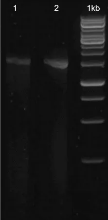
The identification of two strains of endophytic bacteria that produce antibiotic compounds, namely STG-1 and OOH-1 isolates was performed based on OOH-16S rRNA gene analysis via polymerase chain reaction using the universal 27 F primer (AGAGTTTGATCMTGGCTCAG) and 1492R Primer (TACGGYTACCTTGTTACGACTT). Then the reaction was checked using gel electrophoresis. There was only a single band on the outcome of PCR in a measure about 1500 bp indicating that it was a 16S band.
Fig 1. Gel electrophoresis on the outcome of PCR STG-1 (1) and OOH-1 (2).
T h e m o l e c u l a r i d e n t i f i c a t i o n o f STG-1 and OOH-1 isolates was based on
16S rRNA sequence analysis. The DNA sequences obtained were further used to
each isolate was assigned a species name.
The results are summarized in Table 1.
Janda and Abbott (2004) reported that the identity of a bacterial strain is the same if it has a similarity value with the type strain of bacteria at least 99%.
These sequences were compared with previously published sequences of two
bacterial groups. Group 1 consisted of Pseudomonas migulae strain CIP 105 470, Pseudomonas panacis strain C62010 and
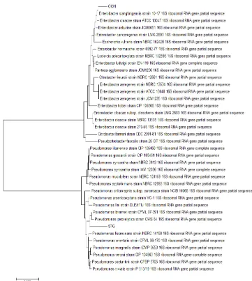
Pseudomonas proteolitica strain CMS 64, 3913. Group 2 consisted of Enterobacter Fig 2. Phylogenetic tree based on 16S rRNA gene sequences of Pseudomonas and Enterobacter
bacteria isolate groups.
Table 1. Results of the analysis of 16S rRNA gene
sequences using the BLAST-N program.
Isolates code
Homolog Bacteria Species
Access Number
Similarity (%)
STG-1
Pseudomonas
brenneri strain
CFML 97-391 NR.126208.1 99
OOH-1
Enterobacterxiang
fangensis strain
10-17
sequence comparisons are visualized in a
phylogenetic tree (Fig 2).
STG-1 isolate was in one group with Pseudomonas brenneri strain CFML 97-391 and Pseudomonas proteolytica strain CMS 64, whereas OOH-1 isolate was in one group with Enterobacter xiangfangensis strain 10-17. Therefore, STG-1 is closely related to Pseudomonas brenneri strain CFML 97-391 and OOH-1 is closely related to Enterobacter xiangfangensis strain 10-17.
Puriication and Separation of Antibiotic
Compounds
T h e i d e n t i f i c a t i o n o f a n t i b i o t i c compounds was performed only on STG-1 as a production model isolate, which was based
on the nature and speciic reagents that can be
detected by fractionation of phytochemicals and thin layer chromatography (TLC).
Antibiotics produced by STG-1 had an Rf value of 0.82 after elution with NH4Cl and Rf value of 0.90 on eluent water that was saturated with butanol. To obtain the antibiotics, these eluent can be used based on the observed spot, which is the purple colored spot beside the standard solution of stigmasterol. The compound was then
found belong to a group of steroids. The eluate was dissolved with a solvent mixture of chloroform:methanol (1:1) and incubated between 15–20 minutes to saturate and provide a bridge to enable the selection of active compounds and their derivatives into the chloroform phase. This study also aimed to reduce the number of components to be fractionated (El Shohly et al., 1999).
The fractions were then spotted on the preparative plate and eluted with the mobile phase consisting of n-hexane:ethyl acetate
(93:7). The elution results were seen under
visible light and the yellow spots were found.
The results of subsequent fractionation poured
in sintered glass and the eluate was evaporated in an evaporator (Rinanto et al., 2008).
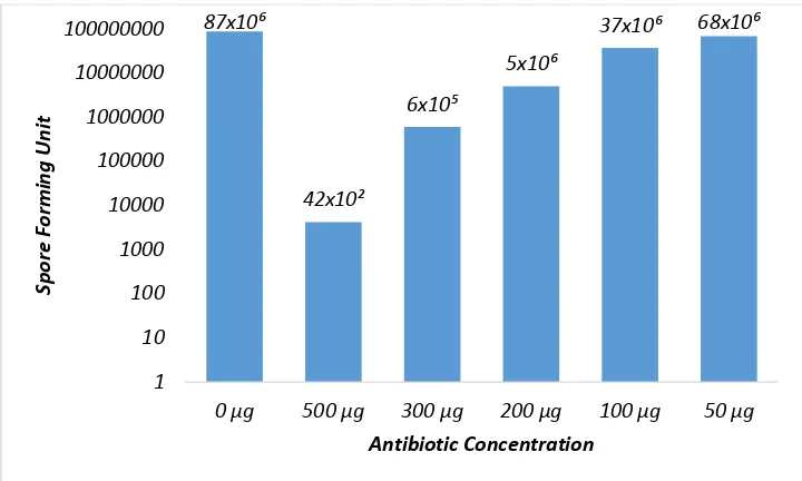
Activity Analysis of Antibiotics with Lethal Concentration (LC50)
The LC50 of antibiotic compounds in various concentrations was determined according to Tebbets et al. (2013). With a little modification, the determination of the concentration of antibiotics performed through some dilution concentrations, namely 10-1,10-2, 10-3, 10-4, 10-5, and 10-6.The microbial indicator, Fusarium oxysporum, was grown on PDA media for 48 hours. Cultures were mixed with various antibiotic concentrations, grown on agar medium using the pour plate method, and the observations were made after 24 hours. The results showed that the concentration that kills half of the
microbial indicator ranged from 100–200 µg/ ml to 273 µg/ml (Fig 4).
Based on the study of antibiotics toxicity from endophytic microbe was known that amphoterisin-B and voriconazole with Fusarium oxysporum and LC50 was 250
µg/ml (Wiyakrrutta et al., 2004), whereas
fluconazole was 64 µg/ml and mancozeb
with phenylmercury acetate was effective to kill Fusarium oxysporum pathogen. The results
were compatible with other inding that the
toxicity of antibiotics isolates from STG -1 can be used as a reference in determining new antibiotics with similar compounds.
The results on antibiotic compound production of the Pseudomonas genus as well as Pseudomonas viridiflava endophytyc bacteria showed that the antibiotic compounds contained surfactin agents that could act as biocontrol agents of Fusarium oxysporum, were highly biodegradable and had low toxicity (Snook et al.,2004). This antibiotic compound was identified as the major lipopeptida for the toxic component. The antibiotic activity is accomplished on concentration dependent actvity, low concentrations, and its product comformational changes within the phospolipids. At intermediate concentrations, it causes segregation within the bilayers, resulting in channels that are permeable to cations. At high concentrations, it produces a detergent effect resulting in complete membrane disruption. The results of this study revealed that Pseodomonas brenneri produced a steroid antibiotic compound against the pathogenic fungus Fusarium oxysporum.
Conclusion
The results of molecular identiication
showed that isolate STG-1 had a 99% level
xiangfangensis strain 10-17. The results of this study showed that STG-1 isolate produced antibiotic compounds belong to steroid group and had an LC50of 0.01–0.02% against the microbial indicator of Fusarium oxysporum.
References
Bicchi, C.and Rubiolo, P. 1996. High
performance liquid chromatographic
particle mass spectrometri analysis of
sesquiterpeneLacton with different carbon
sceletons. Journal of Chromatography 727: 211-221.
El Sohly, H. N., Croom, E.M., El Feraly, F.S. and El Sherey M.M. 1990. A Large-scale
extraction technique of artemisin from
Artemesiaonnua. Journal of Nat. Prod.
53(6):1560-1565.
Joko T. and Kusumandari N. 2014. Deteksi molekuler bakteri penyebab penyakit busuk lunak pada anggrek menggunakan teknik Polymerase Chain Reaction. Pengembangan dan pemanfaatan IPTEKS untuk kedaulatan pangan. Seminar Nasional Dies Natalis Faperta UGM ke-6
Fig 4.Effect of the antibiotic concentration on the growth of Fusarium oxysporum.
87x10⁶
42x10²
6x10⁵
5x10⁶
37x10⁶ 68x10⁶
1 10 100 1000 10000 100000 1000000 10000000 100000000
0 μg 500 μg 300 μg 200 μg 100 μg 50 μg
S
po
re Forming
Uni
t
development in Streptomyces coelicolor. Journal of Microbiological Methods. 10: 10-16. He, R.I., Wang, G.P., Liu, X.H., Zhang, C.L and Lin.F.C. 2009. Antagonistic bioactivity of an endophytic bacterium isolated from Epimedium brevicornu Maxim African Journal of Biotecnology. 8(2): 191-195.
Lestiyani, A., Wibowo, A., Subandiyah, S. 2014. Uji metode inokulasi pada bawang merah dengan Fusarium spp. Pengembangan dan Pemanfaatan IPTEKS untuk Kedaulatan Pangan. Seminar Nasional Dies Natalis Faperta UGM ke-68.
Lockhart, S.L., Kiederma, D.J., Pfaller, M.A. 2009. The Epidemiology of Fungal Infection InAnaissie, EJ, McGinnis MR, Pfaller MA. (eds). Of Clinical Mycologv. 2nded oxford V K ElseviSp Inc.
Margino, S. 2008. Produksi metabolit
sekunder (antibiotik) oleh jamur endoit.
Majalah Farmasi Indonesia 19: 86-94 Meliawati, R., Widyaningrum, D.N., Djohan,
A.C., Sukiman, H. 2006. Pengkajian bakteri endofit penghasil senyawa bioaktif untuk proteksi tanaman. Jurnal Biodiversitas. 7(3) :221-224.
Muhammed, M., Fuchs, 13. B., Wu, M.P.,
Breger. J., Coleman, J.J., Mylonakis, E. 2012. The role of mycelium production and a MAPK-mediatedimune response in the C. elegans – Fusarium model system. Journal of Medical Mycology. 50: 488-496. Nasution, A., Usyati, N., and Santosa. 2014.
Varietas lokal padi sebagai sumber ketahanan penyakit blas daun dan blas leher. Pengembangan dan pemanfaatan IPTEKS untuk kedaulatan pangan. Seminar Nasional Dies Natalis Faperta UGM ke-68.
Patel, H.A., Khristi, S.M., Perikh, K. and Rajeram G. 2012. Isolation and
Characterization of Bacterial endophytic
from Lycopersicumesculentum plant and their plant promoting characteristics. Nepal. Journal of Biotechnology 2(1): 37-52. Rinanto, Y., Sumarni, T., Iskamto, B. 2008. Fraksi aktif jamur Candidaalbicans dar
iekstrak metanalik bunga kamilen (Matricamachamommilla L.) Majalah Farrnasi Indonesia. 19 (2): 65-69.
Silverstein, R.M., Bassler, G.C. and Morrill, T.C. 1991. Spectometric identification organic compounds. Fifth Edition. John Willey and Sons Inc. Singapore 1-410. Smitha, S.L. and Philip, R.K. 2014. Antibiotik
Organic Compound production by a marine fungus Pencilliumcirinum
S36 through solid state fermentation: optimization by response surface
methodology. International Journal of Research Biomedicine and Biotechnology
4(1): 6-13.
Snook, M.E., Mitchel, T. Hinton D.M. and Bacon, C.W. 2009. Isolation and
characterization of leu7-surfactin from the
endophytic bacterium Bacillus mojavensis RRC 101, a biocontrol agent for Fusarium verticillioides. Journal of Agriculture and Food Chemical. 57: 4287-4292.
Song, I., Ye.C., Zhang, Z., Lu, Y. and Ind Jing, K. 2014. Daptomycin antibiotic production processes in fed-batch fermentation by Streptomyces roseosporus
NRRL. 11379 with precusor effect and medium optimization. Journal of Bioprocess Biosynthesis Enginering (37): 415-423
Strobel, G., Daisy, B., Castillo, U. and Harper J. 2004. Natural Product from Microorganism.Journal of Natural Product. 67: 257-269.
Tebbets, B., Yu, Z., Stewart, D., Zhao, L.X., Jiang, Y.I., Qu, L.H., Andes, D., Shen,
B. and Klein, B. 2013. Identification
of antitungal natural products via Saccharomyces cerevisiaebioassay : insight in to macrotetrolide drug spektrum, potency and mode of action. Journal of Medical Mycolou. 51: 280-289.
Widiastuti, A. and Ningtyas, O.H. 2014.
Identiikasi jenis jamur penyebab busuk