Linda S. Costanzo
Virginia Com m onwea lth University, Medica l College of Virginia , Richm ond, Virginia 23298
Teaching Ca21and phosphate homeostasis in a physi-ology survey course for medical or graduate students is done very effectively with an integrated approach. The material can be taught in the endocrine section of the course, where the renal and gastrointestinal (GI) handling of Ca21and phosphate and bone remodeling can be linked under the umbrella of teaching parathy-roid hormone (PTH), vitamin D, and calcitonin. This allows one teacher to pull the whole story together (and it really does hang together!).
cov-We must emphasize that only the free ionized Ca21is biologically active; stated another way, any Ca21 bound to proteins or complexed to anions is inactive. We can then demonstrate several conditions in which the free ionized Ca21changes as follows.
Changes in total protein concentration are associated with changes in total Ca21concentration. However, it is difficult to predict, without other knowledge, whether the ionized Ca21will change in the expected direction. We are probably wise to leave these subtle-ties for inclusion in subsequent pathophysiology courses rather than entering these muddy waters in an introductory physiology course.
It is quite simple to explain what happens to ionized Ca21 when there is an increase or decrease in the plasma concentration of a complexing anion such as phosphate, citrate, or sulfate. As the concentration of the anion increases, a greater fraction of the Ca21is complexed and the ionized Ca21 concentration de-creases.
Finally, we discuss the effects of acid-base changes on ionized Ca21, which are easily explainable with a simple diagram of a plasma albumin molecule (Fig. 2). Plasma proteins have negatively charged sites that can bind either H1 or Ca21. In acidemia, for example, when there is excess H1in blood, more H1is bound to plasma proteins and less Ca21 is bound; thus the
free ionized Ca21 increases. On the other hand, in alkalemia, when there is a deficit of H1in blood, less H1is bound and more Ca21is bound; thus the ionized Ca21 decreases. Symptomatic hypocalcemia is com-monly seen in acute respiratory alkalosis, and students may already know about the tingling and numbness that accompanies psychogenic hyperventilation.
Overall Ca21Homeostasis
Ca21 balance is maintained by the interplay of three organ systems: GI tract, bone, and kidney (Fig. 3). This type of diagram allows the student to appreciate the ‘‘big picture’’ of Ca21 homeostasis and superimpose the important hormonal actions. Once the material is learned in more detail, the student can return to this diagram for summary and review. An adult ingesting 1,000 mg of elemental Ca21 daily maintains Ca21 balance as follows. Approximately 35%, or 350 mg, of the ingested Ca21is absorbed in the small intestine by a mechanism that is stimulated by vitamin D (more correctly, its active form, 1,25-dihydroxycholecalcif-erol). However,netabsorption of Ca21is only 200 mg per day, because pancreatic and other GI secretions contain Ca21. Bone turnover, or bone remodeling, is a continuous process involving both resorption of old bone and deposition of new bone; the bone resorptive processes are stimulated by the synergistic actions of 1,25-dihydroxycholecalciferol and PTH and are inhib-ited by calcitonin. Logically, to maintain perfect Ca21 balance, the kidneys must excrete the same amount of Ca21that the GI tract absorbs, or 200 mg per day; this
FIG. 1.
For ms of Ca21in blood [adapted with per mission fr om
L. S. Costanzo.Physiology. Philadelphia, PA: Saunders, 1998]. Per centages ar e per centage of total Ca21
concen-tration in each for m.
FIG. 2.
Effects of acid-base disturbances on Ca21binding to
is accomplished by a combination of filtration of Ca21 across the glomerular capillaries and subsequent reab-sorption of most of the filtered Ca21.
Whereas Fig. 3 demonstrates the situation of perfect Ca21balance (in which there is no net gain or loss of Ca21 from the body), one can easily visualize other conditions of positive or negative Ca21balance. For example, in positive Ca21balance (e.g., the growing child), net Ca21absorption from the GI tract exceeds urinary excretion and the difference is deposited in the growing bones. In negative Ca21 balance (e.g., pregnancy, lactation), net Ca21absorption from the GI tract is less than excretion and the difference comes from the maternal bone. (In the case of lactation, there is a second ‘‘excretory’’ route via the breast milk.)
At this point in the lecture, a large table can be constructed on the board with the following
head-ings: hormone, stimulus for secretion or production, actions (on bone, kidney, and intestine), and overall effect on serum Ca21concentration and serum phos-phate concentration (Table 1). The students will see how the lecture will be organized and where it is headed. We will teach the material by filling in the table and elaborating on points as we proceed. Such a table gives the students all of the salient points about Ca21and phosphate homeostasis and is a lasting way to review. (I have never been in favor of including such summary tables in the lecture syllabus—the students will be more actively involved in the lecture if they construct their own table with us!)
PARATHYROID HORMONE
The overall role of PTH is regulation of the serum ionized Ca21concentration. This regulation employs a simple negative feedback system. When the serum
FIG. 3.
Ca21homeostasis in an adult in Ca21balance [adapted with per mission fr om L. S. Costanzo.Physiology. Philadelphia,
ionized Ca21concentration decreases below normal, PTH secretion is stimulated. The hormone then has coordinated actions on bone, kidney, and intestine that raise the serum Ca21concentration and, simulta-neously, decrease the serum phosphate concentra-tion.
Structur e, Synthesis, and Secr etion of PTH
In humans, there are four parathyroid glands, which are located in the neck under the thyroid gland. The chief cells of the parathyroid glands synthesize and secrete PTH, which is an 84-amino acid single-chain polypeptide. The biologic activity of PTH resides entirely in the 34 NH2-terminal amino acids. Thus it is
important that radioimmunoassays for PTH be di-rected to the NH2 terminus rather than the COOH
terminus, particularly for use in parathyroid disorders in which hormone fragments may be circulating.
PTH is synthesized on the ribosomes as preproPTH, which has 115 amino acids. A 25-amino acid signal peptide is cleaved as synthesis is being completed on the ribosomes, forming a 90-amino acid proPTH, which is transported to the Golgi apparatus. There, 6 more amino acids are cleaved, yielding the 84-amino acid peptide, which is stored in secretory vesicles for subsequent secretion.
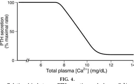
PTH secretion is regulated by the plasma Ca21 concen-tration (Fig. 4). When the total Ca21concentration is in the normal range (10 mg/dl) or higher, PTH is
secreted at a low, basal rate. However, when the plasma Ca21 concentration decreases to ,10 mg/dl, PTH secretion is strongly stimulated, reaching maxi-mal rates when the Ca21 concentration falls to 7.5 mg/dl. The students should note that although PTH secretion is plotted on the graph as a function oftota l Ca21concentration, actually it is theionizedCa21that regulates secretion by the chief cells. The response of the chief cells to a decrease in ionized Ca21 is remarkably prompt, within seconds, and the faster the ionized Ca21 falls, the greater the PTH secretory response.
Students may be puzzled initially that the chief cells would secrete PTH in response to adecrea sein Ca21,
TABLE 1
Summary of hor mones that r egulate Ca21and phosphate homeostasis
Hormone
PTH <[Ca21] >Bone resorption
(synergy with
>Ca21reabsorption, distal
tubule (hypocalciuric effect)
>Ca21absorption (via
activa-tion of 1a-hydroxylase, vitamin D)
>Ca21reabsorption
>Ca21absorption (calbindin
D-28K)
>Phosphate absorption
> >
Calcitonin >[Ca21] < Resorption <
*In the case of vitamin D, the stimuli for ‘‘secretion’’ are the factors that activate the 1a-hydroxylase enzyme in kidney that converts 25-hydroxycholecalciferol to 1,25-dihydroxycholecalciferol, thus increasing production of the active form of vitamin D. PTH, parathyroid hormone; P, inorganic phosphate.
FIG. 4.
Relationship between PTH secr etion and plasma Ca21
because they may recall that many endocrine cells (e.g.,b-cells of the pancreas) secrete their hormones in response to an increa se in intracellular Ca21. Actually, there is no paradox, because the chief cells sense a decrease inextra cellula rCa21, not intracellu-lar Ca21.
The mechanism of PTH secretion is explained as follows. The chief cell membrane contains a Ca21 sensor, which detects a decrease in extracellular ionized Ca21 concentration. This Ca21 sensor is coupled, via a Gsprotein, to adenylyl cyclase. Thus,
when extracellular Ca21concentration decreases, ad-enylyl cyclase is activated and catalyzes the conver-sion of ATP to cAMP. After a series of phosphorylation steps, there is exocytosis of PTH from secretory vesicles into the bloodstream.
These events describe the response of the chief cells to an acute decrease in Ca21concentration. However, there are additional effects of chronic hypocalcemia (e.g., due to chronic renal failure or vitamin D deficiency). Chronic hypocalcemia causes secondary hyperparathyroidism, which is characterized by in-creased transcription of the gene for preproPTH, increased synthesis and storage of PTH, and hyperpla-sia of the parathyroid glands. Conversely, chronic hypercalcemia causes decreased synthesis and storage of PTH and increased breakdown of stored PTH.
Mg21has effects parallel to those of Ca21on the chief cells. Hypomagnesemia stimulates PTH secretion, and hypermagnesemia inhibits PTH secretion. An excep-tion is the case of severe hypomagnesemia associated with chronic Mg21 depletion (e.g., alcoholism), in which PTH synthesis and secretion are inhibited, leading to hypoparathyroidism and hypocalcemia.
Actions of PTH
To introduce the actions of PTH, it is helpful to reemphasize that the actions of PTH on bone, kidney, and intestine are coordinated to produce an increase in plasma ionized Ca21concentration.
At this point in the lecture it is tempting to discuss the cellular mechanisms of action of PTH on bone and kidney, which occur via a Gsprotein and activation of
adenylyl cyclase. However, in the interest of
effi-ciency, this is best briefly noted and deferred to the discussion of the phosphaturic action of PTH in the renal proximal tubule (where it naturally fits).
Actions of PTH on bone. PTH has actions on all three cell types in bone: osteocytes, osteoblasts (re-sponsible for bone formation), and osteoclasts (respon-sible for bone resorption). Initially, PTH stimulates osteolysis in osteocytes, which results in dissolution of surface bone. As a result of this action, Ca21moves from bone canalicular fluid into the osteocytes and then into the extracellular fluid. In a second, slower action, PTH stimulates osteoclasts to increase resorp-tion of previously mineralized bone, releasing both Ca21 and phosphate into extracellular fluid. The organic portion of bone matrix, primarily type I collagen, also is resorbed, and a major component of collagen, hydroxyproline, is released and then ex-creted in urine. (Thus urinary hydroxyproline excre-tion is an indicator of bone resorpexcre-tion.) In a still later phase, PTH inhibits osteoblasts and bone formation. Interestingly, receptors for PTH are found on osteo-cytes and osteoblasts, but not on osteoclasts. An interpretation of this observation is that the hormonal effects on osteoclasts are mediated by paracrine sig-nals from osteocytes and osteoblasts.
The overall effect of PTH on bone, therefore, is to promote bone resorption, delivering both Ca21 and phosphate to extracellular fluid. However, it is very important to emphasize that these effects on bone a lone cannot account for the ultimate action of the hormone to increase the plasma ionized Ca21 concen-tration. The reason is that both Ca21and phosphate are released from bone—the phosphate will complex with the Ca21and limit the increase in ionized Ca21 concentration. Therefore, an additional mechanism must coordinate with the action on bone to eliminate the phosphate from the body. That takes us to the kidney and the all-important phosphaturic action of PTH.
The renal handling of phosphate is shown in Fig. 5. Phosphate is mainly unbound in the plasma and therefore is almost freely filtered. Subsequent to filtration, ,70% of the filtered phosphate is reab-sorbed in the proximal convoluted tubule and,15% is reabsorbed in the proximal straight tubule. The remainder of the nephron reabsorbs little, if any, phosphate (a debatable issue that need not concern our students). Thus 15% of the filtered phosphate is normally excreted, and it is surely worth mentioning that this excreted phosphate serves as a major urinary buffer for excretion of H1(called titratable acid). PTH inhibits the phosphate reabsorption process and
in-creases the fraction of phosphate excreted (see below for a detailed description).
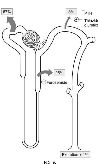
The renal handling of Ca21 is shown in Fig. 6. Ca21 filtration deserves special comment because such a large percentage of Ca21 in plasma is bound to proteins (40%) and, therefore, is not filterable across glomerular capillaries. Sixty percent of the plasma Ca21is ultrafilterable. Ca21differs from phosphate in that several segments of the nephron participate in its reabsorption. In the proximal tubule, Ca21 reabsorp-tion follows Na1and water reabsorption, primarily via a paracellular pathway; water reabsorption initially causes a small increase in luminal Ca21concentration, which then drives Ca21 reabsorption between the
FIG. 5.
Phosphate handling along nephr on [adapted with per mission fr om L. S. Costanzo.Physiology. Philadel-phia, PA: Saunders, 1998]. Arr ows show r eabsorption, and per centages ar e per centage of filter ed load r eab-sorbed.
FIG. 6.
Ca21handling along nephr on [adapted with per
cells. As evidence of this linkage, the percentage of Ca21reabsorbed in the proximal tubule is exactly the same as for Na1(67%). Any maneuver that alters Na1 reabsorption in the proximal tubule (e.g., volume expansion or volume contraction) alters Ca21 reabsorp-tion in the same direcreabsorp-tion and to the same extent. In the thick ascending limb, Ca21 reabsorption also follows Na1reabsorption (again, Ca21is moving by a paracellular path). In this case, Ca21 reabsorption is driven by the normal lumen-positive potential differ-ence that is generated by the Na1-K1-2Cl2 cotrans-porter; lumen positivity drives the passive reabsorp-tion of Ca21, a divalent cation. Loop diuretics such as furosemide inhibit the Na1-K1-2Cl2 cotransporter, eliminate the lumen-positive potential, and, as a conse-quence, inhibit Ca21 reabsorption, an effect that can be exploited in the treatment of hypercalcemia. In the distal tubule, Ca21is reabsorbed independent of Na1 (in contrast to the other nephron segments in which these two ions are linked). In fact, because of the presence of a Na1/Ca21exchanger in the basolateral membrane of distal tubule cells, the reabsorption of these ions is inversely related. As an aside, this inverse relationship is nicely demonstrated by the effect of thiazide diuretics in the distal tubule: Na1 reabsorp-tion is inhibited, whereas Ca21 reabsorption is aug-mented. Because thiazide diuretics increase Ca21
reab-sorption and decrease Ca21excretion, they are used to treat idiopathic hypercalciuria with the intention of preventing urinary stone formation.)
Now, back to the specific actions of PTH on the kidney. The first action of PTH on the kidney is to inhibit the Na1-phosphate cotransporter in the proxi-mal convoluted tubule, which results in inhibition of phosphate reabsorption and phosphaturia (see Fig. 5). The mechanism of this action of PTH is shown in Fig. 7. PTH binds to its receptors on the basolateral membrane. These receptors are coupled, via a Gs
protein, to adenylyl cyclase. When activated, adenylyl cyclase catalyzes the conversion of ATP to cAMP, which activates a series of protein kinases and ulti-mately phosphorylates intracellular proteins, leading to inhibition of the Na1-phosphate cotransporter in the luminal membrane. As a result of this inhibition, a greater fraction of the filtered phosphate is excreted, i.e., phosphaturia. In addition to increased phosphate excretion, the cAMP produced in proximal tubule cells diffuses into the urine; increased urinary cAMP is a hallmark of PTH action and was the basis for bioassay until these were replaced by radioimmunoas-says. The phosphaturic action of PTH is critically important for the overall ‘‘mission’’ of the hormone (to raise ionized Ca21 concentration), because
phos-FIG. 7.
phate that was resorbed from bone (by the action of PTH on osteoclasts) is excreted in urine; without the phosphaturic effect, this phosphate would have com-plexed Ca21in extracellular fluid.
The second action of PTH is to increase Ca21 reabsorp-tion in the distal tubule, also via an adenylyl cyclase mechanism (see Fig. 6). This hypocalciuric action complements the action of PTH on bone and assists in increasing the plasma Ca21concentration.
Action of PTH on intestine.The action of PTH on intestinal Ca21 absorption is indirect. PTH stimulates the 1a-hydroxylase enzyme in the kidney that is responsible for production of the active form of vitamin D, dihydroxycholecalciferol. In turn, 1,25-dihydroxycholecalciferol stimulates intestinal Ca21 ab-sorption.
Pathophysiology of PTH
Disorders of the parathyroid gland beautifully demon-strate the physiology of PTH, its feedback regulation by Ca21, and its actions on target tissues. The patho-physiology falls in three categories: hormone excess, or hyperparathyroidism; hormone deficiency, or hypo-parathyroidism; and hormone resistance, or pseudohy-poparathyroidism. Before embarking on this portion of the teaching, we emphasize once again that, by knowing the physiology well, one can perfectly pre-dict the characteristics of each disorder.
Hyperparathyroidism can be primary (originating in the parathyroid gland) or secondary (secondary to hypocalcemia). Primary hyperparathyroidism is most commonly caused by a parathyroid adenoma that secretes excessive amounts of PTH. The conse-quences of primary hyperparathyroidism are predict-able from the known physiological actions of PTH: increased bone resorption, increased Ca21 reabsorp-tion from kidney and absorpreabsorp-tion from intestine, and decreased phosphate reabsorption (phosphaturia). As a result of these actions, there will be hypercalcemia and hypophosphatemia. Persons with primary hyper-parathyroidism excrete excessive amounts of phos-phate and cAMP in their urine and, perhaps unexpect-edly, also excrete large amounts of Ca21. Does increased Ca21 excretion make sense, in light of the hypocalciuric action of PTH that we just taught? Yes, it does, but we must explain that, whereas the direct
action of PTH is to increase Ca21reabsorption, once the blood Ca21 concentration increases, the filtered load of Ca21 also increases and overwhelms the reabsorptive capacity of the nephron; the Ca21that is not reabsorbed is spilled in the urine. Persons with primary hyperparathyroidism are said to have ‘‘stones,’’ ‘‘bones,’’ and ‘‘groans’’—stones from hypercalciuria, bones from increased bone resorption, and groans from constipation. Treatment of primary hyperparathy-roidism is parathyroidectomy. In secondary hyperpara-thyroidism, the parathyroid glands secrete excessive PTH secondary to hypocalcemia (e.g., from chronic renal failure or vitamin D deficiency). In secondary hyperparathyroidism, circulating levels of PTH are elevated and plasma Ca21levels are either low (hypo-calcemia) or normal, but never high. Thus secondary hyperparathyroidism is distinguished from primary hyperparathyroidism by the absence of hypercalce-mia.
Hypoparathyroidism is a relatively c ommon in-advertent or unavoidable consequence of thyroid or parathyroid surgery. Autoimmune and congenital hy-poparathyroidism are rare. The characteristics of hypo-parathyroidism are, once again, predictable on the basis of the physiology: low circulating levels of PTH, decreased bone resorption, decreased renal reabsorp-tion and intestinal absorpreabsorp-tion of Ca21, and increased phosphate reabsorption. As a consequence of these actions, there is hypocalcemia and hyperphospha-temia.
bone and kidney; PTH cannot have its physiological actions on these tissues because the second messen-ger, cAMP, is not generated. In addition to type 1a, other variants of the disorder are types 1b, 1c, and II, which involve defects at other steps in the second messenger pathway including the PTH receptor, the adenylyl cyclase, and the protein kinases. Given the rarity of these disorders, I cannot imagine any value (other than because ‘‘it’s very interesting’’) in having students learn which variant has which defect.
VITAMIN D
A slide showing a child with vitamin D-deficient rickets makes a nice transition from PTH to vitamin D. We then want to emphatically state the overall role of vitamin D and compare it to that of PTH. PTH is for regulating the ionized Ca21concentration in plasma. Vitamin D is for mineralization of bone, and its actions, therefore, are coordinated to increase both Ca21 and phosphate concentrations in blood so that these elements can be deposited in new bone mineral. Recall that we have a table growing on the board and have now moved to our second hormone, vitamin D. First, we will make a digression to its metabolism (Fig. 8).
Vitamin D Metabolism
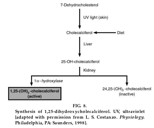
There are two sources of cholecalciferol, or vitamin D3, in the body. It is either ingested in the diet, or it is
synthesized in the skin from 7-dehydrocholesterol in the presence of ultraviolet light. Cholecalciferol itself is physiologically inactive. It is hydroxylated in the liver to form 25-hydroxycholecalciferol, which also is inactive; this hydroxylation step occurs in the endoplas-mic reticulum and requires NADPH, O2, and Mg21, but
not cytochrome P-450. 25-Hydroxycholecalciferol is bound to ana-globulin in plasma and is the principal circulating form of vitamin D.
In the kidney, 25-hydroxycholecalciferol undergoes one of two routes of hydroxylation. It can be hydrox-ylated at C-1 to produce 1,25-dihydroxycholecalcif-erol, which is the physiologically active form, or it can be hydroxylated at C-24 to produce 24,25-dihydroxy-cholecalciferol, which is inactive. C-1 hydroxylation is catalyzed by the enzyme 1a-hydroxylase in the renal mitochondria and requires NADPH, O2, Mg21, and
cytochromeP-450.
Whether the renal cells produce 1,25-dihydroxychole-calciferol (the active metabolite) or 24,25-dihydroxy-cholecalciferol (the inactive metabolite) depends on the Ca21 status of the body. When Ca21 is sufficient, with adequate dietary intake of Ca21 and normal or increased plasma Ca21 concentration, the inactive metabolite is preferentially produced because there is no need for more Ca21. When Ca21is insufficient, with a low dietary intake of Ca21 and decreased plasma Ca21concentration, the active metabolite is
preferen-FIG. 8.
tially synthesized to ensure that additional Ca21will be absorbed from the GI tract.
Production of the active metabolite 1,25-dihydroxycho-lecalciferol is regulated by changing the activity of the 1a-hydroxylase. 1a-Hydroxylase activity is increased by each of the following (which should be listed on the board in the column of factors that increase secretion or production): decreased plasma Ca21 con-centration, increased circulating PTH, and decreased plasma phosphate concentration. (We can recom-mend that students learn the factors in this order because it is easiest to remember that decreased plasma Ca21 leads to increased PTH, which leads to decreased plasma phosphate.)
Actions of Vitamin D
To reemphasize, the overall action of vitamin D (or more correctly, 1,25-dihydroxycholecalciferol) is to increase the plasma concentrations of both Ca21and phosphate to promote bone mineralization. In this effort, vitamin D has the following actions on intes-tine, kidney, and bone.
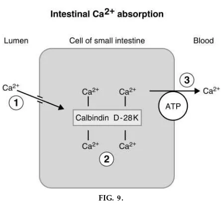
Actions of vitamin D on intestine. The major actions of 1,25-dihydroxycholecalciferol are in the small intestine, where it increases both Ca21 and phosphate absorption. Far more is known about its effect on Ca21absorption, which involves induction of the synthesis of a vitamin D -dependent Ca21 -binding protein called calbindin D-28K (a cytosolic protein with four binding sites for Ca21). The mecha-nism of intestinal Ca21absorption and a proposed role of calbindin D-28K is shown in Fig. 9. Ca21 diffuses from the lumen into the cell down its electrochemical gradient. In the cell, it is bound to or chelated by calbindin D-28K. Subsequently, it is pumped across the basolateral membrane by a Ca21 ATPase. The exact role of calbindin D-28K is uncertain. It may act as an intracellular shuttle, moving Ca21 from the luminal to the basolateral side of the cell, or it may act as a Ca21buffer to keep intracellular Ca21low, thus maintaining the concentration gradient for Ca21 diffu-sion across the luminal membrane.
Actions of vitamin D in the kidney.The actions of 1,25-dihydroxycholecalciferol on the kidney are
paral-lel to its actions in the intestine—it stimulates both Ca21and phosphate reabsorption. These renal actions are clearly distinguishable from those of PTH. (Recall that PTH increases Ca21 reabsorption but inhibits phosphate reabsorption.)
Actions of vitamin D on bone. In bone, 1,25-dihydroxycholecalciferol and PTH act synergistically to stimulate osteoclast activity and bone resorption. This synergistic action of 1,25-dihydroxycholecalcif-erol may at first seem paradoxical, because the overall role of this hormone is to promote bone mineraliza-tion. However, we can explain that, yes, 1,25-dihydroxycholecalciferol promotes the resorption of ‘‘old’’ bone, bringing more Ca21and phosphate in to the extracellular fluid, to facilitate the formation of ‘‘new’’ bone (or bone remodeling).
Pathophysiology of Vitamin D
In children, vitamin D deficiency causes rickets, a condition in which there is insufficient Ca21 and phosphate to mineralize growing bones. Rickets is characterized by growth failure and skeletal deformi-ties. (This condition is rare in parts of the world where vitamin D is supplemented in the diet or where there is adequate exposure to sunlight.) In adults, vitamin D deficiency causes osteomalacia, in which the failure to
FIG. 9.
Role of calbindin D-28K in intestinal absorption of Ca21[adapted with per mission fr om L. S. Costanzo.
mineralize new bone results in bending and softening of the weight-bearing bones.
Vitamin D resistance occurs if the kidney is unable to produce the active metabolite 1,25-dihydroxycholecal-ciferol. These conditions are called ‘‘resistant’’ be-cause, no matter how much vitamin D is supplied in the diet, it will be inactive because the C-1 hydrox-ylation step in the kidney is impaired or is missing. Vitamin D resistance can be caused by congenital absence of 1a-hydroxylase or, more commonly, by chronic renal failure. Chronic renal failure is associ-ated with a constellation of bone abnormalities (called renal osteodystrophy) including osteomalacia (due to the lack of 1,25-dihydroxycholecalciferol).
CALCITONIN
The details of calcitonin physiology usually are deem-phasized because it is unclear what physiological role this hormone plays in Ca21homeostasis. In contrast to PTH, calcitonin does not participate in the minute-to-minute regulation of the plasma Ca21concentration. In fact, neither thyroidectomy (with decreased nin levels) nor thyroid tumors (with increased calcito-nin levels) causes a derangement of Ca21metabolism, as would be expected if this hormone had a key regulatory role.
Synthesis, Secr etion, and Actions of Calcitonin
Calcitonin is a straight-chain peptide with 32 amino acids. It is synthesized and secreted by the parafollicu-lar or C cells (‘‘C’’ for calcitonin) of the thyroid gland. The calcitonin gene directs the synthesis of preprocal-citonin, a signal peptide is cleaved to yield procalcito-nin, other peptide sequences are cleaved, and the final hormone, calcitonin, is stored in secretory granules for subsequent release.
Conceptually, calcitonin is the ‘‘mirror image’’ of PTH. The major stimulus for calcitonin secretion is an increased plasma Ca21 concentration. The major ac-tion of calcitonin is to inhibit bone osteoclasts and to inhibit bone resorption. As a consequence, calcitonin decreases the plasma Ca21concentration acutely and, in theory, can be used to treat hypercalcemia.
CASE STUDY OF RENAL OSTEODYSTROPHY
If time permits, a case study of renal osteodystrophy (the bone disease that accompanies chronic renal failure) illustrates a significant amount of the physiol-ogy of Ca21and phosphate homeostasis.
For example, a 30-year-old female with advanced renal failure is receiving peritoneal dialysis while awaiting transplantation. She is admitted to the hospital for evaluation because, in the preceding month, she experienced severe bone pain and pruritus (itching). Upon admission, she had increased blood phosphate (hyperphosphatemia), decreased blood Ca21 (hypocal-cemia), increased circulating PTH, and decreased circulating 1,25-dihydroxycholecalciferol. Radiologi-cal examination revealed increased bone resorption, osteomalacia, and soft-tissue calcification.
The key points that illustrate the physiology in this case are as follows. Her chronic renal disease, with decreased renal mass and decreased GFR, led to decreased filtration of phosphate and phosphate reten-tion. The increased blood phosphate then complexed Ca21and caused a decrease in ionized Ca21 concentra-tion. The decreased renal mass also led to decreased production of 1,25-dihydroxycholecalciferol and, as a consequence, decreased intestinal Ca21 absorption and a further decrease in plasma Ca21concentration. The decrease in plasma Ca21caused increased secre-tion of PTH and hyperplasia of the parathyroid glands (i.e., secondary hyperparathyroidism). The increased circulating levels of PTH caused increased bone resorp-tion, and this, coupled with osteomalacia caused by the decreased 1,25-dihydroxycholecalciferol, is ‘‘renal osteodystrophy.’’ Calcification and pruritus resulted from deposition of Ca21-phosphate salts in soft tissues and skin (i.e., the complexation of Ca21 and phos-phate already mentioned). Treatment includes attempt-ing to limit the increase in blood phosphate by dietary phosphate restriction or phosphate-binding in the intestine, administration of synthetic 1,25-dihydroxy-cholecalciferol to replace what the kidneys fail to produce, and, if necessary, a parathyroidectomy.