Probing quantum coherence in a biological system by means
of DNA amplification
Erhard Bieberich *
Institute of Molecular Medicine and Genetics,Medical College of Georgia,1120 15th Street CB-2803,Augusta,GA30912,USA Received 25 March 2000; received in revised form 12 June 2000; accepted 20 June 2000
Abstract
As a result of rapid decoherence, quantum effects in biological systems are usually confined to single electron or hydrogen delocalizations. In principle, molecular interactions at high temperatures can be guided by quantum coherence if embedded in a dynamics preventing decoherence. This was experimentally investigated by analyzing the thermodynamics, kinetics, and quantum mechanics of the primer/template duplex formation during DNA amplifica-tion by polymerase chain reacamplifica-tion. The structures of the two oligonucleotide primers used for amplificaamplifica-tion of a cDNA template were derived either from a repetitive motif or a fractal distribution of nucleotide residues. Contrary to the computer-based calculation of the primer melting temperatures (Tm) that predicted a higher Tm for the
non-fractal primer due to nearest-neighbor effects, it was found that theTmof the non-fractal primer was actually
2°C lower than that of its fractal counterpart. A thermodynamic analysis of the amplification reaction indicated that the primer annealing process followed Bose – Einstein instead of Boltzmann statistics, with an additional binding potential ofm=500 J/mol or 10−21J/molecule due to a superposition of binding states within the primer/template
duplex. The temporal evolution of the Bose – Einstein state was determined by enzyme kinetic analysis of the association of the primer/template duplex to Taq polymerase. Assuming that collision with the enzyme interrupted the superposition, it was found that the Bose – Einstein state lasted fortdec=0.7×10⁻¹²s, corresponding to the energy
dispersion (DE) of quantum coherent states (m=DE]h/tdec). A quantum mechanical analysis revealed that the
coherent state was stabilized by almost vanishing separation energies between distinct binding states during a temperature-driven shifting of the two DNA strands in the primer/template duplex. The additional binding potential is suggested to arise from a short-lived electron tunneling as the result of overlapping orbitals along the axis of the primer/template duplex. This effect was unique to the fractal primer due to the number of binding states that remained almost constant, irrespective of the size of shifting. It is suggested that fractal structures found in proteins or other macromolecules may facilitate a short-lived quantum coherent superposition of binding states. This may stabilize molecular complexes for rapid sorting of correct-from-false binding, e.g. during folding or association of macromolecules. The experimental model described in this paper provides a low-cost tool for simulating and probing quantum coherence in a biological system. © 2000 Elsevier Science Ireland Ltd. All rights reserved.
Keywords:Quantum mechanics; Quantum biology; Polymerase chain reaction; Enzyme kinetics; Fractal; Thermodynamics www.elsevier.com/locate/biosystems
* Tel.: +1-706-7219113; fax:+1-706-7218727. E-mail address:[email protected] (E. Bieberich).
1. Introduction
The use of quantum computation for imple-mentation of a fast search algorithm raises the question whether the quantum coherent superpo-sition of different binding states can be exploited for molecular recognition in biological systems (Monroe et al., 1996; Ahn et al., 2000). The classical model describes the association/ dissocia-tion of molecular binding partners as a single collision, the association of which is energy cost intensive and diffusion-rate limited. In various biological systems, e.g. complementary base pair-ing in DNA, formation of a substrate – enzyme complex, or protein folding, molecular recogni-tion is based on the simultaneous binding of several molecular sites resulting from a non-cova-lent interaction of distinct electron orbitals. A superposition of several possible binding states, e.g. by overlapping electron orbitals, could rapidly trace out the optimal complex conforma-tion and sort correct-from-false binding. The di-rect experimental observation of a quantum coherent superposition in a biological system, however, is only reported in very rare cases such as single electron or hydrogen dispersion (Cha et al., 1989; Kohen et al., 1999; Ringe and Petsko, 1999). Most recently, it has been found that hy-drogen tunneling due to a quantum superposition of two localized binding states of a hydrogen atom occurs independent of temperature between 30 and 65°C (Kohen et al., 1999). The tunneling effect was ‘catalyzed’ by the particular enzyme structure of thermophilic alcohol dehydrogenase. The experimental investigation of a superposition consisting of more than one or two electrons or atoms in a mesoscopic system is difficult due to rapid decoherence of the quantum coherent state at high temperatures. Conservative estimations predict decoherence times of 10−13
to 10−20
s as the life-time for a superposition in a biological system (Tegmark, 2000; Seife, 2000). Even if these states exist, they are indistinguishable from classi-cal association/dissociation kinetics. In order to investigate a quantum coherent state of a biologi-cal system in vitro, it is necessary to stabilize a superposition of several binding states between two molecules. Furthermore, it must be possible
to experimentally determine the effect of the su-perposition for tracing out true/false binding states by quantization of the correct complex over time. Finally, the thermodynamics of the reaction must reveal a deviation from classical Boltzmann statistics in order to indicate a quantum coherent state during complex formation (Hill, 1986). Note that it is not intended to suggest a superposition of molecules themselves. However, a delocaliza-tion of binding electrons could contribute to an additional stabilization of a complex by forming overlapping orbitals in a quantum coherent state. This may be used for molecular recognition by ‘survival’ of most the stable state.
Sup-posing that mismatches can arise between the primer and template by shifting the two sequences along each other, the fractal structure will still match with a certain number of residues at each scale of shifting. It was expected that this topol-ogy rendered the F2/template duplex more stable than a duplex with R2. Note that the two primers differed only by a permutation of the fifth and sixth residue (Fig. 2). This permutation, despite conferring a self-similar structure to F2, resulted in a nearest-neighbor effect due to an additional gg pair in R2 (Aboul-ela et al., 1985; Breslauer et al., 1996; Rychlik et al., 1990; Rychlik, 1995). Thus, in contrast to the effects expected from a fractal topology, conventional programs for PCR primer design predicted a more stable duplex for R2 (Rychlik, 1995). The predicted melting tem-perature (Tm) was calculated to be 39.9°C for R2
and 39.1°C for F2. In order to evaluate these two contradictory predictions, PCR amplification was performed with a template of 500 base pairs (bp) at various annealing temperatures and the stabil-ity of the primer/template analyzed by determina-tion of the amount of amplified DNA. The amplification reaction was performed with differ-ent concdiffer-entrations of Taq polymerase in order to control the association kinetics of the enzyme
with the primer/template duplex. The thermody-namics and enzyme kinetics of the amplification reaction were analyzed in order to investigate Boltzmann versus Bose – Einstein statistics and classical versus quantum coherence in a biological model system.
2. Materials and methods
2.1. Materials
Platinum Taq polymerase, buffer, and deoxynu-cleotides were from Gibo BRL (Gaithersburg, MD). Oligonucleotide primers were synthesized by an in-house facility, and their purity analyzed by polyacrylamide gel electrophoresis and high-performance liquid chromatography before use. The PCR amplification was performed with a Crocodile III thermocycler (Appligene/Oncor, Gaithersburg, MD). The sialyltransferase II (ST-II) specific template DNA was cloned from a library of mouse cDNA as described previously (Bieberich et al., 2000). All other reagents were of analytical grade or freshly redistilled before use. The sequence of the primers used was:
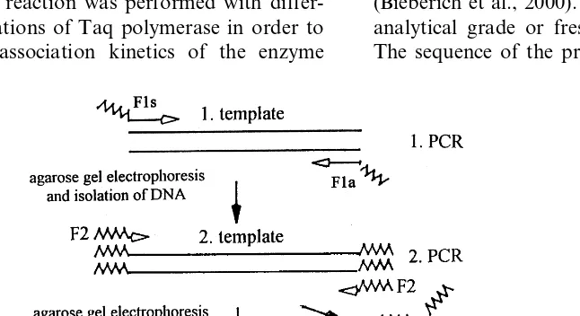
Fig. 2. Primer/template duplex formation and temporal evolution. (A) and (B), dissociation of the non-fractal primer occurs rapidly after shifting due to a short time for temporal evolution of the binding or duplex state. (C) and (D), the duplex formed with the fractal primer withstands rapid decoherence due to a sufficient number of binding states invariant to the extent of shifting. The binding state of a fractal sequence lasts longer than that of a non-fractal sequence. (D) The temporal evolution of the binding state is prematurely terminated due to collision with Taq polymerase fixing a particular state. This state can be binding and the DNA is amplified or anti-binding and the duplex dissociates.
F1s(forward):
5%gaagagaggaaggagttccatatcaggatgcggctagac3%
F1a(reverse):
5%gaagagaggaaggagttaggggctgaacctccacac3%
F2: 5%gaagagaggaaggag3%
R1s(forward):
5%gaaggaaggaaggagttccatatcaggatgcggctagac3%
R1a(reverse):
5%gaaggaaggaaggagttaggggctgaacctccacac3% R2: 5%gaaggaaggaaggag3%
2.2. Methods
All PCR reagents were first prepared as master mixes following the instructions given by the man-ufacturer (Gibo BRL). Thermocycling was per-formed using a reaction volume of 25 ml and the following amplification reaction: 2 min at 95°C; 35 cycles (30 s at 94°C, 30 s at various annealing temperatures (Ta), 30 s at 68°C); followed by final
the ST-II specific cDNA was first amplified with the primer combinations R1s/R1a or F1s/F1a at
Ta=58°C, giving rise to an amplification product
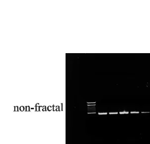
of 500 bp endowed with the fractal or non-fractal primer sequence at both ends of the DNA strand. The amplification product was then purified by means of agarose gel electrophoresis and an amount of 1 ng DNA was used as a template for the second round of PCR with the primer R2 or F2 (Fig. 1). The annealing temperatures were chosen as indicated in Fig. 3. The amount of amplification product was determined by ethid-ium bromide staining of the gel electrophoreti-cally separated DNA followed by densitometric analysis. The concentration of Taq polymerase was determined by a Lowry protein assay follow-ing a modification by Wang and Smith (1975).
3. Results and discussion
3.1. Thermodynamic analysis of the primer/template duplex stability by PCR
In order to determine the stability of the primer/template duplex, the PCR amplification was performed under identical conditions except that the annealing temperature was raised step-wise for each amplification reaction consisting of 35 thermocycles, as shown in Fig. 3. The amplifi-cation product was then separated by agarose gel electrophoresis and stained with ethidium bro-mide. Densitometric analysis of the staining inten-sity was used for product quantification. Reduction of the staining intensity by 50% indi-cated that the amplification reaction was per-formed at the melting temperature (Tm) of the
primer/template duplex. In Fig. 3, it can be seen that Tm is 2°C higher for the fractal primer (Tm
(F2)=58°C; Fig. 3, lane 8) than for the non-frac-tal primer (Tm(R2)=56°C; Fig. 3, lane 6). This
result contrasted with the Tm calculated on the
basis of the oligonucleotide sequence according to the method by Rychlik (1995), predicting
Tm(R2)=39.9°C and Tm(F2)=39.1°C. The
Rychlik method is generally accepted for calcula-tion of the primer/template duplex stability and predicts a higher melting temperature for an oligonucleotide sequence with identical purine residues as nearest neighbors (Petruska et al., 1988; Rychlik et al., 1990; Rychlik, 1995; Breslauer et al., 1996). As shown in Fig. 2, this was achieved in the non-fractal primer by permu-tation of the fifth and sixth residue. It should be noted that this permutation was the only differ-ence between the sequdiffer-ence of the fractal (F2) and non-fractal (R2) primer. The observation that the melting temperature of the two different primer/
template duplexes was about 20°C higher than calculated is common to short oligonucleotide sequences and due to the specific conditions of the PCR reaction. This, however, does not affect the prediction arising from the nearest-neighbor effect inherent to the non-fractal primer sequence.
Apparently, the amplification reaction was af-fected by additional factors, which are inherent to the primer sequence, but different from nearest-neighbor effects. It was assumed that these effects result from an attempted separation of primer and template due to a temperature-driven increase of kinetic energy. This separation can be initiated by shifting between template and primer upon
bilization of the DNA duplex. Fig. 2 depicts the temporal evolution of a stable binding state t(8) as compared with a temperature-induced decoher-ence at time tdec. Any stable binding state is
assumed to collapse rapidly upon shifting for the non-fractal primer (t(8)Btdec) but may persist
longer for the fractal primer (t(8)\tdec). This
assumption is best explained by the observation that shifting of the fractal primer sequence along the template generates a more stable base pairing
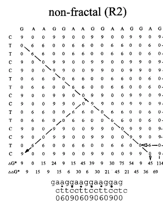
than shifting of the non-fractal sequence. As shown in Fig. 4, this can be estimated by calcula-tion of the binding energies contributed by base pairing upon shifting. The diagonals of the ma-trices represent a shift of the row (primer) se-quence along the column (template) sese-quence by one base residue to the right (upper half) or left (lower half). A matching base pair containing three hydrogen bonds (c/g) is stabilized by the apparent binding energy of DG*=9 kJ/mol, and
Fig. 4. (Continued)
a matching base pair containing two hydrogen bonds (a/t) by DG*=6 kJ/mol (Aboul-ela et al., 1985). These calculations were derived by a sub-traction of the binding energies contributed by non-matching base pairs (a/g, a/c, t/g, t/c), which were then assigned to have a binding strength of 0 kJ/mol. It can be calculated that, upon shifting, the fractal and non-fractal primers have the same combined binding strength of DG*=492 kJ/mol for matching base pairs, as indicated at the bot-tom of the matrices (Fig. 4). However, the differ-ence between the binding strengths upon two shifts by one base residue is lower for the fractal
primer, suggesting that the probability for consec-utive or reversible shifts is higher. This is indi-cated by the sum of the differences between the binding energies of two consecutive shifts, which was calculated to be 249 kJ/mol for the fractal and 357 kJ/mol for the non-fractal primer (Fig. 4). A thermodynamic analysis will take into ac-count all permutations caused by random shifting of binding between two matching bases according to a Boltzmann distribution of binding energy. The partition function (Q) for the distribution of all accessible energy states (oj) of a system withN
Q= 1
N!q
N with
q=%e−oj/kT (1)
where kis the Boltzmann constant.
In a Bose – Einstein state, the independence of single states vanishes in favor of the sum of all binding energies (Ej):
Q=%e−Ej/kT (2)
It is assumed that a probability of shifting without losing matching base pairs facilitates a transition from classical Boltzmann to Bose – Ein-stein statistics. This would yield an additional binding potential (m) arising from the superposi-tion of potential shift operasuperposi-tions resulting in a coherent binding state. In other words, the coher-ent state is characterized by a cooperative effect of binding states on the primer/template duplex for-mation. As discussed later, this may arise from electron tunneling between base pairs. By replac-ing Ej with oj−m (less than ground state energy o0=0) in Eq. (2), the binding energy between
primer and template is always higher in a Bose – Einstein state than with classical thermodynamics. The additional binding potential m can be deter-mined by comparing the free enthalpy of the primer/template duplex formation with the energy dispersion of a potential Bose – Einstein state.
The calculation of the free enthalpy for primer/
template duplex formation with dependence on the template concentration (cT) is derived fromTm
according to (Aboul-ela et al., 1985):
DGo(T
m)=RTmln(cT/4) (3)
The difference between the fractal and non-fractal primer binding strengths (DG* in Fig. 4) may then account for the additional binding po-tentialmof the fractal primer due to the
superpo-sition of potential shifts. However,
thermodynamic analysis cannot clearly demon-strate that this superposition originated in a quan-tum coherent state. One may argue that the shifting does occur, but the primer/template du-plex stays partially unseparated between two shifts. A potential quantum coherence without adopting classical (single) binding states can only be evaluated by a kinetic analysis of the temporal evolution of the primer/template duplex formation.
3.2. Kinetic analysis of primer/template duplex formation
The temporal evolution of the primer/template duplex formation was analyzed by evaluating the Taq polymerase concentration dependent velocity of the amplification reaction. If the duplex forma-tion proceeded without quantum coherent super-position of binding states, the concentration of the amplification product (cA) increased over time
in a linear correlation to the concentration of the duplex (cD)and the enzyme (cE) in a second-order
reaction (Rychlik et al., 1990):
dcA
dt =cDcEkII (4)
Fig. 5 shows the amount of amplification product obtained with the fractal and non-fractal primers with increasing concentrations of Taq polymerase as analyzed by agarose gel elec-trophoresis (Fig. 5A) and densitometric analysis (Fig. 5B) of the ethidium bromide stained DNA. The melting temperatures determined in Fig. 3 (Tm(R2)=56°C and Tm(F2)=58°C) were chosen
Fig. 5. PCR amplification in dependence on enzyme concentration. The PCR amplification was performed at the annealing temperatureTa=56°C for the non-fractal (top) andTa=58°C for the fractal (bottom) primer with different concentrations of Taq
polymerase. The amplification product was separated by agarose gel electrophoresis, visualized by staining with ethidium bromide (A), and the amount of DNA determined by UV-densitometric analysis (B). Lane 1, standard; lanes 2 – 7, 10, 25, 50, 100, 200 nM Taq polymerase. Each amplification reaction was repeated five times. Standard variations are indicated as bars.
compensating for the linear increase of the reac-tion velocity. As shown in Fig. 2D, this may have arisen from a premature dissociation of the primer/template duplex due to binding of the enzyme.
In the following analysis, it is assumed that the formation of the duplex with the fractal primer involves a temporal evolution of superimposed or quantum coherent binding states. In the case of primer/template duplex formation, this can be described by a superposition of rotational states due to helical winding of the two DNA strands around each other. Helical unwinding by 35° re-sults in shifting of primer and template byDN=
1. The time evolution (U(8)) of the quantum coherent superposition of two rotational states with a separation energy of DE can be described by (Ioffe et al., 1999):
U(8)=e−i8(t) with 8(t)=DEt/h (5)
As shown in Fig. 2, this evolution would be disturbed by two different environmental effects resulting in decoherence, either driven by temper-ature or induced by binding to Taq polymerase. The first process is obvious since the uptake of kinetic energy by heating will separate the primer
and template DNA. The second process can be explained by the assumption that binding of the enzyme to the primer/template duplex collapses the superposition by fixing a particular binding state. This is comparable with a demolition mea-surement terminating a quantum coherent state. The thermally induced random shifting of primer and template DNA is no longer reversible, but fixed as a result of binding to the enzyme (Fig. 2D). Since the superposition involves binding and anti-binding states, it is very likely that the num-ber of duplexes decohering to anti-binding states will not be amplified, but will dissociate after collision with the enzyme. The dissociation kinet-ics are then correlated to the concentration of enzyme due to a higher probability of enzyme binding with increasing enzyme concentration. The time interval between two collisions of the enzyme with the primer/template duplex will be lowered to an extent that it terminates the tempo-ral evolution of the binding state (8(t)) with
t(8)=tcoll=tdec(Fig. 2D). The dissociation
duringn cycles proceeds by duplication of a frac-tion cTk%1=cD of the template concentration in
each cycle according to:
cA=cD2n=cT(1+k%1)n (6)
Note that the prime indicates a normalization of time onto the period of one amplification cycle. It follows from Eq. (5) that ifcEcT, the
amplifi-cation reaction proceeds as a pseudo-first-order reaction with:
k%1=cEk%II (7)
In Fig. 5B, it can be seen that at the shoulder of the plot using 25 – 100 nM Taq polymerase, with
cT=1 ng/25ml, andn=35,k%Iis 0.14/cycle for the
fractal and 0.17/cycle for the non-fractal primer. This can be explained by a faster dissociation of the fractal primer/template duplex induced by binding of Taq polymerase. According to the model already discussed, association with the en-zyme entails decoherence of the primer/template superposition into single binding or anti-binding states. The statistics of the reaction converts in-stantaneously from Bose – Einstein to Boltzmann, resulting in an increase of the dissociation rate. The free enthalpy for dissociation can be calcu-lated from the pseudo-first-order reaction, with (Stryer, 1995):
DG(dis)=RTln(kT/hk1) (8)
whereR=8.314 J/K,k(Boltzmann’s constant)=
1.38×10⁻²³ J/K, and h (Planck’s constant)=
6.63×10⁻³⁴ J s.
The contribution of the binding state superposi-tion in the fractal primer/template duplex towards a reduction of the dissociation energy will then be approximated by:
mdec=RTln[k%1(fractal)/k%1(non-fractal)] (9)
UsingT=329 K, Eq. (9) gives a free enthalpy of
−531 J/mol for primer/template dissociation due to decoherence induced by binding of the enzyme. From Eq. (3), it follows that the increase ofTm
due to quantum coherence by binding state super-position within the primer/template duplex can be approximated by:
mcoh=R[Tm(fractal)−Tm(non-fractal)] ln(cT/4)
(10) Using cT=1.2×10⁻¹⁰ M as the initial template
concentration (equals 1 ng DNA of 500 bp length in a reaction volume of 25ml) andDTm=2 K, Eq.
(10) yields a free enthalpy of −403 J/mol, at-tributed to the formation of a Bose – Einstein state.
Taking into consideration that these calcula-tions are based on indirect measurement, the two values for free enthalpy are still sufficiently close to indicate an additional binding potential m re-sulting from a quantum coherent process for frac-tal primer/template duplex formation. If we take 500 J/mol or 10−21 J/molecule as an approximate
binding potential mcontributed by the Bose – Ein-stein state, the quantum process will be run in the range of the thermal energy (4×10⁻²¹
J/
molecule) of the reaction.
3.3. Quantum mechanical analysis of the primer/template duplex formation
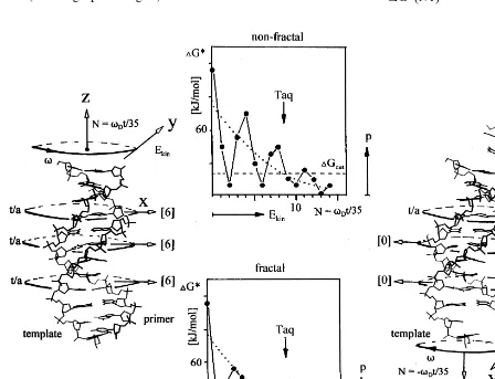
The thermodynamic and kinetic effects are sug-gested to arise from a superposition of binding states due to a potential well with mdec=500
J/mol or 10−21
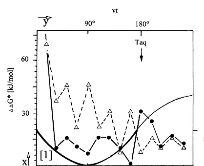
J/molecule. This potential well emerges at the very low DDG* values or separa-tion energies (Fig. 4) for step-wise primer/ tem-plate shifts with the fractal primer sequence. As shown in Fig. 6, the width of the well is depends on the shift size N=vDt/35° (vD is the rotation
angle for unwinding of the primer/template du-plex in B-conformation of the DNA) with suffi-ciently high binding energy DG* to withstand decoherence at DGcut. The angle of 35°
corre-sponds to the unwinding by one base pair (DN=
1). The rotation and shifting motion is described by three vectors for the momentum direction (x,
y,z) withxthe axis for base pairing with DG*,y
the clockwise (+) or counter-clockwise (−) rota-tion byvD, and zthe axis for shifting by N(Fig.
6). As discussed previously, since the time for collision with Taq polymerase (tcoll) is dependent
on the enzyme concentration, there will be a threshold value for t(8)=tcoll=tdec when the
critical concentration is reached when the enzyme collides faster than the time needed for the undis-turbed evolution of the superposition (tcoll5
t(8)). The Bose – Einstein state will then collapse to a probability distribution of binding and anti-binding states revealing Boltzmann statistics due to the distribution of kinetic energy in the heat bath (dotted graph in Fig. 6).
The decoherence time,tdec, will be derived from
the summation of the binding energies for base pairing after shiftN, as given by the diagonals of the matrix shown in Fig. 4, e.g.
diag(N1)fractal=[06000009060900]=30 kJ/mol
=DG*(N1)
Fig. 6. Model mechanism for the primer template shifting. Shifting of the DNA by size N is induced by temperature-driven unwinding of the primer/template duplex by the anglevDt=N · 35° (35 corresponds to the angle upon rotation by one base pair
orDN=1 on condition of a B-conformation of the duplex). The plots in the center show the summed binding energiesDG* for each base pairing upon shifting byNand are taken from Fig. 4. The arrow indicates the evolution of consecutive shifts. The probability (p) to find a particular duplex with the kinetic energy (Ekin) is an approximation and follows Maxwell – Boltzmann statistics (dotted
graph). The duplex dissociates whenEkinis higher thanDGcut, the minimum binding energy for a stable duplex. The duplex can be
diag(N2)fractal=[0069690000009]=39 kJ/mol
=DG*(N2) etc.
A different notation with the separation energies
DDG*(i)=DG*(i+1)−DG*(i) between two consecutive shifts of sizeDN=1 (other shift sizes are neglected) is given by the following column matrix:
M(frac or non-frac)=
DDG*(1) 0 0
0 DDG*(2) 0
0 0 DDG*(3) etc.
It is understood that this matrix is not complete since shift sizes of DN\1 are not included. The
complete matrix would contain 2n instead of n
diagonal values. Furthermore, the off-diagonal values arising from the imaginary term in exp (−i8) with ‘incomplete’ shifts ofDNB1 (see Eq. (5)) are neglected in favor for a real-valued solu-tion of the wave funcsolu-tionU(8). These boundary conditions have been introduced in order to allow more intuitive access to the quantum mechanics of superimposed binding states by a numerical solu-tion forU(8) derived from analysis of the matrix in Fig. 4. The arrow in Fig. 4 indicates the direction for the step-wise evolution of the separation ener-gies DDG* between two consecutive shifts that appear as on-diagonal values inM. The summed values or traces of the two matrices M(frac) and
M(non-frac) are correlated with the binding poten-tial contributed by a superposition of binding states within the fractal duplex (the non-fractal duplex is assumed to be non-coherent) according to:
mdec=c
%DDG*(i)frac−%DDG*(i)non-fracn
(11)For example, in Fig. 4, the difference of the diagonals yields aDDG*(N2,N1) value of 9 kJ/mol for the shift of the fractal and 36 kJ/mol for that of the non-fractal primer/template duplex. As shown in Fig. 6, this results in a binding energy
DG*(N2) of 39 kJ/mol for the fractal and 9 kJ/mol for the non-fractal primer. Since DG*(N2)non-frac
falls below an approximatedDGcutfor stable
bind-ing, we will assume that the non-fractal primer/
template duplex at N=2 represents an
anti-binding state. The coherent binding state is only possible if the separation energies DDG* are small. The summed values for DDG*(N1 … Nn) give a good approximation for the shift reversibil-ity which is inversely correlated to the decoherence time or stability of the primer/template duplex. As shown in Fig. 4, the sums of the separation energies or traces ofMare calculated to be 357 kJ/mol for the non-fractal and 249 kJ/mol for the fractal primer/template duplex. According to Eq. (11), the difference of the traces (108 kJ/mol) is then corre-lated to the binding potentialmof 500 J/mol for the escape from thermal decoherence due to superim-posed binding states. The difference of the traces corresponds to the overall separation energy gap between the binding state [1]=primer/template duplex stays (binding) or [0]=dissociates (anti-binding). The constant (c) for calculation ofmhas been introduced as empirical coefficient for linear correlation and can be approximated to 0.005. From Eq. (5), it can be concluded that the binding states [1] or [0] evolve by (mexpressed as 1×10⁻²¹
J/molecule):
U(8)=%e−i8(N)
with 8(N)=mtdec/h (12)
The ground state80of the potential well is derived
from a real-valued solution of Eq. (12) in a one-di-mensional box, as shown in Fig. 7:
U(80)=sinntdec (13)
with 1/tdec=n, the frequency of the wave function,
equal to m/h=1.5×1012/s.
The potential well extends over approximately
N=10 shifts consistent with DNA/primer unwind-ing by about vD=350° or a phase shift80([1]
[0]) for binding to anti-binding of 0.5=180°=p. From this shift, a decoherence time oftdec=1/n=
0.7×10⁻¹²
s can be calculated. The quantum mechanical interpretation of U(80), which
corre-sponds to the amplitude of the sine function, is given by the square root of the probability of finding the primer/template duplex in a superposi-tion within the window of N=10. The value for 1/tdeccorresponds to that of the pseudo-first-order
rate constant kI for dissociation induced by
Fig. 7. Quantum superposition of binding and anti-binding states. The plot indicates the formation of a potential well for superposition of binding states due to low separation energies between consecutive shifts of primer and template DNA. The potential well is approximated by a sine wave function for superposition of a binding [1] and an anti-binding [0] state with a frequencynof 1012/s. The arrow indicates the evolution
of the binding state upon shifting. A phase shift of 8 by p/2=90° corresponds to a DNA shift of N=5 or helical unwinding of vD=175°. A rotation of the DNA by vD=
350° (N=10) within tdec=0.7×10⁻¹² s corresponds to a
phase shift of the wavefunction byp(180°). The phase shift by p from binding to anti-binding can be induced by collision with Taq polymerase as indicated.
According to Fig. 4B, decoherence and dissoci-ation resulting from anti-binding states induced by collision with the enzyme occurs at approxi-mately cE=10
−7M of Taq polymerase. The
dis-sociation rate calculates to dc/dt=10−8 to 10−9
M/s, or in a reaction volume of 25ml, 1012to 1013
molecule collisions/s. The average collision time of 10−13 to 10−12 s/molecule corresponds to the
decoherence time calculated from Eq. (12). Thus, it can be consistently concluded that the decoher-ence of the coherent binding state is induced by collision with Taq polymerase. This explains why, in a certain concentration range, the fractal primer/template is less efficiently amplified than the non-fractal primer despite annealing at a higher melting temperature.
4. Conclusions
PCR amplification of DNA as an in vitro model for the detection of quantum processes in a biological system is useful for the following rea-sons: (i) the pairing of nucleotides provides a digital computation process of binding and anti-binding states that can be programmed by the sequence of the oligonucleotide primers; (ii) the amplification due to repetitive thermocycling al-lows for a very sensitive analysis of the binding reaction thermodynamics by determination of the efficacy of the product formation; (iii) the reaction kinetics can be directed by the concentration of enzyme; and (iv) the primer/template complemen-tarity is a suitable model for molecular recogni-tion that can be applied to other biologically significant molecular surfaces. In addition, the helical structure of the primer/template duplex allows for a translation of base-shifting into a rotation operation providing a convenient method for the computation of potential quantum processes.
The evolution U(8) of the primer/template du-plex formation as given in Eq. (12) can be rewrit-ten on the basis of a binding state superposition contributed by each base pairing after successive shifting steps (a and b coefficients for normaliza-tion of [1]=binding state and [0]=anti-binding state; a2+ib2=1):
This is calculated from Eq. (8) withm=500 J/mol for the potential contributed by quantum coherence:
kI=
kT
em/RTh=6×10
12/s (14)
If the primer/template dissociation is induced by binding to Taq polymerase, then the concen-tration dependence can be approximated by Eq. (4) (dc/dt=cDcEkII) with dc/dtthe rate of DNA –
enzyme complex formation, cD the concentration
of primer/template duplex=1.7×10⁻¹¹
M (=
cTkI; see Eq. (6) with n=1), cE the enzyme
con-centration, and kII the diffusion-controlled rate
constant for DNA/enzyme complex formation, approximated on the basis of average collisions calculated for a variety of molecules in biological systems :109 to 1010 (M/s)−1 (Duke and Bray,
U(8)=1/2(a0+ib1) (15)
In a classical system, there is only a transition from 1 to 1 or 1 to 0. A shift to the binding state 1 entails a stable DNA duplex, whereas 0 results in an irreversible dissociation of primer and template. In a quantum coherent system, however, the ‘bra-ket’ term ib1 (with
b=exp(−i8(t))) contributes to the additional binding potential arising from a reversible rota-tion of the two DNA strands around each other. The binding states are superimposed, ‘in between’ complementary base pairings in the x direction (see Fig. 6), and ‘compose’ the wave function
U(8) in the z direction (see Figs. 6 and 7). The binding state vector rotates in the complex xy or Argand plane (Fig. 6) by 8=nt (Fig. 7). Note that the rotation of the binding state vector arises from the rotation of the DNA but is not equiva-lent to it. In the Argand plane, the y component corresponds to the imaginary term ib1, whose projection on the x axis yields the amplitude
U(8)=sin(nt) (real-valued solution, see Eq. (13)). This quantum mechanical interpretation, of course, demands for the identification of a physi-cal substrate that can actually undergo a binding state superposition. Since the DNA molecules are too heavy for a superposition upon shifting, we will assume in the following analysis that a bind-ing state superposition results from an electron delocalization. Accordingly, the wave function
U(8) will describe the actual binding orbital in the z direction corresponding to the axis of the double helix (Fig. 6). The amplitude of this func-tion (sin(nt)) is then equivalent to the square root of the probability for finding a ground state elec-tron at a certain location within this orbital. The electron dispersion arises from sufficiently low separation energies between two consecutive bind-ing states that allow for an electron tunnelbind-ing in thezdirection. The spatial limit for this tunneling is derived from the uncertainty relationDzDp]h
(p is the momentum of the electron in the zaxis,
h is Planck’s constant). Assuming that the elec-tron dispersion is driven by the thermal energy of the PCR reaction at 58°C, it can be calculated from the thermal deBroglie wavelength le for a
single electron that:
Dz=le=h/(2pmekT)
1/2 (16)
A single electron with the mass me=9.1×
10−31 kg will be dispersed by approximately 4
nm. This easily extends the length of a DNA stretch of ten stacked base pairs (3.5 nm) and is consistent with the potential well calculated from the wave function U(8). It should be noted that the classical electron orbitals for base pairing result from hydrogen bonding in the x direction (Fig. 6). In stable base pairings, these orbitals do not overlap due to Coulomb repulsion. The molecular orbital in thezdirection is hypothetical and emerges from an electron excitation due to thermally induced shifting or rotation of the DNA strands. The kinetic energy conferred by the angular momentum in theydirection may be high enough to ‘loosen’ the hydrogen bonds in the x
direction to a transition state and to excitate electrons into a potential binding orbital in the z
direction (Fig. 6). The energy band for this orbital or ‘z-band’ should be above an energy gap of
cDGcut/6×1023 (c is an empirical constant,
esti-mated to be 0.005 – 0.025; see also Eq. (11)) as illustrated in Fig. 6. A fractal periodicity as de-signed within the primer sequence F2 is suggested to sustain the binding orbital in the z-band for 0.7×10– 12 s, corresponding to an energy
disper-sion of 10−21J
/molecule (=DE=h/tdec]cDGcut/
6×10²³). Accordingly, shielding of the
Bose – Einstein state against thermally induced de-coherence is a dynamic process counteracting a separation of primer and template by an ongoing binding potential arising from electron delocaliza-tion/tunneling. In the present study, the experi-mental approach by PCR amplification was sufficiently sensitive to detect this additional bind-ing potential. It remains to be elucidated whether the tunneling effect can be determined by other experimental approaches, e.g. spectroscopic meth-ods with a heated primer/template sample at a resonance frequency of 1.5×10¹² Hz (
=DE/h). What is the biological significance of a binding state superposition? It is certainly not a critical contribution to overall binding in DNA or other molecules. A binding potential of 500 J/mol or 10−21J/molecule is way too small to be crucial in
in a biological system. However, biological sys-tems are based on mutual molecular recognition. This is relying on finding a matching binding state upon single molecular collisions. The collisions are the time-dependent steps rate-limited by their association kinetics. A binding state superposition due to quantum coherence, however, may stabi-lize a molecular complex for a sufficiently long time that fast sorting mechanisms can discrimi-nate correct-from-false binding. Most recently, Patel has suggested that quantum coherent effects can be utilized for molecular recognition of the genetic code (Buchanan, 2000). He argues that the evolutionary optimization of using three bases for the coding of 20 amino acids emerges from a particular application of a quantum search al-gorithm introduced by Grover (1997). One out of
N=20 matching states can be found by superpo-sition of Q=3 binding states with Q=pN1/2/4.
This is suggested to accelerate a search fromN/2 (classical) to N1/2 (quantum) steps. It is assumed
that the environment created by binding with an enzyme, in this case aminoacyl tRNA synthetase, maintains shielding against a heat bath for undis-turbed quantum evolution of the superimposed binding states. Previously, this shielding effect was demonstrated for hydrogen tunneling in other enzymatic reactions (Ringe and Petsko, 1999).
In this study, however, molecular recognition of complementary surfaces takes place in an open system that is difficult to shield against decoher-ence effects induced by the environment. This is not only observed with binding of DNA strands, but also with molecular interactions of protein surfaces. In contrast to Patel’s assumption, inter-action with the enzyme Taq polymerase induces decoherence of the superimposed binding states resulting in a fixed position of the primer/template duplex. Only the fixed position allows for DNA amplification by enzyme-catalyzed chain elonga-tion of the primer. However, the duplex stability prior to binding of Taq polymerase could be enhanced due to a potential quantum coherence of binding states. The generation of the quantum coherent superposition is suggested to arise from a specific geometry of binding states that can be generally described as self-similar or fractal distri-bution (Peitgen et al., 1992; Barnsley, 1993).
Ide-ally, the distance ratio between two binding states is the same at all scales. This leads to a scale-in-variant probability for matching states regardless of the size for shifting of the molecules. The separation energy between two binding states may then be low enough to induce tunneling effects between electron orbitals as the result of ther-mally induced loosening of hydrogen bonds in the primer/template duplex. The thermal deBroglie wavelength of 4 nm is long enough to sustain an electron delocalization over several base pairs. This may be sufficient to overcome the separation energies for consecutive binding states in a fractal primer/template duplex but not in a non-fractal duplex.
The low additional binding energy of this bind-ing state superposition, however, does not justify the implication of Grover’s quantum search al-gorithm for single molecular recognition. As clearly demonstrated by the PCR reaction, the quantum effect was only detectable after exponen-tial amplification. Therefore, Patel’s suggestion to utilize a quantum search algorithm as a general mechanism for molecular recognition upon single molecule collisions is not realistic. Nevertheless, short-lived quantum coherence may play a role for sorting mechanisms that discriminate correct-from-false binding by fast recognition. Fast sort-ing has been described for proof-readsort-ing dursort-ing DNA replication or protein translation at ribo-somes (Stryer, 1995). Besides these specific proof-reading mechanisms, there are fast recognition events during protein or DNA folding that are common to all biological systems. It is still poorly understood how the search for protein folding can accomplish a fast correct-from-false sorting of one conformation within an average of 1035 possible
this implies the implementation of quantum ef-fects for molecular recognition. The model system presented in this paper may provide fundamental insight into the generation of quantum coherent binding state superposition in biological systems and may also be used for specific programming of the primer/template sequence for simulation of other binding state distributions.
Acknowledgements
The author wishes to thank Sarah MacKinnon for critically reading the manuscript. Part of the work was supported by an A.D. Williams grant.
References
Aboul-ela, F., Koh, D., Tinoco, I., Jr., Martin, F.H., 1985. Base – base mismatches. Thermodynamics of double helix formation for dCA3XA3G+dCT3YT3G (X, Y=A, C, G,
T). Nucleic Acids Res. 13, 4811 – 4824.
Ahn, J., Weinacht, T.C., Bucksbaum, P.H., 2000. Information storage and retrieval through quantum phase. Science 287, 463 – 465.
Barnsley, M.F., 1993. Fractals Everywhere. Academic Press, Cambridge, MA.
Bieberich, E., Tencomnao, T., Kapitonov, D., Yu, R.K., 2000. Effect ofN-glycosylation on turnover and subcellular dis-tribution ofN-acetylgalactosaminyltransferase I and sialyl-transferase II in neuroblastoma cells. J. Neurochem. 74, 2359 – 2364.
Breslauer, K.J., Frank, R., Blo¨cker, H., Marky, L.A., 1996. Predicting DNA duplex stability from the base sequence. Proc. Natl. Acad. Sci. USA 83, 3746 – 3750.
Buchanan, M., 2000. Life force. Do quantum computers make us what we are? New Sci. 2234, 20 – 24.
Cha, Y., Murray, C.J., Klinman, J.P., 1989. Hydrogen tunnel-ing in enzyme reactions. Science 243, 1325 – 1330. Duke, T.A.J., Bray, D., 1999. Heightened sensitivity of a
lattice of membrane receptors. Proc. Natl. Acad. Sci. USA 96, 10104 – 10108.
Goetze, T., Brickmann, J., 1992. Self similarity of protein surfaces. Biophys. J. 61, 109 – 118.
Grover, L.K., 1997. Quantum computers can search arbitrary large databases by a single query. Phys. Rev. Lett. 79, 4709 – 4712.
Hill, T.L., 1986. An Introduction to Statistical Thermodynam-ics. Dover, New York.
Ioffe, L.B., Geshkenbein, V.B., Feigel’man, M.V., Fauchere, A.L., Blatter, G., 1999. Environmentally decoupled sds-wave Josephson junctions for quantum computing. Nature 398, 679 – 681.
Kohen, A., Cannio, A., Cannio, R., Bartolucci, S., Klinman, J.P., 1999. Enzyme dynamics and hydrogen tunnelling in a thermophilic alcohol dehydrogenase. Nature 399, 496 – 499. Monroe, C., Meekhof, D.M., King, B.E., Wineland, D.J., 1996. A ‘Schro¨dinger Cat’ superposition state of an atom. Science 272, 1131 – 1136.
Peitgen, H.-O., Ju¨rgens, H., Saupe, D., 1992. Chaos and Fractals. New Frontiers of Science. Springer-Verlag, New York.
Petruska, J., Goodman, M.F., Boosalis, M.S., Sowers, L.C., Cheong, C., Tinoco, I.J.r., 1988. Comparison between DNA melting thermodynamics and DNA polymerase fidelity. Proc. Natl. Acad. Sci. USA 85, 6252 – 6256. Ringe, D., Petsko, G.A., 1999. Tunnel Vision. Nature 399,
417 – 418.
Rychlik, V., Spencer, W.J., Rhoads, R.E., 1990. Optimization of the annealing temperature for DNA amplification in vitro. Nucleic Acids Res. 18, 6409 – 6412.
Rychlik, W., 1995. Selection of primers for polymerase chain reaction. Mol. Biotechnol. 3, 129 – 134.
Seife, C., 2000. Cold numbers unmake the quantum mind. Science 287, 791.
Stryer, L., 1995. Biochemistry. Freeman, New York. Tegmark, M., 2000. The importance of quantum decoherence
in brain processes. Phys. Rev. E 61, 4194 – 4206. Wang, C.-S., Smith, R.L., 1975. Lowry determination of
protein in the presence of Triton X-100. Anal. Biochem. 63, 414 – 417.