Diagnostic and Prevention Approach in Post Endoscopic
Retrograde Cholangiopancreatography Pancreatitis
Stella Ilone*, Achmad Fauzi**
*Faculty of Medicine, Universitas Indonesia/Dr. Cipto Mangunkusumo General National Hospital, Jakarta
**Division of Gastroenterology, Department of Internal Medicine, Faculty of Medicine Universitas Indonesia/Dr. Cipto Mangunkusumo General National Hospital, Jakarta
Corresponding author:
Achmad Fauzi. Division of Gastroenterology, Department of Internal Medicine, Dr. Cipto Mangunkusumo General 1ational +ospital. -l Diponegoro 1o. -akarta Indonesia. 3hone Facsimile: +62-21-3142454. E-mail: [email protected]
ABSTRACT
Obstructive jaundice (icterus) was an emergency situation in gastroenterology. Endoscopic retrograde cholangiopancreatography (ERCP) was a nonsurgical approach to release obstruction, mostly in common bile duct. Nowadays, this procedure was become frequently used in daily practice, but several complications also emerging. One of the severe complication was Post-ERCP Pancreatitis (PEP). Since it has a high mortality and morbidity, and also reduce patient quality of life, several approaches have been developed to reduce its incidence. In general, approaches consist of patient identi¿cation, ef¿cient procedure, until pharmacological agent prevention. Although there were still contradiction among these, careful approach should be considered for each patients for a better outcomes.
Keywords: post-endoscopic retrograde cholangiopancreatography (post-ERCP), pancreatitis, prevention
ABSTRAK
Ikterus obstruktif merupakan masalah darurat di bidang gastroenterology. Endoscopic retrograde cholangiopancreatography (ERCP) merupakan prosedur non-bedah untuk melepas sumbatan yang sering ditemukan pada duktus biliaris. Saat ini, prosedur tersebut menjadi rutin dilakukan pada praktik keseharian, namun beberapa komplikasi juga terjadi. Salah satu komplikasi yang parah adalah pankreatitis post-ERCP. Oleh karena tingginya mortalitas dan morbiditas, juga menurunkan kualitas hidup pasien, beberapa upaya telah dikembangkan untuk mengurangi insidensi pankreatitis ini. Secara umum, upaya dilakukan melalui identi¿kasi pasien, tindakan prosedur yang e¿sien, serta medikamentosa. :alaupun masih ada kontradiksi di antara hal tersebut, upaya yang hati-hati seharusnya dapat dipertimbangkan untuk setiap pasien untuk hasil yang lebih baik.
Kata kunci: post-endoscopic retrograde cholangiopancreatography (post-ERCP), pankreatitis, pencegahan
INTRODUCTION
Obstructive jaundice (icterus) was still an important emergency in gastroenterology. Both acute and chronic bile obstruction could lead to several complications
in daily practice, but severe complication was still present from mild to severe. Among the complications was acute pancreatitis that raise mortality and morbidity in patients.
Post ERCP pancreatitis was related to dif¿culty, technique, and skills of the endoscopist. Post ERCP pancreatitis (PEP) incidence was about 2-9% and in several center could as high as 30% because of different de¿nition of pancreatitis.1 PEP could be mild (90%) to severe (pancreatitis necroticans, multiorgan failure, and death). <et, there were several de¿nition of PEP with different severity of pain and duration as its factors.
Some therapy was used to prevent the developing of PEP, such as right patient selection, pancreatitis prophyla[is, and more advanced skills during procedure. Early detection of PEP was also studied to develop a better outcomes in patients management. This review will discuss about diagnosis and prevention of post ERCP pancreatitis comprehensively from pharmacological to various ERCP technique.
(SLGHPLRORJ\ DQG 'LDJQRVLV RI 3RVW(5&3 Pancreatitis
Pancreatitis was a severe complication of ERCP procedure in obstructive jaundice patients. In general, pancreatic enzymes increase after procedure but could not be classi¿ed as pancreatitis. Based on Atlanta Criteria, PEP could be diagnosed if (1) New and progressive abdominal pain; (2) New or longer hospitalization; (3) Increase serum amylase by 3 times or more from upper norma limit in 24 hours after ERCP procedure.2
Based on diagnostic criteria above, PEP incidence in a Meta Analysis study involving 21 prospective study was about 1,6-15% (mean 3,5%).3 On the other hand, Cotton et al proposed another diagnostic criteria: (1) Increase serum amylase more than 3 times from upper normal limit; (2) Typical abdominal pain for pancreatitis; (3) Abdominal pain more than 24 hours; (4) A severe pain that need hospitalization.4 Cotton et al also classi¿ed patients into several pancreatitis grading based on its severity, as shown in Table 1. Pancreatitis severity based on Atlanta Criteria was shown in Table 2.
3RVW(5&33DQFUHDWLWLV3DWKRJHQHVLV
8ntil nowadays, the e[act mechanism of PEP development was still unknown. Several hypothesis have been proposed by e[perts, such as mechanical trauma in papillae orificium, hydrostatic damage, and enzymatic damage caused by proteolytic enzyme activated form duodenum.4 Mechanical trauma
7DEOH3RVWHQGRVFRSLFUHWURJUDGHFKRODQJLRSDQFUHDWRJUDSK\
(PEP) severity grading4
Mild Moderate Severe
– Elevation in serum amylase concentration more than three times upper normal level
– At least 24 hours after the procedure
– Requiring admission or prolongation of planned admission to two to three days
– Hospitalization
– Patients with hemorrhagic pancreatitis,
– Patients with newly developed organ failure and local or systemic complications
– Transient organ failure (organ failure < 2 days)
– Local
*Local complications include acute peripancreatic Àuid collections, pseudocyst, acute pancreatic or peripancreatic necrotic collection, and walled-off necrosis
damaging papilla ori¿cium that lead to Oddi sphincter spasm or pancreatic ori¿cum edema, so that pancreatic duct obstruction happened and lead to increase in pancreatic enzyme and finally inflammation. This damage could occur as a result from dif¿ cult-canulation ERCP that need a longer time, or a recanulation procedure. Beside that, thermal trauma from electrocautery during spinchterectomy could also lead to those damage.5
Hydrostatic damage was possible in overinjection in pancreatic duct that disrupt cell membrane and tight junction. As a result, a backÀow of intraductal secrete to interstitial tissue cause pancreatitis.6 Chemical trauma from ionic contrast with high osmolarity was another cause of pancreatic tissue damage. Otherwise, there were no meta analysis yet to show the relation between contrast and non-contrast media. This chemical trauma cause premature proteolytic enzymes activation that cause pancreatic cell autodigestion with reduced protective effect of acinar cell secretion, so that pancreatitis was developed.4
5LVN)DFWRUVRI3RVW(5&33DQFUHDWLWLV
on meta analysis by Masce et al from 15 studies, it can be concluded that those risk factors was:7 (1) patient factors: women (RR = 2.23), SOD (RR = 2.23), previous pancreatitis (RR = 2.46), young age (< 60 years old), normal bilirubin (OR = 1.89); (2) Procedural factors: sphinchterectomy precut (RR = 2.71), pancreatic duct injection (RR = 2.21), balloon dilatation in intact sphincter , pancreatic sphicnterectomy, dif¿cult cannulation (6-15 [ try, OR = 3.41), papilla minor sphicnterectomy (OR = 3.82), pain during ERCP (OR = 1.95), and ampulectomy; and (3) operator factor: trainee involvement. The more risk factors present, the highest PEP incidence possibility. High risk patient identi¿cation was an important thing to do in PEP prevention. Patient with more than one risk factors should be given detailed e[planation and counseling before the procedure. Other approach such as MRSP or EUS to reduce PEP risk was prioritize to replace diagnostic ERCP procedure.
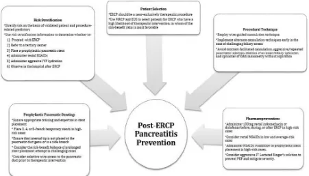
3UHYHQWLRQRI3RVW(5&33DQFUHDWLWLV3(3
In daily practice, there were so many treatments developed to reduce PEP in obstructive jaundice patients, from careful patients identification, pharmacological therapy, until various technique during ERCP. Unfortunately, there were no strong evidence which approach has been successful and effective to prevent PEP.
Endoscopist Prevention
This risk of pancreatitis was strongly related to endoscopist skill during ERCP. In referral center, which ERCP was done more routinely, dif¿culty during cannulation was commonly found and impact on the higher pancreatitis incidence. The presence of trainee was also an important factors of PEP incidence. Based on prospective study by Williams et al and Testoni et al, it can be concluded that PEP incidence was not correlate to cases number done by endoscopist in a referral center.7
7(&+1,&$/ 35(9(17,21 2) 3267(5&3 PANCREATITIS
Standard Canulation
Standard biliary cannulation was using catheter with or without soft tip guidewire. Contrast then injected through catheter could be unintendedly enter pancreatic duct. A guidewire insertion to pancreatic and common bile duct could reduce the risk of contrast
injection entering pancreatic duct with Àuoroscopy. A systematic review study by Cheung et al from 7 RCTs showed a reduced pancreatitis risk compared to contrast injection using guidewire (RR = 0.38; 95% CI: 0.19-0.76).7
Pancreatic Stent as Prophylaxis
The rational use of pancreatic stent was based on the principal that mechanical trauma will cause a Àow obstruction and and inÀammation in pancreas. A Meta Analysis showed a reduced risk in 13.3% and NNT of 8 to prevent 1 patient develop PEP with this technique.4 Menawhile, pancreatic stent placement has a higher risk of occlusion, migration, perforation, infection, erotion, and the need of re-endoscopy for evaluation and removal of the stent. A pancreatic duct stricture was also a possible complication. Therefore, this approach was not yet been choosen as an alternative for PEP prevention.
Pancreatic Duct Injection
Pancreatic duct injection was a multiple injection that correlate to the risk of PEP. Until now, ERCP was also done as diagnostic tool for neoplasms, such as pancreatic cyst, mainly to know any connection between cyst and biliary tract. Low osmolarity contrast was more recommended than high omsolarity. By reduce injection frequency, risk of PEP was predicted to be lower.7
3DQFUHDWLF*XLGHZLUH$VVLVWHG%LOLDU\&DQQXODWLRQ
Pancreatic guidewire placement was effective to ensure biliary tract access by straightening ampula and avoid pancreatic duct cannulation. This technique was mainly used in hard-cannulated patients or coincidentally recannulated pancreatic duct. Pancreatic stent placement after guidewire was recommended to reduce PEP incidence.7
Perendoscopic Sphincterectomy
Thermal damage caused by cauter could lead to ampula edema and obstruct pancreatic duct. Endocut was related to reduced risk of PEP by reducing edema in patients. Eventhough, bleeding risk was signi¿cantly increased. Therefore, there were still some pro and contra to done endocut.7
Balloon Sphincteroplasty
Oddi sphincter was preserved, especially for young patients, and also lower bleeding risk. Instead, several studies showed an increased risk of PEP in patients underwent this balloon sphincteroplasty. Yet, this technique was not concluded as a risk of PEP, e[ept combined with perendoscopic sphincterectomy.7
1HHGOHNQLIH3UHFXW
Precutting using needle knife was needed in a condition where standard cannulation was unsuccessful. This technique was strongly correlate to PEP. Meanwhile, this technique was also overcome with its less multiple cannulation possibility. Manes et al studied 151 patients with hard-cannulating after 10 minutes and compared to precut or standard cannulation with incidence was found lower in precut group (2,6% vs. 14.9%; p = 0.0008). But, a study by Cennamo et al investigate precut and standard cannulation result during hard-cannulation for 5 minutes showed no difference among two groups in PEP incidence.7
Oddi Sphincter Mannometry
Oddi Sphincter Mannometry was a gold standard for sphincter dysfunction e[amination. This sphincter dysfunction was correlate to high PEP incidence in patient. Pancreatitis risk could be reduced if pancreatic manometry was done before to know any SOD in patient, so that early pancreatic stenting could be done.
Pharmacological Prevention
Ideal pharmacological therapy was effective to reduce PEP incidence and could be administerd in short time, well-tolerated, and low side effect.
AntiinÀammatory drugs was a potent inhibitor for inÀammatory mediators, mainly prostaglandin and phospholipase A2 which related to acute pancreatitis. Elmunzer et al showed in a meta analysis involving 4 RCTs that NSAIDs per rectum was effective to reduce PEP incidence. Rectal indometachin before procedure or diclofenac after the procedure was effective. Oral administration of the drugs was not effective since it has a longer time to reach highest blood concentration. Besides, first-pass metabolism also reduce its bioavailability. ESGE recommended rectal diclofenac 100 mg to be administered just before the procedure.8
Glyceryl trinitate (GTN) was a smooth muscle rela[ant thant could reduce basal pressure of Oddi sphincter, administered sublingually or as a transdermal patch. An evaluation by Kaffes et al showed that there were no signi¿cant difference between therapy
and placebo groups on PEP incidence, although Moreto et al showe a signi¿cant improvement in 144 patients administered GTN. Until present, this drug administration was not recommended yet in PEP prevention.7
Ceftazidime was also used hypothese as its antimicrobial effect would prevent PEP development. Administration of 26 Ceftazidime 30 minutes before ERCP procedure could reduce PEP incidence in 160 patients of a trial, but this accuracy was stilll questionable.
Somatostatin and octreotide was also a potent inhibitor of pancreatic e[ocrine secretion that have an important rote in PEP pathogenesis. There were 2 meta analysis that investigate the role of both drugs. Andriulli et al studied 9 study in his meta analysis and showed a nonsigni¿cant effect of Somatostatin in PEP prevention (OR = 0.73; 95% CI: 0.54-1.006). There also studies to investigate both short-term Somatostatin infusion (< 6 hours) and long-term infus¿on (! 12 hours) in patients, but unfortunately it also give an insignificant result. Therefore, ESGE has not yet recommended ocreotide as PEP prevention, but further studies was developed to investigate its ef¿cacy on > 0,5 mg dosage.8
Protease inhibitor was also used as assumed that acute pancreatitis was happened because of intracelullar tripsin activation, and could be precented. Several known protease inhibitor drugs was Gabe[ate, Ulinastatin, and Nafamostat mesylate. Several RCTs given an contradiction result. Xiong et al showed a less signi¿cant effect of PEP in 97 patients given gabe[ate
)LJXUH3KDUPDFRORJLFDOUHJLPHQWXVHGWRSUHYHQWSRVW(5&3
30 minutes before ERCP, while Manes et al showed a signi¿cant result in gabe[ate given 1 hours before or after the procedure. Meanwhile, further meta analysis showed an insigni¿cant result of Gabe[ate. This was possible because of its very short half-life time (55 seconds) while there were need a continuos infusion for prevention. Differ to Gabe[ate, Ulinastatin has a longer half-life time (35 minutes) and could be given as bolus intravenous. Administration of Ulinastatin 150.000 Unit before ERCP was signi¿cantly reduce PEP incidence (2.9% vs. 7.4%, p = 0.041).9
Nafomastat showed a signi¿cant reduction in PEP if given 1 hours before ERCP and continued for the ne[t 24 hours (p = 0.018). Yet, protease inhibitor still rarely used and have a higher cost to be given routinely in pre-ERCP patients.
Allopurinol, a [hantine o[idase inhibitor, would catalyze hypo[hantine to [hantine that its end product was free radical. This free radical would trigger capillary endothelial damage and result in acute pancreatitis. Although animal studies showed a good result, some clinical trial did not showed the same result. Administration of 600 mg Allopurinol 15 hours before ERCP and 3 hours before ERCP showed a good result (2.3% vs. 9.4%; p = 0.04), while and administration at 4 and 1 hours before ERCP showed no differences to placebo.7
Oral prednisone with 40 mg dose did not reduce PEP incidence in 1115 patients at a clinical trial. Heparin have a protease inhibiting effect in both plasm and tissue, but studies showed that LMWH administration before ERCP did not reduce PEP incidence (8.1% vs. 8.8%; p = 0.87).7
N-acetylcystein was an anti-free-radical that have an important role in pancreatitis pathogenesis. A study by Katsinelos et al and Milewski et al showed no difference of PEP incidence in patients administered N-acetylcystein compared to placebo (12.1% vs. 9.6%; p > 0.05 and 7.3% vs. 11.8%; p = NS).7 The summary of all pharmacologic therapy in PEP prevention was shown in Table 4.
In general, prevention of PEP could be divided into 5 main area: (1) Correct patients identi¿cation; (2) Risk strati¿cation for patients undergo ERCP; (3) Atraumatic and ef¿cient procedure technique, 4) pancreatic stent as prophyla[is; (5) Pharmacological prevention.11
CONCLUSION
Pancreatitis was still a main complication of ERCP and further could reduce patient quality of life, morbidity, and mortality. Several approaches have been developed to reduce its incidence from
7DEOH3KDUPDFRORJLFDJHQWVDQGLWVHIIHFWRISRVW(5&3SDQFUHDWLWLV3(3SUHYHQWLRQ7
Drug Suggested way of action Effective in prospective RCT
Calcium channel blockers
Platelet activating factor inhibitors Interleukin-10
InÀammation cascade
No
ConÀicting data No
ConÀicting data, need for more trials ConÀicting data
ConÀicting data No
ConÀicting data No
No
ConÀicting data ConÀicting data ConÀicting data
Yes in only one study, need for more trials
NSAIDs non-steroidal anti-inÀammatory drugs RCT randomized control trial
7DEOH0HFKDQLVPRIDFWLRQRIVHYHUDOGUXJVIRUSRVW(5&3SDQFUHDWLWLV3(3SUHYHQWLRQ10
Postulated mechanism of action Agents
Interruption of inÀammatory cascade
Reduction of pancreatic enzyme secretion Inhibition of protease activity
Reduction of sphincter of-Oddi pressure
Prevention of infection Anti-oxidants
Anti-metabolites
NSAIDs, steroids, interleukin-10, allopurinol, adrenaline spray, pentoxifylline, platelet-activating factor-acetylhidrolase, semapimod, aprepitant, risperidone
Octreotide, somatostatin, calcitonin
Gabexate mesilate, heparin, ulinastatin, nafamostat, magnesium sulphate
Nitroglycerin, nifedipine, botulinum toxin, lidocaine, secretin, phosphodiesterase inhibitor type 5
Antibiotics
Beta-carotene, N-acetylcysteine, sodium selenite 5-Àuorouracil
patient identification, efficient procedure, until pharmacological agent prevention. Since several studies did not showed an absolute signi¿cant result, every case should be identified carefully, so that pancreatitis incidence as ERCP complication in obstructive jaundice patients could be reduced.
REFERENCES
1. Lazaraki G, Katsinelos G. Prevention of post ERCP pancreatitis: an overview. Annals of Gastroenterology 2008;21:27-38.
2. American Society for Gastrointestinal Endoscopy. Complication of ERCP. Gastrointest Endosc 2012;75:1-7. 3. Cotton PB, Garrow DA, Gallagher J, Romagnuolo J. Risk
factors for complicationsafter ERCP: a multivariate analysis of 11,497 procedures over 12 years. Gastrointest Endosc 2009;70:80-8.
4. Zouhairi ME, Swartz D, Shah T. Post-ERCP pancreatitis: mechanisms, risk fators, and prevention. Pancreatic Dis Ther 2013;2:1-4.
5. Rafani RS, Milis TN, Ainley CC, Swain CP. Electrophysical factors inÀuencing endoscopic sphincterectomy. Gastrointest Endosc 1999;49:43-52.
6. Sherman S, Hawes RH, Trolano FP, Lehman GA. Pancreatitis
following bile duct sphincter of Oddi manometry: utility of the aspirating catheter. Gastrointest Endosc 1999; 38:347-50. 7. Donnellan F, Byrne MF. Prevention of post-ERCP pancreatitis.
Hindawi Publishing 2012.p.1-12.
8. Dumonceau JM, Andriulli A, Deviere J, Mariani A, Riqau[
J, Baron TH, et al. European Society of Gastrointestinal
Endoscopy (ESGE) Guideline: prophyla[is of post-ERCP
pancreatitis. Endoscopy 2010;42:503–15.
9. Tsujino T, Komatsu Y, Isayama H, Hirano K, Sasahira N, Yamamoto N, et al. Ulinastatin for pancreatitis after endoscopic retrograde cholangiopancreatography: a randomized, controlled trial. Clin Gastroenterol Hepatol 2005;3:376–83.
10. Wong LL, Tsai HH. Prevention of post-ERCP pancreatitis. World J Gastrointest Pathophysiol 2014;5:1-10.
11. Elmunzer BJ. Prevention of ERCP-induced pancreatitis. American Pancreatic Association 2015;6:1-20.