ORLI Vol. 44 No.1. Tahun 2014 ORLI Vol. 44 No.1. Tahun 2014 Woakes syndrome Woakes syndromeORLI Vol. 44 No.1.
Laporan Kasus
Woakes syndrome
Retno Sulistyo Wardani, Ika Dewi Mayangsari, Trimartani Koento, Ervin Amouzegar Otorhinolaryngology Head and Neck Surgery Department
Faculty of Medicine University of Indonesia – Dr. CiptoMangunkusumo Hospital
Jakarta
ABSTRACT
Background: Extensive nasal polyp growth in the paranasal sinuses can lead to bone erosion of
the sinus walls and cause facial disfigurement due to continuous pressure or chronic inflammation. This extremely rare phenomenon is called Woakes syndrome. This syndrome consist of several symptoms
include the destruction of ethmoid sinus that cause broadening of the bridge of the nose, frontal sinus aplasia and bronchiectasis. Purpose: To give complete information about the diagnosis and management
of Woakes Syndrome. Case: A 16-year-old boy with deformity of the left nose, nasal obstruction and frequent episodes of rhinorrhea since 4 months before admission. Nasoendoscopic evaluation showed
huge nasal polyps filling the left nasal cavity, pushing the septum and narrowing the right nasal cavity.
Histopathology result was edematous polyp with necrosis and massive bleeding without signs of malignancy. Management: Patient was managed in two stages operations. First, nasal polyp removal by FESS technique in general anesthesia, and the second stage four months later, was septorhinoplasty for aesthetic bridge reconstruction. Conclusion: Nasal polyps could be related to Woakes syndrome, characterized by broadening of nasal bridge which needs functional and aesthetic surgery.
Keyword: Woakes Syndrome, nasal polyps, Functional Endoscopic Sinus Surgery,Septorhinoplasty
ABSTRAK
Latar Belakang: Polip hidung besar yang meluas dalam sinus paranasal dapat menyebabkan erosi dinding sinus dan menyebabkan cacat wajah akibat tekanan terus-menerus atau peradangan kronis. Fenomena ini sangat langka dan disebut sebagai sindrom Woakes. Sindrom ini terdiri dari beberapa gejala termasuk kerusakan dinding sinus etmoid yang menyebabkan hidung melebar, aplasia sinus frontal dan bronkiektasis. Tujuan:Untuk memberikan informasi yang lengkap tentang diagnosis dan penatalaksanaan Woakes Syndrome. Kasus: Seorang anak laki-laki 16 tahun dengan deformitas hidung kiri, hidung tersumbat dan pilek berulang sejak 4 bulan. Evaluasi nasoendokopi menunjukkan polip hidung masif mengisi rongga hidung kiri, mendorong septum dan menyempitkan rongga hidung kanan. Pemeriksan histo-patologi memperlihatkan polip edematosa dengan nekrosis dan perdarahan masif tanpa tanda-tanda keganasan. Penatalaksanaan: Pada pasien dilakukan dua tahap tindakan. Pertama, dilakukan Bedah Sinus Endoskopik Fungsional (BSEF) dan polipektomi dalam anestesi umum, dan empat bulan kemudian pasien menjalani septorinoplasti untuk rekonstruksi wajah. Kesimpulan: Polip hidung pada kasus ini kemungkinan terkait dengan sindrom Woakes, ditandai dengan pelebaran pyramid hidung yang membutuhkan tindakan operasi fungsional dan estetika.
Kata kunci: sindroma Woakes, polip hidung, Bedah Sinus Endoskopik Fungsional, Septorinoplasti.
Correspondence address: DR. Dr. Retno S. Wardani, SpTHT-KL(K), Rhinology Division ENT
Department Faculty of Medicine University of Indonesia, [email protected]
76
ORLI Vol. 44 No.1. Tahun 2014 ORLI Vol. 44 No.1. Tahun 2014 Woakes syndrome Woakes syndrome Woakes syndrome
INTRODUCTION
Woakes syndrome or ethmoiditis was first described by Woakes in 1885. The clinical
hallmark of this entity with severe recurrent growth of nasal polyposis is broadening of the nose and hypertelorism. Polyps in Woakes’ syndrome do not differ histologically from nasal polyps but are more often caused by a
non-eosinophilic dominated inflammation.1
Woakes syndrome is a very rare case. No
more than 10 cases had been reported in the
published journals. Abud-Neme et al in 1986 reported one case of 17-year-old girl with Woakes syndrome who underwent resection
of polyps and fibrous tissue and reconstruction
of the nasal pyramid. Kellerhals et al in 1977 reported a 5-year-old-girl with Woakes syndrome managed with polyp resection and planned for reconstruction after puberty.2, 3
The occurrence of nasal polyps is a
multifactorial disease resulting from chronic
immune inflammatory processes within the paranasal sinuses. The pathophysiological processes of the underlying inflammation
causing chronic hyperplastic sinusitis and nasal polyposis are still unclear, and the patho-etiology of this syndrome is also still obscure. Some authors have suggested genetic factors, as the syndrome has occurred more frequently among sisters, as mentioned by Groman et al,1,4 and others implied infectious origin,
primarily syphilitic. External noxious agents and allergies, could accelerates the growth of polyps. However, in many cases, no agents or
allergy could be found. This indicates that the
syndrome with its deforming and recurrent
polyps is only a clinical entity. The extreme
broadening of the nose is explained as the result of the chronic pressure of the polyps.
The chronic pressure also leads to a bolding
of the lamina papyracea with progressive protrusiobulbi to the lateral side and to a risk of slowly progressing blindness due to compression of the optic nerve.1
The etiology of nasal polyps is unknown.
Some theories consider polyps as consequence
of conditions which cause chronic inflammation
in the nose and nasal sinuses characterized by
stromal edema and variable cellular infiltrate.
While many aspects have been documented to support this theory, the initiating cause remains unknown and might be different in many cases.1-4 Medical conditions commonly
associated with polyps include asthma,
bronchiectasis, and cystic fibrosis There is
a well recognized subgroup of patients with
Samnter’s Triad comprising polyposis, asthma,
and aspirin hypersensitivity which makes up
almost 10% of cases of nasal polyps.5-8
Extensive polyp growth in the paranasal
sinuses and facial disfigurement due to continuous pressure or chronic inflammation and ethmoiditis.The clinical hallmark of
this entity with severe recurrent growth of nasal polyposis is broadening of the nose and
hypertelorism. The syndrome has also been
described with bronchiectasis in children.1
Caversaccio et al,1 reported following four characteristics (1) bilateral nasal polyps in the
middle meatus beginning during childhood,
(2) ethmoiditis, (3) hypertrophic process with nasal pyramid deformation and (4) therapeutic
failure with constant and rapid recurrences. Patient with Woakes syndrome can complaint about abundant nasal discharge and the secretions being of a rubber-like viscosity. Further examination revealed frontal sinus
aplasia, bronchiectasis with clubbed fingers,
deformation of the nasal bridge and no sign of allergies.3,4
Laboratory findings showed no signs of
allergic disease. AlI blood examination seems
to be normal. CT scan showed a complete
obliteration of the paranasal sinuses on both sides with a bulging of the ethmoid walls into the orbits.1
Management of Woakes syndrome consists
of two stages. The first is by complete
77
ORLI Vol. 44 No.1. Tahun 2014 ORLI Vol. 44 No.1. Tahun 2014 Woakes syndrome Woakes syndromeORLI Vol. 44 No.1.
resection of the nasal polyps, best executed by
Functional Endoscopic Sinus Surgery (FESS)
followed by the second stage of reconstruction. Septorhinoplasty on Woakes syndrome has a goal in restoring nasal function by maximizing nasal airflow and improving cosmetic appearance.9
The aim of publishing this case is to remind
us about the nature of chronic polypoid
inflammatory disease that may cause facial disfigurement as distinctive as broadening of
nasal pyramid and its management.
CASE REPORT
A 16 year-old-boy was referred from a district hospital with a diagnosis of sinonasal tumor. Patient complained of recurrent common
colds with nasal blockage since 3 years before
admission. Six months earlier, patient could not
breathe through the nose. The last four months
patient felt impaired sense of smell and found that his left nose became enlarged. He had moderate headache especially when the nasal
blockage become severe. There was no history
of facial pain, allergy or trauma. Patient did not complain of nosebleeds, hearing loss, double vision, or neck lymph node enlargement.
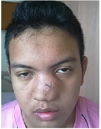
the nose. The last four
There was no h
Fig. 1
External nose enlargement
left frontal sinus. There were signs of
Physical examination revealedpolypoid mass filling the entire nasal cavity
with enlargement of the nose. Frontal view appearance showed nose enlargement with no distinctive border of the left nose. From the basal view appearance, the tip of the nose is deviated
to the right side. Sinus paranasal CT Scan taken
on December 19th, 2012 showed opacities on
the right and left nasal cavities, both choana and nasopharyngeal space, bilateral maxillary sinuses, bilateral ethmoid sinuses, left sphenoid
sinus and left frontal sinus. There were signs
of erosion of the nasal septum, bilateral medial maxillary sinus walls, left posterior maxillary sinus wall, left orbital inferomedial wall, and
left inferior sphenoid sinus wall. There was
thickening of right sphenoid sinus mucosa.
the nose. The last four
There was no h
Fig. 2
Anterior rhinoscopy
side. Sinus paranasal CT
2012 showed
left frontal sinus. There were signs of
sphenoid sinus wall. There was thickening
78
ORLI Vol. 44 No.1. Tahun 2014 ORLI Vol. 44 No.1. Tahun 2014 Woakes syndrome Woakes syndrome
Paranasal CT
, 2013 with the
, 2013. Polypoid mass was
mass. The bleeding from the
prescribed coamoxiclav 3x625 mg, mefenamic acid 3x500 mg and
2013 and nasoendoscopy
ENT Division for rhinoplasty correction.
2013. Graft augmentation was
Fig. 3 Paranasal CT Scan of the patient with WoakesSyndrome
Patient underwent biopsy of the nasal mass in January 15th, 2013 with the result of
chronic rhinitis and nasopharyngitis. FESS was performed in March 8th, 2013. Polypoid mass
was extracted with forceps. Inferior and medial
turbinate was difficult to recognize due to the expansion of the polypoid mass. The bleeding
from the sphenopalatine artery was cauterized. Uncinectomy was performed with backbiting forceps, followed by middle meatal antrostomy
finding no polyp inside the maxillary antrum.
Histo-pathology examination showed edematous polyps with necrosis and massive bleeding and no signs of malignancy.
Patient was discharged on the fifth day
after hospitalization and was prescribed
coamoxiclav 3x625 mg, mefenamic acid 3x500 mg and normosaline solution for nasal
rinsing. First post operative follow up was done on March 14th 2013 and nasoendoscopy
examination revealed wide left nasal cavity, there was clotting and no mass. Right nasal cavity still narrow due to septal deviation. Patient did not complain of nasal blockage anymore and could breathe through the nose. Four months after surgery, patient
prepared for second stage surgery by Plastic
Reconstruction ENT Division for rhinoplasty
correction.
Fig.4 Polypoid tissue of surgical specimen.
Septorhinoplasty was performed on July 8th 2013. Graft augmentation was taken from left auricle cartilage. Transcollumelar incision was
carried out, followed by infracartilage incision. Septal cartilage was incised sparing superior and posterior part. Septal extension graft was done with interdomal suture. Osteotomy was performed at right and left side. Nasal splint was
maintained for 2 weeks and removed on July 25th, 2013. Patient was satisfied with the result.
79
ORLI Vol. 44 No.1. Tahun 2014 ORLI Vol. 44 No.1. Tahun 2014 Woakes syndrome ORLI Vol. 44 No.1.
Fig.5 patient 16 years old boy with Woakes syndrome after septorinoplasty
DISCUSSION
According to Slavin et al as quoted by Casale,6 nasal polyps are present in approximately 1% to 4% of the population, more often in males than females. This is
similar with the case reported in this paper.
Case reports in published journals showed
age incidence of Woakes syndrome under 10
years old.3 Our case was a male teenager. Patient in this case presented with nasal blockage and frequent episodes of rhinorrea
since 3 years previously. He also complained
loss of smell and moderate headache.
According to Tritt et al,10 patients with nasal polyposis may present clinically with complaints of nasal obstruction, congestion, hyposmia, rhinorrhea, epistaxis, postnasal drip, headaches, and snoring. Patient also complained of enlargement of his left nose. Broadening of the nose is a characteristic of Woakes syndrome as described by Caversaccio et al.1
T
splint was maintained for 2 weeks and
removed on July 25 , 2013. Patient was
.5
patient 16 years old boy with W
approximately 1% to 4% of the
females. This is similar with the case
syndrome under 10 years old
3rhinorrea since 3 year
moderate headache. According to Tritt et
10
The
pressure of the polyps. This is
CT s
Physical examination revealed clearpolypoid mass filling the left nasal cavity.
Nasal polyps typically present bilaterally but can present unilaterally. Unilateral nasal masses may be benign unilateral nasal polyps in relation with chronic rhinosinusitis. Biopsy from nasal and nasopharyngeal region showed chronic rhinitis and nasopharyngitis, which conclude that the patient has unilateral nasal polyp.7
The extreme broadening of the nose in
our patient is explained by the ongoing
pressure of the polyps. This is in accordance
with hypertrophic process and nasal pyramid deformation, one of the four characteristics of Woakes syndrome.1
Paranasal sinus CT scan is needed to prove the frontal sinus aplasia. These findings added to the biopsy result suggest a
Woakes Syndrome. Woakes’ syndrome was
defined as necrotizing ethmoiditis and nasal
polyps with broadening of the nose. The
syndrome was defined by the following four
characteristics: (1) bilateral nasal polyps in the middle meatus beginning during
childhood, (2) ethmoiditis, (3) hypertrophic
process with nasal pyramid deformation
and (4) therapeutic failure with constant
and rapid recurrences.1 In our case, frontal
sinus aplasia was not found, but there was
full opacification of left frontal sinus without any polps inside it. The left frontal sinusitis
is caused by obstruction of the frontal recess. We extirpate the bulk of nasal mass by nasoendoscopic procedure and sent it for histo-pathological examination. Functional
Endoscopic Sinus Surgery (FESS) is
considered the gold standard for the surgical treatment of chronic rhinosinusitis with or without nasal polyps. It is performed with a rigid endoscope which allowed a complete visualization of the nasal and paranasal
cavities. The aims of this surgical technique
is to restore normal sinus ventilation and
80
ORLI Vol. 44 No.1. Tahun 2014 ORLI Vol. 44 No.1. Tahun 2014 Woakes syndrome Woakes syndrome
drainage removing polyps or other tissue obstructing the osteomeatal complex. FESS could minimized the recurrence rate of the
disease. The extent of surgery varies with
the extent of disease, the surgeon’s expertise, the available equipments and technology, and patient’s condition and expectation.11-13
Four months afterwards septorhinoplasty was performed. The goal of septorhinoplasty and rhinoplasty are to restore nasal function by maximizing nasal airflow and to improve cosmetic appearance. There are two completely different methods of rhinoplasty for correcting nasal pyramid deformations; the first is in which the osteo-cartilaginous resection of the nasal dorsum is carried out and the second is based on preservation of the dorsum.9,14 The two techniques were never combined because osteo-cartilaginous resection of the nasal hump prevents complete disarticulation of the septal cartilage from its attachment on the perpendicular plate of the ethmoid bone and the vomer, an essential step in Cottle septoplasty. Indeed, once the septal cartilage is completely detached from its posterior and inferior edges, as performed in Cottle septoplasty, it can no longer be separated from the upper lateral cartilages as performed in nasal hump resection. This would involve total detachment with major risk of post-operative saddling and, moreover, stenosis of the internal nasal valve due to the collapse of the upper lateral cartilages.2,14
In the literatures reviewed, there was only one case of Woakes syndrome which underwent septorhinoplasty.2
In this paper, a case of Woakes syndrome
was reported. The patient underwent two stages surgery, the first was FESS to clear out
the polyp mass and the second surgery was septorhinoplasty to restore physiologic function
and aesthetic appearance of the nose.
REFERENCE
1. Caversaccio M, Baumann A, Helbling A. Woakes’ syndrome and albinism. Auris Nasus
Larynx 2006; 34:245-8.
2. Abbud-Neme F, Reynoso VM, Reiss ED. Woake’s syndrome. A case report in a teenager. Int J Pediatr Otorhinolaryngol
1987; 12:327-33.
3. Kellerhals B, Utheman Bd. Woakes’ syndrome: the problem of infantile nasal polyps. Int J Pediatr Otorhinolaryngol 1979; 1:79-85.
4. Groman JD, Bolger W, Brass-Ernst L, Macek M Jr, Zeitlin P, Cutting G. Recurrent
and destructive nasal polyposis in 2 siblings:
a possible case of Woakes’ syndrome.
Otolaryngol Head Neck Surg 2004; 131:1009-11.
5. Walsh WE, Kern RC. Head & neck surgery.
4 ed. Bailey BJ, Johnson JT, Newland SD, editors: Lippincott Williams & Wilkins; 2006.
6. Casale M, Pappacena M, Potena M, Vesperini E, Ciglia G, Mladina R, et al. Nasal polyposis: from pathogenesis to treatment,
an update. Inflamm Allergy Drug Targets 2011; 10:158-63.
7. Akarcay M, Ekici N, Miman MC, Firat Y,
Bayindir T, Selimoglu E. Do comorbidities influence objective and subjective recovery
rates of nasal polyposis? J Craniofac Surg
2010; 21:71-4.
8. Newton JR, Ah-See KW. A review of nasal
polyposis. Ther Clin Risk Manag 2008; 4(2):507-12.
9. Chisholm E, Jallali N. Rhinoplasty and septorhinoplasty outcome evaluation. Ear
Nose Throat J 2012; 91:10-14.
10. Tritt S, McMains KC, Kountakis SE. Unilateral
nasal polyposis: clinical presentation and pathology. Am J Otolaryngol Head and Neck
Med Surg 2008; 29:230-2.
81
ORLI Vol. 44 No.1. Tahun 2014 Woakes syndrome ORLI Vol. 44 No.1.
11. Ajaml. Nasal polyp-etiology-clinical finding. Available from: http://medilinks.blogspot.
com/2011/07/nasal-polyp-etiology-clinical-findings.html%5D.
12. Bechara Y. Ghorayeb M. Available from:http:// www.ghorayeb.com/antrochoanalpolyp.
html%5D.
13. Fokkens WJ, Lund VJ, Mullol J, Bachert C, Alobid I, Baroody F, et al. Rhinology
Official Journal of the European and
International Societues European Position Paper on Rhinosinusitis and Nasal Polyps
2012; 50:1-329.
14. Boulanger N, Baumann C, Beurton R, Elueque H, Gallet P, Grosjean R, et al. Septorhinoplasty by disarticulation: early assessment of a new technique for morphological correction of
crooked noses. Rhinology 2013; 51:77-87.
82