THESIS
THE EFFECTS OF PROPOLISON
HISTOLOGICALFEATURESONSMALL INTESTINE
OF MICE (
Musmusculus
)
By:
KARINA RATNANINGRUM 061111037
FACULTY OFVETERINARY MEDICINE
UNIVERSITAS AIRLANGGA SURABAYA
vi ABSTRACT
The purpose of this research was to determine the side effect of propolis on histology of small intestine of mice (Musmusculus). 25 male mice (Musmusculus) aged 12-week-old with average 25-30 gram were used. Mice divided randomly into five groups; each group consisted of five mice. P0 as control group, and other groups; P1, P2, P3 and P4 were given propolisethanolic extract orally with doses as follows; 1.6 mg/0.5ml/day, 3.2 mg/0.5ml/day, 6.4 mg/0.5ml/day and 12.8 mg/0.5/day. Treatment were given for two weeks, animal were euthanized and the organs (small intestine) were taken for making the histological specimen using H.E staining. Objects observed were epithelial villi damage, congestion &oedema and neutrophil infiltration. Data were analyzed using Kruskall-Wallis method, continued using Mann-Whitney test if there’s significant difference. The result showed that treatment using different dose of ethanol extractedpropolis did not give any significant difference effect toward histological features of small intestine of mice.
vii
ACKNOWLEDGEMENT
First of all, my greatest gratitude for Jesus Christ for the blessing and
mercy so my thesis entitled THE EFFECT OF PROPOLIS ON HISTOLOGICAL
FEATURES ON SMALL INTESTINE OF MICE (Musmusculus) has been
completed.
In arranging this thesis, I would like to give my sincere gratitude to:
Dr. PudjiSrianto, M. Kes.,drh. as the dean, Prof. Dr. Fedik Abdul Rantam,
drh. as the first vice dean, and Dr. Rr. Sri Pantja Madyawati, drh., M.Si. as the
head of academic division of the Faculty of Veterinary Medicine, Universitas
Airlangga.
My supervisor committee, SuzanitaUtama, drh., M. Phill., and Ph. D Prof.
Hj. RomziahSidik, drh., Ph.D. for the guidance, advices, motivations, and
patience from the beginning up to the completion of this thesis. The advisor chief,
Dr. EkaPramyrthaHestianah, drh.,M.Kes., who also act as creator of my research,
and the advisor committee, Dr.Hani Plumeriastuti, drh., M.Kes and drh.
HardanyPrimarizky, MVM and for all the advices and corrections.
Prof. Dr. M.Lazuardi, drh., M. Si. as my academic counselor for all
experience, help, patience, and guidance throughout the years. All the lecturers for
the knowledge, information, help, and encouragement.
My lecturer Djoko Legowo, drh.,M.Kes and my friends Bilqisthi Ari Putra
and Lucky Ramadhan who are very helpful to do my observation of specimens
viii
me. My sisters and brothers, Ajeng and Rio for all the love and motivations.
To my precious college best friends, Dinar, Elsa, Gita and Riri, my
beloved classmates in D class 2011, Dea, Gheby, Benda, Kemala, Inanda, Diana,
Dona, Belga, Tika, Hadi, Sony, Vidi and the others for all motivations,
informations, help, energy, prayers, and encouragement.
To my friends in UKM Taekwondo Universitas Airlangga who give me
encouragement and cheer me up to finish my thesis. Also, thanks to everyone that
can’t be mentioned one by one who was very helpful during my study.
I, as the author, understand that this writing is still lacking in several parts
and far from perfection. However, I sincerely hope that this research will be useful
for the advancement of science and may contribute to the veterinary medicine
world and the society.
Surabaya, February,17th 2016
ix
LIST OF APPENDICES ………xi
ABBREVIATIONS AND SYMBOLS ……….xii
CHAPTER 1 INTRODUCTION ……….…….1
1.1 Background of Problem………..1
1.2 Identification of Problem………2
1.3 Theoritical Base ……….3
1.4 Objectives of Research………...………5
1.4.1 Spesific objectives ……….5
1.4.2 General objectives ……….5
1.5 Outcomes of Research ………6
1.6 Hypothesis of Research ………..6
CHAPTER 2 LITERATURE REVIEW ………..……….7
2.1 Characteristic of Propolis ………...………6
2.2 The advantage of Propolis ……….………9
2.3 Histological Features of Small Intestine……...………10
2.4 Mice (Musmusculus) as Experimental Animal …..………13
CHAPTER 3 MATHERIAL AND METHODS ………16
3.1 Place and Period of Research ……..………16
3.2 Design of Research ………..16
3.3 Research Material ………..…..17
3.4 Research Method ………..18
3.4.1 Preparing extract of propolis ………..…18
3.4.2 Treatment in experimental animal ……….…18
3.5 Preparing Histological Specimen of Small Intestine…..……..…20
3.5.1 Organs collection ………...……….20
3.5.2 Histological specimenmaking process ………..…….20
3.6 Examination of Histological Specimen of Small Intestine ……..20
3.7 Data analysis ………..……….21
3.8 Research Framework ………22
CHAPTER 4 RESULT OF RESEARCH ………..……23
4.1 Epithelial Villi Damage ………25
4.2 Congestion and Oedema ………..26
4.3 Neutrophil Infiltration ………..27
CHAPTER 5 DISCUSSION ……….………28
5.1 Epithelial Villi Damage ………..……….28
x
xi
LIST OF TABLE
Table Page
2.1 Chemical Composition of Propolis ………... 9 4.1 Scoring of epithelial damage, congestion & oedema, and neutrophil infiltration (Mean±SD) of small intestine of mice (Mus
xii
2.1 Apismellifera ... .7
2.2 Histological picture of small intestine ... 12
2.3 Musmusculus ... 14
4.1 Villi of small intestine intended to visualize epithelial damage ... 25
4.2 Red blood cells accumulation in venule and oedema in muscular layer of the small intestine ... 26
xiii
LIST OF APPENDICES
Appendix Page
1. Propolis Extraction Process ... 40
2. Propolis Dilution Process ... 42
3. Dosage Convertion Table ... 43
4. Determination of the Number of Minimal Replications ... 45
5. Determination of the Dose of Propolis ... 46
6. Dosage Calculation ... 47
7. Procedure of Making Histological Specimen ... 49
8. Scoring Result... 52
9. SPSS Windows Statistic Result ... 43
xiv Ca : Calcium
CMCna :Carboxymethyle Cellulose Natrium CRD : Completely Randomized Design
Cu : Cuprum
et al : et alli
Fe :Ferum
GDC :Gedung Diagnostic Center H+ : Ion Hidrogen
HE :Hematoxilin Eosin
I :Iodin
K+ : Ion Kalium Mg :Magnesium Mn : Manganese Na+ : Ion Natrium NaCl :NatriumClorida
pH : potential of Hidrogen
SPSS : Statistical Product and Service Solutions
1
CHAPTER 1 INTRODUCTION
1.1 Background of Problem
It has been known that Indonesia has many kinds of local bees. One of the
local bee propolis producer is Trigona sp. and Apis sp. (Angraini, 2006). The
genuses of Apis has several species, namely Apis cerana, Apis koschevnikovi,
Apis nigrocincta, and Apis mellifera. Apis mellifera honeybees are the most
productive and able to withstand all weather, makes this species became the most
widely farmed bees (Suranto, 2010).
Bee propolis is a mixture of compounds collected by honey bees from
various plant sources and used by bees to seal holes in their honey combs, smooth
out the internal walls and protect by entrance against intruders (Kamel, 2007).
Propolis is not only a building material, it is the most important “chemical
weapon” of bees against pathogen microorganisms and has been used as a remedy
by humans since ancient times (Bankova, 2005). Etymologically, the Greek word
propolis means “pro”, means indefence, and “polis” means the city, so that the
meaning is “defence of the hive” (Sforcin and Bankova, 2007).
In general, propolis is composed of 30% wax, 50% resin and vegetable
balsam, 10% essential and aromatic oils, 5% pollen, and other substances
(Burdock, 1998). Propolis contains some minerals such as Mg, Ca, I, K, Na, Cu,
Zn, Mn and Fe as well as some vitamins like B1, B2, B6, C and E, and a number
of fatty acids (Thikonov, 1987 in Lotfy, 2006). Halim et al. (2012) in their study
comparing propolis products from Indonesia (Apis Melifera) and propolis
products from Brazil and the result said that Indonesian Propolis has a content of
vitamins and mineral nutrients (vitamin B1, B2, B6, C, and E and minerals Na,
Ca, Mg, Cu, Zn, Mn and Fe) is higher than the Brazilian propolis, but its levels of
vitamin A are lower.
There are so many benefits of propolis. In 1963, a rat was found dead for 5
years inside a bee hive and does not rot. This happens because the rat carcass
wrapped by propolis which proved that propolis contains an antimicrobial agent.
In addition of its antibacterial effect, some research indicates that effective
propolis as anticancer, antiviral, antifungal, antioxidant, immune-boosting,
strengthen, anti-inflammatory, and accelerate cell regeneration (Angraini, 2006).
Plants and plant extracts are effective mainly on the digestive system of
animals. Their function either by wiping out the pathogenic microflora in the
digestive system or increasing the concentration of microbial population in the
digestive system that contributes to improved digestion and absorption of
nutrients (Wenk, 2000 in Tekeli et al., 2010).
1.2 Identification of Problem
Based on the proposed formulation of the problem has been described as follows:
1. Does propolis administration affect on the histological features of small
intestine of male mice (Musmusculus)?
2. Does different dose of propolis affect on the histological features of small
3
1.3 Theoritical Base
Small intestine of mice consisting of the duodenum, jejunum and ileum.
Small intestine has functions such as doing food digestion and absorption
(Samuelson, 2007). Villi is the most responsible part of nutrition absorbtion
because it has absortive cell in its mucosa which is a single layer of columnar
epithelial cells with striated border (Leeson, 2012).
Propolis contains of many nutrition values, like vitamin A, B, C, minerals
(Ca, Mg, Na, Fe, Mn, Cu, Zn). Active substances known in propolis are poliferol
(flavonoid, fenolat acid, and its esters), terpenoid, steroid, and amino acid.
Flavonoids are substances found in many plants and has an antioxidant effect on
decreasing free radical (Halim et al., 2012). Flavonoids and phenolic acids is
known as a substance that is capable of enhance the immune system. Propolis as
an immunomodulator is increasing the number of macrophage cells as a cellular
immune response when administered in a short period of time (Mustafiah et al.,
2011).
Propolis extract and other substance Z. officinale supplemented in the
ration both separately and in combination proved to stimulate lactic acid bacteria
and significantly decrease pathogenic bacteria such as total mesophilic aerobicm
coliform and E. Coli (Tekeli et al., 2010). Propolis showed a significant protective
effect on ileal mucosa and reduced bacterial translocation by enhancing mucosal
barrier function, supporting generalized immune function, and reducing bacterial
antibacterial, antifungal, antiviral, hepatoprotective, immunomodulatory
anti-inflammatory, and anti-ulcer properties (Castaldo, 2002 in de Barros etal., 2007).
Naturally, propolis contains of wood chips, sand and leaves. To separate
propolis from those material, the used method is using ethanol solvent extraction.
Ethanolic extract of Apis mellifera propolis had a potent antioxidant activity
(Radiati et al., 2008).
The excess of propolis as a natural antibiotic compared with synthetic
material is safer and side effects are minor. Previous studies stated the only side
effects occured and rarely happened was allergic reaction when it used locally,
whereas given orally did not cause resistance. (Winingsih, 2004 in Angraini,
2006).
Research about the side effect of propolis were still minimum thus
propolis aministration to mice or to humans does not seem to have side effects
(Sforcin and Bankova, 2007). The knowledge of propolis volatile oils is far from
being exhaustive. The flora that bee used to collected porpolis determines the
chemical composition of propolis, including volatile compounds. Volatile
compound, which is the secondary plant metabolites are produced by different
plant species and are not the same in all over the countries. The term “propolis”
does not have any specific chemical connotation, unlike the scientific name of a
plant species (Bankovac, 2014). The research are going to determine whether the
area of local Apis mellifera to collecting propolis will give the side effect the
5
In acute inflammation of small intestine, there was transient
vasoconstriction followed by vasodilatation increased capillary permeability and
decrease in blood flow. Thus happened congestion of blood vessels, oedema plus
the presence of fibrin network, and also infiltration of leucocytes such as
neutrophils, lymphocytes, macrophages, eosinophils (Chauhan, 2010). Ulcer are
the loss of mucosa and deeper tissue. It happened when cells repaired from acute
inflammation (Kemp et al., 2008).
Therefore, the research would examine the effect of propolis ethanolic
extract based on three objects, which were: villi epithelial damage, congestion and
oedema, and neutrophil infiltration.
1.4 Objectives of Research 1.4.1 Specific objectives
1. To observe the effect of propolis on the histological features of small intestine
of male mice (Mus musculus).
2. To observe the effect of different dose of propolis on the histological features
of small intestine of male mice (Musmusculus).
1.4.2 General objective
This study aimed to determine the provision of propolis can be used as
1.5 Outcomes of Research
The results of this study were expected to provide information to the
public that propolis is a natural remedy that is safe for the intestinal organs.
Propolis can be useful for human and veterinary medicine, and also to contribute
knowledge to medical and veterinary science.
1.6 Hypothesis of Research
Hypothesis proposed in this study are:
1. Administration of propolis affect on the histological features of small intestine
of male mice (Musmusculus).
2. The different dose of propolis affect on the histological features of small
7
CHAPTER 2 LITERATURE REVIEW
2.1 Characteristic of Propolis
Bees have been in existence for 125 million years and their evolutionary
success has allowed them to become perennial species that can exploit virtually all
habitats on earth. This success was largely because of the chemistry and
application of the specific products that bees manufacture: honey, beeswax,
venom, pollen, royal jelly and propolis. (Wollenweber, 1990 in Bankova, 2005).
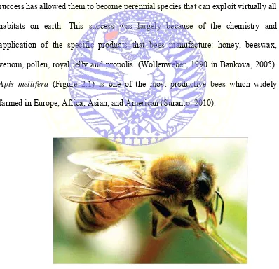
Apis mellifera (Figure 2.1) is one of the most productive bees which widely
farmed in Europe, Africa, Asian, and American (Suranto, 2010).
Figure 2.1 Apismellifera (Suranto, 2010)
Propolis is not only a building material, it is the most important “chemical
weapon” of bees against pathogen microorganisms and has been used as a remedy
by humans since ancient times. It was applied for wounds and burns treatment,
soar throat, and stomach ulcer (Wollenweber, 1990 in Bankova, 2005).
Percentage of workers who are in charge of collecting bee propolis is very
low but it is done all the time. At the source location, bee propolis collector bite
the material with their mandibulae (lower jaw) and with the help of a pair of first
leg. Propolis is transferred to the pollen basket. This activity takes 15-60 minutes.
In the hive, hive guards bee move propolis from collector bee‟s arm and
dispensing propolis with their mandibulae, sometimes add a little wax. Then
propolis transfered to where it‟s needed or stored as a backup. (Gojcmerac, 1993
in Angraini, 2006).
Honeybees collect propolis from plants and use it in their hive. They apply
it to seal the walls, to strengthen the borders of combs, to embalm dead invaders.
(Wollenber, 1990 in Bankova, 2005). It is dense, sticky, blackish brown, has a
typical smell, and a bitter taste (Radiati et al., 2008). Propolis has a characteristic
smell and shows adhesive properties because it strongly interacts with oils and
proteins of the skin (Burdock, 1998 in Sforcin and Bankova, 2007).
Propolis in natura is composed of 30% wax, 50% resin and vegetable
balsam, 10% essential and aromatic oils, 5% pollen, and 5% other substances
(Sforcin and Bankova, 2007). Krell (1996) in Radiati et al. (2008) describes the
9
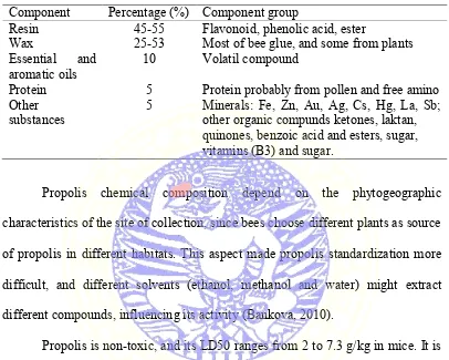
Table 2.1 Chemical Composition of Propolis (Source: Krell, 1996). Component Percentage (%) Component group
Resin 45-55 Flavonoid, phenolic acid, ester
Wax 25-53 Most of bee glue, and some from plants Essential and
aromatic oils 10 Volatil compound
Protein 5 Protein probably from pollen and free amino Other
substances 5 Minerals: Fe, Zn, Au, Ag, Cs, Hg, La, Sb; other organic compunds ketones, laktan, quinones, benzoic acid and esters, sugar, vitamins (B3) and sugar.
Propolis chemical composition depend on the phytogeographic
characteristics of the site of collection, since bees choose different plants as source
of propolis in different habitats. This aspect made propolis standardization more
difficult, and different solvents (ethanol, methanol and water) might extract
different compounds, influencing its activity (Bankova, 2010).
Propolis is non-toxic, and its LD50 ranges from 2 to 7.3 g/kg in mice. It is
suggested that the safe concentration for humans could be 1.4 mg/kg and day, or
approximately 70 mg/day (Burdock, 1998 in Sforcin and Bankova, 2007).
2.2 The advantage of Propolis
The ethanolic extract of propolis has been reported to possess various
biological activities, such as antibacterial, antiviral, antiinflammatory,
local-anesthetic, antioxidant, immunostimulating and cytostatic (Silici and Kutluca et
al., 2005).
The pharmacologically active molecules are flavonoids, phenolic acids,
and their esters. These components have multiple effects on bacteria, fungi and
immunomodulatory activities, and antitumor activity. Moreover, propolis has
been shown to lower blood pressure and cholesterol levels (Lotfy, 2006).
Tekeli et al. (2010) found that propolis extract and other substance Z.
officinale supplemented in the ration both separately and in combination proved
to stimulate lactic acid bacteria and significantly decrease pathogenic bacteria
such as total mesophilic aerobicm coliform and E. coli. This effect might be due
to the the ability of essential oils in terms of inactivication of extracellular
enzymes and their antibacterial properties which lead to death of bacteria by
decreased pH of the medium and damage to cell wall structure.
Phenolic structures denature the proteins in the cell wall of the bacteria
and increase cell wall permeability. The corrupted permeability of the cell wall
induces the release of the intracellular fluid, which consequently kills the bacteria.
Phenolic substances that include cinnamic acid derivatives and some flavonoids
are detected in honey and propolis (Kutlu, 1999 in Tekeli et al., 2010).
2.3 Histological Features of Small Intestine
There are three parts of small intestine: duodenum, jejunum and ileum,
while the caecum, colon and rectum are part of the large intestine. Intestinal
epithelium covered by one layer of columnar epithellium with numerous goblet
cells. (Leeson et al., 2012). Though the submucosal Brunner‟s glands are ussually
confirmed that a section were taken from duodenum parts (Scudamore, 2014).
The small intestine has features that enhance food digestion and
11
modifications plus the development of microvilli along the apical surface of the
luminal cells are designed to greatly expand the surface area for absorption
(Samuelson, 2007).
The mucosal folds or plicae, are prominent semicircular extensions project
into the lumen and as a result more than double the surface area of the epithelial
lining. Along the entire inner mucosa, extensions smaller than the plicae project
into the lumen forming the villi. The villi tend to be more developed within the
proximal portion (duodenum) than those located distally (ileum). The shape of the
villi in animals vary: short and conical in rodents. The presence of villi easily
increases the surface area by 10 times or more. (Samuelson, 2007).
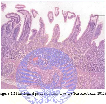
The mucosa of small intestine is thrown up into finger like projecting villi
separated by crypts and confined by single layer of columnar epithelial cells with
goblet cells (Figure 2.2) Goblet cells are found along the small intestine but tend
to be more numerous in the ileum (Scudamore, 2014). Although their heights
exceeds their widths, these secretory cells become round as their cytoplasm
becomes filled with mucigen. When mucigen is released as mucin into the
intestinal lumen, it becomes hydrated and better known as mucus. Mucus protects
the mucosal epithelium as a whole and facilitates the movement of nonabsorbed
Figure 2.2 Histological picture of small intestine (Kierszenbaum, 2012)
Between the base of adjacent villi the epithelium invaginates into the
adjacent lamina propia, forming simple tubular glands, called intestinal crypts or
crypts of Lieberkuhn. These glands composed of a absorptive and goblet cells in
the upper half. Regenerative cells which provided the replenishment of the
absorptive and goblet cells lie more within the lower halves of the glands. Toward
the bottom of each are found the acidophilic granule cells or Paneth cells. Those
cells are typically pyramidal and have been shown to have antimicrobial
capabilities (Samuelson, 2007).
The form of lamina propria is a loose connective tissue, which is the center
of the villi and surrounding intestinal glands, composed of collagen and elastic
13
lymph vessels, leukocytes, fibrocyte, smooth muscle, plasma cells and mast cells
(Eurell et al., 2007). Intestinal glands extend from the base of the villi into the
underlying lamina propia. Undifferentiated epithelial cells located in the glands
divide and migrate up to renew the glandular and surface epithelium every 4-5
days (Leeson, 2012).
Tunica submucosa consists of loose connective tissue containing collagen
and elastic fibers, located between the lamina muscularis mucosa and tunica
muscularis (Eurell et al., 2007). The myenteric plexus is located between the
Submucosal glands or Brunner‟s glands and Aggregated lymphatic nodules
(GALT) are present in submucosa layer. The nodules are particularly numerous in
the ileal region. Specialized M cells which present antigens to the lymphoid tissue
are present in the epithelium which overlies the nodules (Leeson, 2012).
Tunica muscularis is consists of two layer of smooth muscle, the inner
layer of circular muscle and outer layer of longitudinal muscle. The myenteric
plexus (Auerbach) is located between the inner and outer layers of the tunica
muscularis and functions to direct the outer longitudinal muscle. (Samuelson,
2007). Tunica serosa lies outside the tunica muscularis as the outermost layer of
the small intestine (Leeson, 2012).
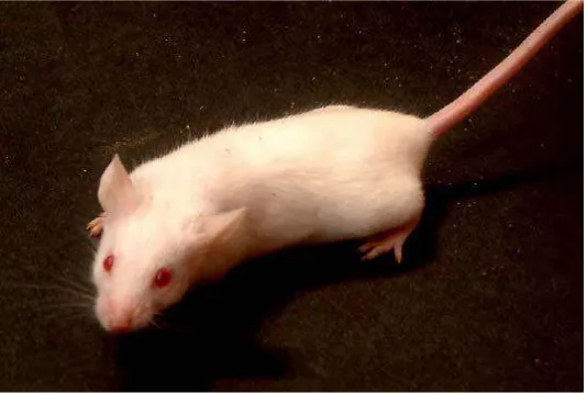
2.4 Mice (Musmusculus) as Experimental Animal
Mice (Mus Musculus) are rodentia animal which easy to breed, also easy
to mantain in large scale, have many various genetics and have good anatomic and
Mice or white mouse are frequently used as a experimental animals for
research. This laboratory mice strains have many good inbread (DDY, Balb / c,
DBA, and B6) and outbred like webster. Ballenger (1999) classified the
laboratory mice are as follows:
Kingdom : Animalia Phylum : Chordata Subphylum : Vertebrata
Class : Mammalia
Order : Rodentia
Family : Muridae
Subfamily : Murinae
Genus : Mus
Species : Mus musculus
Mice (Mus musculus) have characteristics such as small and white body.
(Figure 2.1). Cage condition for maintenance of mice (Mus musculus) should
always be clean, dry and away from the noise. Room temperature should be
maintained range between 18-19ºC and relative humidity between 30-70%.
15
Adult mice long life of 1-2 years, can reach 3 years. Male or female mice
can be bred at the age of 8 weeks. Long gestation 19-20 days. The average
number of giving birth was 6-15 mice, with birth weight between 0.5- 1.5 g
CHAPTER 3
MATERIALS AND METHODS
3.1 Place and Period of Research
This study were held at the Laboratory of Experimental Animal, Faculty
of Medicine, Universitas Airlangga for the treatment of experimental animals and
organs collection. The extraction were carried out in the Laboratory of
Phytochemistry, Faculty of Pharmacy, Universitas Surabaya. The making of
histological specimens were performed at the Gedung Diagnostic Center (GDC)
Dr. Soetomo Hospital. The examination of histological specimens of small
intestine were held at Laboratory of Histology, Veterinary Medicine Faculty,
Universitas Airlangga. Implementation of this research are performed from
November 2014 to January 2015.
3.2 Design of Research
The research of effect of propolis ethanolic extract on small intestine (Mus
musculus) mice changes was using experimental with CRD (Completely
Randomized Design) patterns. The type of research is experimental research with
25 male mice (Mus musculus) used as the experimental animal. Experimental
animals divided into five group with five repetition, P0 as control group (0.5 ml
Tween80/head/day), P1 (1.6 mg propolis/kgBW/head/day), P2 (3.2 mg
propolis/kgBW/head/day), P3 (6.4 mg propolis/kgBW/head/day), and P4 (12.8 mg
propolis/kgBW/head/day). Examination were carry out by looking at the changes
occured in the small intestine organ with post-test design.
17
The independent variable of this research is propolis dose given to
experimental animals, while the dependent variable is the histological features of
small intestine after administration of propolis ethanolic extract. The control
variable are the type of mice, feed, age of mice, and tools for research.
3.3 Reasearch Material
Experimental animals used in this study were 25 of 12-week-old male
mice (Mus musculus) with an average weight of 25-35 grams. Mice (Mus
musculus) were obtained from PUSVETMA Surabaya and developed in
Laboratory of Experimental Animal of Medicine Faculty, Universitas Airlangga.
The equipment used in this study include stomach tube (1 ml), water and
feed box, plastic cage with length 50 cm, width 50 cm, height 40 cm, and husk.
The other tools are digital scales, plastic bags, scrap paper, scissors, pipette,
bottles, knives, syringes, forceps, scalpel, cutting tools, plastic pot, measuring
tube, object glasses and cover glasses, microtome, hot plate, microscope, and lens
micrometer.
Propolis that used in this research was the result of extraction and dilution
in Phytochemistry Laboratory of the Faculty of Pharmacy, Universitas Surabaya.
Material used for extraction is ethanol 70%. Materials used for dilution are
Tween80 and E-pure. Materials required for making the histological specimens
are formalin 10%, alcohol, or ethanol (70%, 80%, 90%, absolute), xylol,
balsam, and paraffin block. (Muntiha, 2001). Mice were given shaped pellet feed
3.4.1 Preparing extract of propolis
The basic ingredients of propolis is a mixture of beeswax, resin and soil
attached to the honeycomb Apis mellifera. Material samples used are raw propolis
from local Apis mellifera obtained from Ranch Bees Rimba Raya, Lawang,
Malang, East Java, which was extracted using the method of maceration with 70%
ethanol. Extraction was carried out in the Laboratory of Phytochemistry, Faculty
of Pharmacy, Universitas Surabaya. The extraction process desribed in Appendix
1.
3.4.2 Treatment in experimental animals
The second stage of research, the treatment on mice (Musmusculus) males
held in cage experiments. Mice (Mus musculus) males (age 12 weeks) with a
weight of 25-35 grams, was developed in the Laboratory of Experimental Animal,
Medicine Faculty of Airlangga University and randomized by table.
Determination of the number of minimal replications are listed in
Appendix 3. Experimental animals were divided into five groups, P0 as control
group (0.5 ml Tween80/head/day), P1 (1.6 mg propolis/kgBW/head/day), P2 (3.2
19
mg propolis/kgBW/head/day), given environment adaptation for one week.
Determination of the dose of propolis are listed in Appendix 4. The treatment of
3.5 Preparing Histological Specimen of Small Intestine 3.5.1 Organs collection
Collection of the small intestine will be start by anasthesized the male
mice using chloroform and then dissect the body to get the small intestine.
3.5.2 Histological specimen making process
The dissected small intestine will be kept in a pot which filled with
formaline. One specimen of small intestine was made with horizontal slice from
every male mice. The making of histological specimen and staining will be done
at GDC Dr. Soetomo Hospital, Surabaya. The procedure of histological specimen
will be explained in Appendix 4.
3.6 Examination of Histological Specimen of Small Intestine
Examination of intestine specimens that have been stained by HE
performed under a light microscope with a magnification of 400x to see epithelial
tissue damage, hemorrhagic congestion and mucosal oedema, and neutrophil
infiltration. Each specimens were observed in five field. The results of each field
are summed and then average were calculated.
The assessment of the level of damage in a single field can be seen below
(Pothoulakis et al., 1994):
1. Epithelial damage
Score 0: None of the villus are damaged.
Score 1: Destruction of tips of villi.
21
Score 3: Complete destruction of villi.
2. Congestion and Oedema
Score 0: None.
Score 1: Rare venules with Red Blood Cells .
Score 2: Up to half of venules with Red Blood Cells.
Score 3: More than half of venules with Red Blood Cells.
3. Neutrophil Infiltration
Score 0: None
Score 1: Rare of Polymorphonuclear cells which mainly marginated.
Score 2: Marginated and extravasated Polymorphonuclear cells (up to 5
each vessels).
Score 3: Marginated and extravasated Polymorphonuclear cells throughout
lamina propia and epithelium.
3.7 Data analysis
The form of data obtained stated in scores of histopathology changes
level in the small intestine of mice that arranged in table for later statistically
analyzed using the Kruskal-Wallis test. If there is a real difference, then the
analyzing using Mann-Whitney test (Mehotcheva, 2008). The whole process of
3.8 Research Framework
Environment adaptation for 7 days
P0
23
CHAPTER 4
RESULTS OF RESEARCH
Histological speciments of small intestine of mice (Mus musculus) of
control or P0 (Tween80 at a dose of 0.5 ml/day), P1 (propolis dose of 1.6
mg/kgBW/day), P2 (propolis dose of 3.2 mg/kgBW/day), P3 (propolis dose 6.4
mg/kgBW/day), P4 (propolis dose of 12.8 mg/kgBW/day) were stained by HE
and observed under a light microscope with a magnification of 400x to see
epithelial tissue damage, hemorrhagic congestion and mucosal oedema, and
neutrophil infiltration (Figure 4.1). The specimenswere observed in fivefield
each.Epithelial damage, congestion andoedema, and neutrophil infiltration of
small intestine of mice (Mus musculus) were scored using Pothoulakis method
(Pothoulakis, 1994). (Table 4.1).
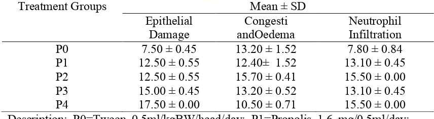
Table 4.1 Scoring of epithelial damage, congestion and oedema, and neutrophil infiltration (Mean ± SD) of small intestine of mice (Musmusculus).
Treatment Groups Mean ± SD
Epithelial
Damage andOedema Congesti Neutrophil Infiltration
P0 7.50 ± 0.45 13.20 ± 1.52 7.80 ± 0.84
P1 12.50 ± 0.55 12.40± 1.52 13.10 ± 0.45
P2 12.50 ± 0.55 15.70 ± 0.41 15.50 ± 0.00
P3 15.00 ± 0.45 13.20 ± 0.52 13.10 ± 0.45
P4 17.50 ± 0.00 10.50 ± 0.71 15.50 ± 0.00
Description: P0=Tween 0.5ml/kgBW/head/day; P1=Propolis 1.6 mg/0.5ml/day; P2=Propolis 3.2 mg/0.5ml/day; P3=Propolis 6.4 mg/0.5ml/day; P4=Propolis 12.8 mg/0.5ml/day.
Data analyzing using Kruskal-Wallis test resulted p=0.119 (>0.05) for
epithelial damage, meanwhile the result for congestion and oedemawas p=0.842
25
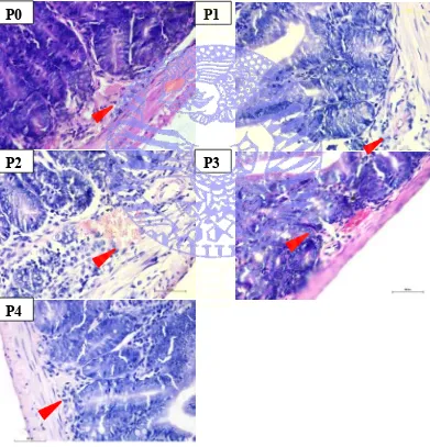
4.1 Villi Epithelial Damage
The histological features of epithelial damage of small intestine of mice
(Mus musculus) can be seen in Figure 4.1.
Figure 4.1 Villi of small intestine intended to visualize epithelial damage (HE staining; 400x magnification; P0=Tween 0.5ml/kgBW/head/day; P1=Propolis 1.6 mg/0.5ml/day; P2=Propolis 3.2 mg/0.5ml/day; P3=Propolis 6.4 mg/0.5ml/day; P4=Propolis 12.8 mg/0.5ml/day).
P4
P2
P0
P1
4.2 Congestion andOedema
The histological features of congestion and oedema of small intestine of
mice (Mus musculus) can be seen in Figure 4.2.
Figure 4.2 Red blood cells accumulation in venule and oedema in lamina propia and submucosa layer of small intestine. Red arrows shows red blood cells accumulation. (HE staining; 400x magnification; P0=Tween 0.5ml/kgBW/head/day; P1=Propolis 1.6 mg/0.5ml/day; P2=Propolis 3.2 mg/0.5ml/day; P3=Propolis 6.4 mg/0.5ml/day; P4=Propolis 12.8 mg/0.5ml/day).
P4
P3
P0
P1
27
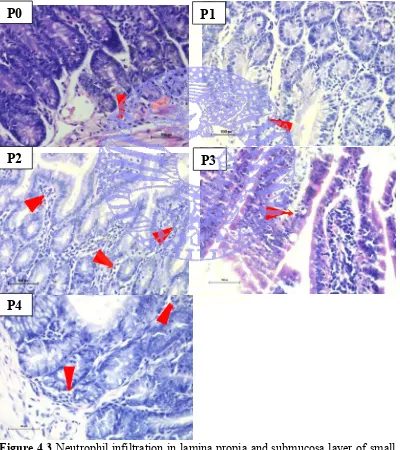
4.3 Neutrophil Infiltration
The histological features of neutrophil infiltration of small intestine of
mice (Mus musculus) can be seen in Figure 4.3.
Figure 4.3 Neutrophil infiltration in lamina propia and submucosa layer of small intestine. Red arrows shows PMN infiltration. (HE staining; 400x magnification; P0=Tween 0.5ml/kgBW/head/day; P1=Propolis 1.6 mg/0.5ml/day; P2=Propolis 3.2 mg/0.5ml/day; P3=Propolis 6.4 mg/0.5ml/day; P4=Propolis 12.8 mg/0.5ml/day).
P4
P2
P0
P1
CHAPTER 5 DISCUSSION
Bee propolis is a mixture of compounds collected by honey bees from
various plant sources and used by bees to seal holes in their honey combs, smooth
out the internal walls and protect by entrance against intruders (Kamel, 2007).
Plants and plant extracts are effective mainly on the digestive system of animals.
Their function either by wiping out the pathogenic microflora in the digestive
system or increasing the concentration of microbial population in the digestive
system that contributes to improved digestion and absorption of nutrients (Wenk,
2000 in Tekeli et al., 2010).
The observed effects were the epithelial damage, congestion and oedema,
and neutrophil infiltration in small intestine of mice.
5.1 Epithelial Villi Damage
The loss of the mucosa and deeper tissues are called ulcer. If only the
mucosa is lost, the correct term is erosion. The observation of small intestine
specimens, it was suspected that mice were suffered from erosion. Erosion
happened in several pathologic condition in duodenal, such as exudative diarrhea,
mucosal exposure to gastric acid, bacterial invasion. Ulcer and erosion happened
when body cannot riditself of the inciting agent.Macrophages, mast cells,
dendritic cells, granulocytes (neutrophils, eosinophils and basophils) and the
innate lymphocytes are the cells of the innate immune system converge at the site
of damage (Kemp et al., 2008).
29
The result told that propolis ethanolic extract treatment gave no significant
effect on epithelial damage of small intestine of mice. Control group had the
lowest mean rank which is 7.50 meant propolis could not act as remedy to
epithelial damage of small intestine of mice (Mus musculus) which in this
research, erosion were happened in every treatment group.
5.2 Congestion and Oedema
Congestion is passive accumulation of blood within vessels, where the
blood vessels are dilated by red blood cells. Due to multiple episodes of acute
passive congestion, red blood cells break down, leaving hemosiderin and
stimulate mild inflammation (Kempet al., 2008). There were production of
chemical factor in inflammation (histamine, serotonin, arachidonic acid
derivatives, quinine) which caused vasodilation and increased capillary
permeability, thus edema were formed (Jankowski, 2011).
The scoring results of treatment using propolis ethanolic extract gave no
significant effect on congestion and oedema of small intestine of mice. All
treatment showed the presence of congestion and oedema in similar condition. P4
treatment group (Propolis 12.8 mg/0.5ml/day) showed lowest mean rank. From
that result could be concluded that propolis ethanolic extract could act as a
5.3 Neutrophil Infiltration
In inflammation, there is transient vasoconstriction followed by
vasodilatation increased capillary permeability and decrease in blood flow.
Circulatory changes are more pronounced in acute inflammation. Thus happened
congestion of blood vessels, oedema plus the presence of fibrin network, and also
infiltration of leucocytes such as neutrophils, lymphocytes, macrophages,
eosinophils (Chauhan, 2010). Since it was the acute symptom that we were
looking for, the observation were focused on neutrophil infiltration.
The scoring result of treatment using propolis ethanolic extract gave no
significant effect on neutrophil infiltration of small intestine of mice. All
treatment showed the presence of neutrophil infiltration in similar
condition.Control group had the lowest mean rank which is 7.80 meant propolis
ethanolic extract could not act as remedy to neutrophil infiltration of small
intestine of mice (Mus musculus).
5.4 General Discussion
As it was already seen, there was erosion occurred in mice‟s histology
picture. Erosion and ulcer is acute inflammation form. (Kemp, 2008).The acute
inflammatory response could be stimulated by exogenous and endogenous agent
which resulted to injury in vascularized tissue. The response began as active
hyperemia, facilitated by various chemical mediator such as prostaglandins,
leuktrienes, nitric oxide caused dilation of arterioles and capillaries. Active
hyperemia is rapidly followed by changes in junctional complexes of endothelial
31
and plasma and plasma proteins can leak directly through breach in the wall of the
capillary or venule. Once activated, endothelial and perivascular cells such as
mast cells, dendritic cells, fibroblasts, and pericytes can produce cytokines and
chemokines that regulate the expression of receptors for inflammatory mediators
and adhesion molecules.
The plasma proteins and fluid that initially accumulate in the extracellular
space in response to injury form transudate. The most common the formation of a
transudate is due to hypertension in veins and capillaries or hypoproteinemia
resulting in oedema; however, transudates occur in the early stages of the acute
inflammatory response when intercellular gaps that open between endothelial
cells are so small that only water and electrolytes can pass through them. In time,
neutrophils and additional protein can enter injured areas resulting in the
formation of exudate (McGavin, 2007).
Propolis ethanolic extract gave no significant effect on histological
features of mice small intestine, specifically on epithelial damage, congestion and
oedema, and neutrophil infiltration. This is contrary to statement that propolis
were active as anti-inflammatory, strengthen and accelerate cell regeneration
(Gojmerac, 1983 in Angraini, 2006). Although higher dose, especially the highest
(Propolis 12.8 mg/0.5ml/day) showed better results in congestion and oedema,
significant differences was still could not be achieved.
It was probably because of mice used were still not SPF
(Spesific-Pathogen Free). According to International Committee of Laboratory Animal,
micro-organisms and parasites, but not necessarily free of others not specific‟.
The nature of animal facility and diets might modulate the outcome of study
(Reliene, 2006). So the mice were probably suffered with unknown pathogen
agent which lead to mild inflammation occurred in every treatment, include the
33
CHAPTER 6
CONCLUSION AND SUGGESTIONS
6.1 Conclusion
Based on the present research that had already been conducted, it showed:
1. Propolis ethanolic extract gave no significant effect on the histological
features on small intestine of mice.
2. Different level of propolis ethanolic extract dose did not affect histological
features of small intestine of mice.
3. Propolis ethanolic extract were safe when given orally to intestinal organ of
mice.
6.2 Suggestion
Based on the research results, the writer suggest:
1. Necessary to conduct a research on the effect of propolis extract using
different dose.
2. Necessary to conduct a research on the effect of propolis extract in other
experimental animal.
3. Necessary to conduct a research using older experimental animal.
4. Necessary to conduct a research using unhealthy animal.
SUMMARY
Karina Ratnaningrum. The Effect of Propolis On Histological Features of Small Intestine of Mice (Mus musculus). This present research was conducted under the guidance of Dr. Eka Pramytha H., M.Kes.,drh, as the creator
of research, Suzanita Utama, drh., MP.Phill., Ph.D as the main supervisor and
Prof. Hj. Romziah Sidik, drh., Ph.D as the co-supervisor.
Indonesia has many kinds oflocal bees Apis mellifera which became the
most widely farmed bees. Bee propolis is a mixture of compounds collected by
honey bees from various plant sources and used by bees to seal holes in their
honey combs, smooth out the internal walls and protect by entrance against
intruders. propolis is composed of 30% wax, 50% resin and vegetable balsam,
10% essential and aromatic oils, 5% pollen, and other substances.Active
substances known in propolis are poliferol (flavonoid, fenolat acid, and its esters),
terpenoid, steroid, and amino acid.
Researchs suggest that propolis has many effects such as antibacterial,
anticancer, antiviral, antifungal, antioxidant, immune-boosting, strengthen,
anti-inflammatory, and accelerate cell regeneration. Small intestine has functions such
as doing food digestion and absorption. Villi is the most responsible part of
nutrition absorbtion because of its absortive cell in mucosa with a single layer of
columnar epithelial cells with striated border.
In acute inflammation, there was transient vasoconstriction followed by
35
happened congestion of blood vessels, oedema plus the presence of fibrin
network, and also infiltration of leucocytes such as neutrophils, lymphocytes,
macrophages, eosinophils.
This study consisted of two stages: the propolis extraction process and
propolis treatment to experimental animals. Basic material used was raw propolis
from Rimba Raya Lawang bees ranch, as much as 500g and extracted using
ethanol 70%. Propolis extraction was conducted in the Phytochemistry
Laboratory Faculty of Pharmacy, Universitas Surabaya in November to
December2014. The second stage is propolis extract administration to
experimental animals using five treatments with five replications. The treatment
given to the inclusion criteria of male mice, aged 12 weeks, weigh 25-35g strain
Balb/c. Provision of propolis was held for 14 days preceded by a period of
adaptation for seven days. Experimental were using 25 mice (Mus musculus).
Plastic enclosure cages were used for maintenance of experimental animals at
Experimental Animal Laboratory, Faculty of Medicine, Universitas Airlangga in
December 2014 to January 2015. The observed parameters in this research were
epithelial tissue damage, hemorrhagic congestion and mucosal oedema, and
neutrophil infiltration. Experimental animals grouping of the present study used
Completely Randomized Design. Data were analyzed with Kruskall-Wallis Test.
The administration of propolis ethanolic extract show the result to
epithelial tissue damage (p=0.119), congestion and oedema (p=0.842), and
neutrophil infiltration (p=0.110), mean the administration of propolis ethanolic
specifically on epithelial damage, congestion and oedema, and neutrophil
infiltration. Suggestions for this research are conduct a research on the effect of
propolis extract using different dose, other experimental animal, the older
37
REFERENCES
Adhityananda, Werstant. 2014. Propolis Potential Toward the Amount of Limphoblast and Spleen Diameter of male mice (Musmusculus).
Akbar, B. 2010. Tumbuhan dengan Kandungan Senyawa Aktif yang Berpotensi sebagai Bahan Antifertilitas. Jakarta: Adabia Press UIN.
Angraini, A.D. 2006. Potensi propolis lebah madu Trigona sp. sebagai bahan antibakteri. Bogor: Fakultas Matematika dan Ilmu Pengetahuan Alam, Institut Pertanian Bogor.
Ballenger, L. 1999. “Musmusculus” (On-line), Animal Diversity Web,
http://animaldiversity.org/accounts/Mus_musculus/. April 2007.
Bankova, V. 2005(a).Chemical Diversity of Propolis and the Problem of Standardization. Journal of Enthopharmacology 100:114 – 117.
Bankova, V. 2005(b). Recent Trends and Important Developments in Propolis Research. Oxford University Press 2:29-32.
Bankova, V., Milena P., Boryana T. 2014 Propolis Volatile Compounds: Chemical Diversity and Biological Activity: a Review. Bankova et al. Chemistry Central Journal 2014, 8:28.
Burdock, G. A. 1998.Review of the Biological Properties and Toxicity of Bee Propolis (Propolis). Food and Chemical Toxicology 36: 343-367.
Chauhan, R. S. 2010. Text Book of Veterinary Pathology Quick Review and Self Assesment. IBDC Publishers.
De Barros, M. P., J. B. P. Sousa, J. K. Bastos, S. F. de Andrade. 2007. Effect of Brazilian Green Propolis on Experimental Gastric Ulcers in Rats. Journal of Ethanopharmacology 110: 567-571.
Eurell, J. A., Frappier, B. L. 2007. Dellman‟s Textbook of Veterinary Histology 6th Edition.
Hadiyah, Z.K. 2009. Local Propolis Extract Has Cytotoxic and Antiproliferative Effect on HeLa Cells. Jurnal Kedokteran Universitas Brawijaya 25: 17-22.
Halim, E., Hardinsyah, Sutandyo N., Sulaeman A., Artika M., Harahap Y. 2012. Bioactive Compounds and Nutrients Content of Indonesian and Brazilian Propolis. Jurnal Gizi dan Pangan 7: 1-6.
Jankowski, J. A. Z. 2011. Inflammation and Gastrointestinal Cancers. Springer Publishers.
Kamel, K. I., El-Hanoun A. M., El-Sbeiy M. S., Gad H. A. M. 2007. Effect of Bee Propolis Extract (Bee Glue) on Some Productive, Reproductive and Physiological Traits of Rabbits Does and Their Progenys. The 5th Inter
Con on Rabbit Prod. In Hot Clim, Hurghada, Egypt: 403-415.
Kemp, Walter L., Dennis K. B., Travis G. B. 2008. The Big Picture Pathology.The McGraw-Hill Companies, Inc.
Kierszenbaum, A. L. 2012. Histology and Cell Biology: An Introduction to Pathology. Elsevier Publishers.
Krell, R. 1996. Propolis. Value Added Products From Beekeeping.FAO Agricultural Service Bulletin. Food and Agricultural Organization of The United Nation, number 124: 157-194.
Leeson, C. Roland., Thomas S. L., Anthony A. P. 2012. Buku Ajar HistologiEdisi ke-5. Jakarta: PenerbitBukuKedokteran EGC.
Lotfy, M. 2006. Biological Activity of Bee Propolis in Health and Disease. Asian Pacific Journal of Cancer Prevention 7: 22-31.
McGavin, M. Donald, James F. Z., 2007. Pathologic Basic Of Veterinary Disease: Fourth Edition. Elsevier.
Mehotcheva, T. H. 2008. The Kruskall-Wallis Test. Seminar in Methodology & Statistics.
Munintha, 2001. Teknik Pembuatan Preparat Histopatologi dari Jaringan Hewan Dengan Pewarnaan Hematoksilin dan Eosin (H&E). Temu Teknis Fungsional Non Peneliti 2001: 156-163.
Mustafiah, S.E.Fatmawati D., Yusuf I. 2011. Indeks Daya Fagosit Makrofag Peritonium setelah Pemberian Propolis pada Mencit (Musmusculus). Propolis sebagai Imunomodulator 3: 121-128.
Novilla, Arina. 2009. Aktivitas Antioksidan Ekstrak Propolis Lebah Madu Lokal Apismellifera. Jurnal Kesehatan Kartika: 64-72.
39
Radiati, L. E., K. U. A.Awwaly, U.Kalsum, F.Jaya.2008. Sistem Kekebalan Seluler pada Tikus Putih (Rattusnorvegicus) Strain Wistar. Jurnal Teknologi Pertanian 9: 1-9.
Reliene, R. 2006. Differences in Animal Housing Facilities and Diet May Affect Study Outcomes-a Plea for Inclusion of Such Information in Publications. DNA Repair 5: 6511-653.
Sabuncuoglu, M. Z. 2007. Propolis Reduces Bacterial Translocation and Intestinal Villus Arthrophy in Experimental Obstructive Jaundice. World Journal of Gastroenterology13: 5226-5231.
Samuelson, D. A. 2007. Textbook of Veterinary Histology. Saunders Company.
Sancheti, G., P. K.Goyal. 2007. Effect of Rosmarinusofficinalis on DMBA-induced Mouse Skin Tumorigenesis a lremilimnary Study. Pharmacologyonline1: 545-556.
Scudamore, C. L. 2014. A Practical Guide to the Histology of the Mouse. Chichester: John Wiley & Sons, Ltd.
Seeley, T. D. 1995. The Wisdom of the Hive : The Social Physiology of Bee Colonies. London: Harvard University Press.
Sforcin, J.M, V.Bankova. 2010. Propolis: Is There a Potential for the Development of New Drugs. Journal of Ethnopharmacology 133:253-260.
Sforcin, J.M. 2007. Propolis and the Immune System: a Review. Botucatu. Department of Microbiology and Immunology, Biosciences Institute.
Silici, S., S. Kutluca. 2005. Chemical Composition and Antibacterial Activity of Propolis Collected by Three Different Races of Honeybees in the Same Region. Journal of Ethnopharmacology 99: 69-73.
Suranto, 2010. Dashyatnya Propolis untuk Menggempur Penyakit. Jakarta: Agromedia Pustaka.
Tekeli, A., H. R.Kutlu, L.Celik, F.Doran.2010. Determination of the Effects of Z. Officinale and Propolis Extracts on Intestinal Microbiology and Histological Characteristics in Broilers. International Journal of Poultry Science Vol. 9(9): 898-906.
Appendix 1
Propolis Extraction Process
Propolis was extracted by soaking 300 grams of raw propolis using 850 ml of 70% ethanol for 4 days, with shaking for 1 hour and conducted a three-day immersion, the filtrate was decanted, the remaining residue was extracted again
Solid residue Repeated 5 times
Raw Apis mellifera propolis
Maseration using ethanol 70%
Filtration
Filtrate
Evaporation
Propolis concentrated
extract
41
with 850 ml of 70% ethanol, shaken 1 hour at 120 rpm, and the filtrate was decanted. Extraction of the residue was repeated up to five times, for a total of 4250 ml of solvent used, and the total time of maceration 7 days. The filtrate is collected in a container, the filtrate was concentrated using a rotary evaporator, the extract forms a paste that is ready for further testing (EEP: ethanol extract of propolis).
Propolis maceration result is a dark red filtrate (red-brown). The amount of yield obtained is closely related to the intensity of the color of the solution extract. Propolis extract solution with a darker color, indicating a higher yield obtained compared with a brighter color. Dark color is due to the high content of flavonoids it contains. Removal of the solvent using a freeze dryer is done to minimize heating. Evaporation of the solvent using a vacuum evaporator, still requires heating to a temperature of about 600C. Removal of the solvent using a
Appendix 2 Propolis Dilution
Propolis thick concentrated with tween 80 and E-pure (Aqua bidest) is inserted into
measuring tube
Dilution using ultrasonic for 3 minutes Weigh all dilution ingredients (extract
43
Appendix 3
Dosage Convertion and Maximum Volum of Solution Administered Table
2) Table of Maximum Volume of Solution Administered into Various Animals
Maximum Volume (ml) according the pathway
i.v. i.m. i.p. s.c. p.o.
Mice (20-30 gr) 0.5 0.05 1.0 0.5-10 1.0
Rat (100 gr) 1.0 0.1 2.5 2.5 5.0
Hamster (50 gr) - 0.1 1-2 2.5 2.5
Guinea Pig (250 gr) - 0.25 2-5 5.0 10.0
Dove (300 gr) 2.0 0.5 2.0 2.0 10.0
Rabbit (2,5 kg) 5-10 0.5 10-20 5-10 20.0
Cat (3 kg) 5-10 1.0 10-20 5-10 50.0
Dog (5 kg) 10-20 5.0 20-50 10.0 100.0
(Suhardjono ,D. 1995).
Explanation
i.v. : intravena
i.m. : intramuscular
i.p. : intraperitoneal
s.c. : subcutan
45
Appendix 4
Determination of the number of minimal replicates
Each treatment was repeated five times so that there are 25 experimental units. Then randomization were performed in 25 experimental units for placement of experimental animals. Determination of the number of minimal replications is as follows :
Explanation :
5(n – 1) ≥ 15 t = number of treatment (1, 2, 3, 4,5)
5n – 5 ≥ 15 n = treatment repeating
5n ≥ 20
n ≥ 4 (Steel and Torrie, 1993).
so the minimum number of repeating which can be done is more than or equal to four.
Appendix 5
Determination of the dose of propolis
47
X=
n-1 %maximum dose range Appendix 6Dosage Calculation
Propolis doses for human = 100 mg (Krell, 1966).
Dosage convertion from human (70 kg) to mice (20 g) in Appendix 2 tabel is 0,0026
Dosage of propolis Extract for 20 g weight mice = 100 mg × 0.0026
Consider the dose range of propolis extract for mice are 1,6 mg for minimum dosage and 16 mg for maximum dosage.
The formulation to determine the dosage constanta √
Explanation : n = treatment (in this calculation the control group are not include, so there are 4 propolis dose treatments)
X= √
X= √
X= 2,15 ~ (constanta integration to be 2 )
Therefore, the dosage calculation is
P1 = 1,6 mg (minimum dose)
P2 = 1,6 X = 1,6×2 = 3,2 mg
P3 = 1,6 X2 = 1,6×22 = 1,6 × 4 = 6,4 mg
P4 = 1,6 X3 = 1,6×23= 1,6 × 8 = 12,8 mg
49
Appendix 7
Procedure of Histological Specimen Preparation
1. Organ Cutting
The first thing to do after the experimental animals euthanized is tissue immersion in preservative agent. Preservatives used are Neutral Buffered Formalin solution (BNF) 10% with a pH 7.0. Comparison between the organ and the solution is 1: 10 so that the fixation of tissue with the solution is complete, while the length of fixation of at least 2 days.
2. Dehydration and Clearing
Drained the organ using filter then cut it using a scalpel blade with a thickness of 0.3 - 0.5 mm and arranged into a tissue cassette, then a tissue cassette is inserted into a special basket. Basket that contains the organ tissue are placed in an automatic processor machine. Furthermore, the network experienced a gradual dehydration process with a lap time as follows: ethanol 70% (2 hours) ethanol 80% (2 hours), 90% ethanol (2 hours) 96% absolute ethanol (2 hours) xylol (2 hours) of liquid paraffin (2 hours). Furthermore, the basket which contain tissue cassette are needed to do the next process.
3. Vacuum
Dehydration process carried out, followed by removal of air from the tissue using a vacuum machine that includes the tube to store basket filled with liquid paraffin with temperature (59-60 ° C) in a vacuum for 30 minutes. Basket removed, the tissue cassette removed and stored at 60 ° C for a while before molding process done with liquid paraffin.
4. Paraffin Block Production
the mold and stored in a freezer (-20 ° C) before cutting. Parafn block containing the tissue will be cut using a microtome machine with thicknesses ranging from 3-4 µm.
5. Tissue Block Slicing
The tissue blocks are carefully placed on the surface of water in a water bath at temperature 46 ° C. On this occasion the tissue sliced orderly then placed on a microscope slide smeared with ewith as an adhesive. Slide with tissue on it are arranged in a rack and put in an incubator at 60 ° C until the specimen is ready for staining process.
6. Hematoxilin – Eosin Staining
Specimen which will be stained are placed in a special rack then dipped in a series into some solutions with time as follows
1. Xylol 3 minutes 2. Xylol 3 minutes
3. Absolute ethanol 3 minutes 4. Absolute ethanol 3 minutes 5. Ethanol 90% 3 minutes 6. Ethanol 80% 3 minutes 7. Rinse with tap water 1 minute 8. Hematoxilin solution 7 minutes 9. Rinse with tap water 1 minute 10. Blue solution 1 minute
51
18. Xylol 3 minute 19. Xylol 3 minute 20. Xylol 3 minute
Appendix 8 Scoring Result
Number Epithelial
Damage and Oedema Congestion Neutrophil Infiltration
53
P3(6) 0 3 3
P4(1) 1 2 3
P4(2) 1 1 3
P4(3) 1 1 3
P4(4) 1 1 3
Appendix 9
SPSS Windows Statistic Result
Ranks perlak
uan N Mean Rank
kerusakan_epitel P0 5 7.50
55
P4 5 15.50
Total 25
Test Statisticsa,b
kerusakan_ep
itel congesti_edema infiltrasi_neurofil
Chi-Square 7.333 1.415 7.535
df 4 4 4
Asymp.
Sig. .119 .842 .110
a. Kruskal Wallis Test
57
neurofil P2 5 3.00 .000 .000 3.00 3.00 3 3
P3 5 2.80 .447 .200 2.24 3.36 2 3
P4 5 3.00 .000 .000 3.00 3.00 3 3
Tot
Appendix 10
Research Documentation
Picture 1: Propolis Extract Picture 2: Propolis dilution
59
Picture 5: Mice weighing
Picture 6: Mice cages