The journal homepage www.jpacr.ub.ac.id p-ISSN : 2302 – 4690 | e-ISSN : 2541 – 0733
Effect of Topical Application of Gel Aloe Vera Extract on The
UVB-Induced Skin Photo-aging in Hairless Rats
Izzatul Lailiyah1, Aulanni’am1,2,*, Sasangka Prasetyawan1
1
Department of Chemistry, Faculty of Science, Brawijaya University
2
Faculty of Veterinary Medicine, Brawijaya University
*Corresponding email: [email protected]
Received 25 January 2017; Revised 11 April 2017; Accepted 11 April 2017
ABSTRACT
UVB generates the production of Reactive Oxygen Species (ROS) and decrease antioxidants enzymatic excessively. Both of these biological effects caused photo-aging. Excessive ROS production afforded overexpression of AP-1 as a major regulator of photoaging. This paper figured out the potential of Aloe vera extract as topical gel treatment on the UVB-induced skin photo-aging in twenty male wistar hairless rats (Rattus novergicus) divided into 2 groups. First group was induced by UVB and the second was induced by UVB and topical gel extract Aloe vera treatment. Each group was given treatment for 4 weeks. The expression of MDA and SOD were measured with immunohistochemistry. The result showed that the topical gel therapy decreased the MDA expression and increased SOD expression significantly (p < 0.01). The conclusion from this study was Aloe vera extract had potentially as an alternative topical treatment of photoaging.
Keywords : photoaging, Aloe vera, malondialdehyde (MDA), AP-1, SOD
INTRODUCTION
In recent years, ultraviolet exposure received by humans is increasing along with the high intensity of sunlight that goes into the earth, whether caused by the outdoor activity or the result of tanning process with the beauty purpose [1]. Ultraviolet rays are continuous spectrum of electromagnetic radiation that is divided according to their wavelengths. UVB is an ultraviolet which most penetrating the atmosphere of Earth and penetrate human skin up to 160-180 μm [2]. UVB causes direct damage to the DNA (formation of cyclobutane-pyrimidone dimers (CDPs) and pyrimidine-cyclobutane-pyrimidone (6-4) photoproduct) and protein (interrupt interactions between amino acids). In addition, UVB also plays indirectly damage in macromolecules, where UVB can increase the production of ROS and induces a decrease in enzymatic antioxidants significantly [3].
Malondialdehyde (MDA) is highly reactive three carbon dialdehyde produced as a byproduct of polyunsaturated fatty acid peroxidation. ROS accumulation in the skin due to UVB exposure affects the antioxidant enzymatic levels and improve lipid peroxidation which characterized by increasing malondialdehyde[5]. The monitoring of MDA levels in biological materials can be used as an important indicator of lipid peroxidation in various diseases. Lipid peroxidation is well established mechanism of cellular injury in both plants and animals and is used as an indicator of oxidative stress in cells and tissue [6].
Aloe vera (Latin: Aloe barbendesis millee) is a family Liliaceae found in many tropical
and subtropical regions with antioxidant potential [7]. Aloe vera extract proven to heal burns, skin irritations, insect bites, and relieves itching and swelling. It is also able to reduce the appearance of wrinkles and repairing damaged skin cells as an early sign of aging. Aloe vera
is a great alternative for detoxifying agent, an antiseptic, and the drug in patients with nervous system [8]. Aloe vera is able to moisturize and act as an anti-aging agent. Muccopolysaccharide in Aloe vera is useful for binding skin moisture. Amino acids are able to soften the hardened skin cells and zinc acts as an astringent to tighten pores. Aloe vera gel gives a cooling effect also acts as a moisturizer. It has a role in the rejuvenation of skin cells that undergo aging. This is due to Aloe vera contains bioactive ingredients, so it is widely used as a raw material for cosmetics and skin tonic [9]. This study aimed to investigate the effect of topical gel with Aloe vera extract ingredients against UVB-induced skin photoaging
in rats by observing the expression of AP-1, MDA, and SOD in white rats (Rattus
norvegicus).
EXPERIMENT
Materials and Instrument
The chemical used in this research were Aloe vera extract, gelatin, aquadest, primary antibody (1)SOD-2 (A-2) Santa Cruz blotechnology (2)Rb pAb to Malondialdehyde
AbCAM, immunohistochemistry KIT, ethanol, xylol, formalin, KCl, KH2PO4, NaCl,
Na2HPO4.7H2O, HCl 37%.
The instrument used in this research were microscope (Olympus BX51), UVB tools (25 watt), micro-pipette, glass object, refrigerator 40C, freezer -200C, glass tools, vortex, falcon tube.
Preparation of Aloe vera Extract
Manufacture extract was conducted in the Udayana University Laboratory, Bali. Extraction process was done by maceration method using methanol and at 90oC for 17 hours. It filtered using filter paper to obtain a filtrate and a residue. Then, the filtrate evaporated using an evaporator at 37oC to separate the solvent methanol and obtain the Aloe vera extract.
Preparation of Photoaging Rats
Preparation of Topical Gel therapy
Topical treatment performed on the skin surface to modulate the epidermis and dermis skin. Topical therapy made by mixed 50 mL extract of Aloe vera with 2.5 g gelatin in 100 mL
beaker glass. The solution was covered with plastic and heated in 700C water bath for 30
minutes. The solution was cooled to room temperature, then transferred and stored in falcon tube during the treatment process.
Immunohistochemistry
Immunohistochemistry was done to detect the expression of malondialdehide and SOD in dermis tissue. The process was started with deparaffinization process for
immunohistochemistry, then washed the slide with PBS pH 7.4 and added hydrogen peroxide 3% for hour then washed with PBS. Slides were drained for a few seconds (do not rinse) and wiped around the sections with tissue paper then applied primary antibody diluted in TBS with 1% BSA. The primary antibody used was SOD-2 (A-2) by Santa Cruz b1otechnology (ratio and Rb pAb to Malondialdehyde by AbCAM (ratio 1:50.000). After that, it was incubated overnight at 40C then washed with PBS. The applied HRP secondary conjugates was incubated in TBS for an hour then washed with H2O. Then it developed with chromagen
for 10 min then counterstained with Mayer’s hematoxylin eosin, dehydrated, cleared, and mounted. The slides were covered with cover glass and observed using microscope.
RESULT AND DISCUSSION
Protein expression of rat skin was measured using immunohistochemistry technique. The result showed that gel topical therapy based on Aloe vera extract decreased the expression of MDA respectively and increased the expression of SOD in rat photoaging skin (table 1). Result of statistical analysis (T-test) showed that gel topical therapy decreased the expression of MDA (p<0.01) and increased expression of SOD (p<0.01).
Table 1. Expression of AP-1, MDA, and SOD on the UVB-induced skin photoaging in rats with gel topical gel topical treatment of extract Aloe vera
Group SOD (%) MDA (%)
Non therapy 0.430 ± 0.184 1.040 ± 0.205
Therapy 1.685 ± 0.425 0.365 ± 0.133
Immunohistochemistry results showed an average expression of SOD in the non-therapy group of 0.430 ± 0.184 compared to non-therapy group that is 1.685 ± 0.425. Statistical analysis showed an increase expression of SOD was significant (p<0.01) of 74.49%. UVB exposure triggers excess production of ROS in skin tissue which reduced epidermal and dermal antioxidants enzymatic. Chronic and acute photo-damage was mediated by depleting antioxidant enzyme expression and increased oxidative protein modification [10]. Topical
Aloe vera gel extract containing bioactive compounds-flavonoid-which acts as non-enzymatic
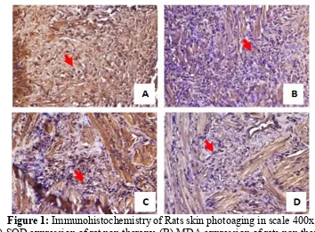
Figure 1: Immunohistochemistry of Rats skin photoaging in scale 400x (A) SOD expression of rat non therapy, (B) MDA expression of rats non therapy
(C) SOD expression of rat therapy, (D) MDA expression of rats therapy.
The expression of SOD and malondialdehyde (blue color) was indicated by red arrow that in Figure 1. UVB exposure on the rat skin led to increased ROS levels in cells. UVB initiated iron-driven Fenton reaction with subsequent generation of hydroxyl radical and lipid peroxidation end products such as malondialdehyde [11]. In non-therapy group, malondialdehyde expression average of 1.040 ± 0.205. This result compared with the expression therapy group average of 0.365 ± 0.133, malondialdehyde expression decreased by 64.90%. High levels of free radicals in the skin were indicated by high levels of malondialdehyde as lipid peroxidation marker. Extract Aloe vera contains flavonoids (isorhamnetin) acts as an antioxidant which prevented overproduction of ROS that detecting by decreasing malondialdehyde expression.
CONCLUSION
Aloe vera extract had potentially used as topical treatment of photoaging by reducing MDA expression and increasing SOD expression excessively on the UVB-induced skin photoaging in hairless rats.
ACKNOWLEDGMENT
The author thanks to Biochemistry Laboratory, Department of Chemistry, Faculty of Mathematics and Natural Sciences and Biochemistry Laboratory of Medicinal Faculty, Brawijaya University.
REFERENCES
12222-[4] Fisher, G. J., Z. Q. Wang, S. C. Datta, J. Varani, S. Kang, J. J. Voorhees, NEJM, 1997,
337(20), 1419-1428.
[5] Feng, X. X., X. T.Yu, W. J. Li, S. Z. Kong, Y. H. Liu, X. Zhang, Y. F. Xian, X. J. Zhang, Z. R. Su, Z. X. Lin, Eur. J. Pharm. Sci., 2014, 63, 113-123.
[6] Janero, D. R., Free Radical Biol Med, 1990, 9(6), 515-540. [7] Qadir, M. I., Int J Nat Ther,2009, 2, 21-26.
[8] R. Rajeswari, M. Umadevi, C. S. Rahale, R. Pushpa, S. Selvavenkadesh, K. P. S.
Kumar, D. Bhowmik, J Pharmacogn Phytochem, 2012, 1(4), 118-124.
[9] Sahu, P. K., D. D Giri, R. Singh, P. Pandey, S. Gupta, A. K Shrivastava, A. Kumar, K. D Pandey, Pharmacol Pharm, 2013, 4, 599-610.
[10] Pandel, R., B. Poljšak, A. Godic, R. Dahmane, ISRN Dermatol., 2013, 2013, 1-11. [11] Wenk, J., P. Brenneisen, C. Meewes, M. Wlaschek, T. Peters, R. Blaudschun, W.Ma, L.