P-ISSN.2503-0817, E-ISSN.2503-0825
CrossMark
Abstract
Objective: Clinically speaking, parotid gland tuberculosis is a rare case. Its diagnostic values are hard to determine as it has clinical similarities with neoplasms. Mistakes during diagnosis happen quite often thus additional layer of examinations are required to be conducted.
Methods: An 18-year-old woman was referred from a privately-owned hospital with initial diagnosis of infected parotic sinistra tumor. Physical examination discovered a swelling on her sinistra parotid gland and a lump at the collisinistra region. Ultrasound scan showed benign hypoechoic areas and cystic regions at the left of submandibular muscle which then expanded towards the neck. Results from the
PPD5TU (-), BTA 3X (1X positive) and FNAB pointed out an existing a/r parotid and collisinistra or tuberculous abscess.
Results: The diagnosis of parotid gland tuberculosis is often established after conducting superficial parotidectomy. The patient managed to be cured with OAT distribution and no recurrences were recorded afterward.
Conclusion: Parotid gland tuberculous abscess is rarely found in clinics. Tuberculosis examination (including PPD5TU and BTA) and FNAB need to be conducted to prevent unnecessary treatment and medical operations.
Keywords: Parotid abscess, Tuberculosis, Neoplasms
Cite this Article: Bustomi AF, Sylvyana M, Syamsudin E, Rizki KA. 2016. Parotid gland tuberculous abscess. Journal of Dentomaxillofacial Science 1(3): 185-189. DOI:10.15562/jdmfs.v1i3.314
Parotid gland tuberculous abscess: a case report
Ahmad F. Bustomi,1*Melita Sylvyana,2Endang Syamsudin,2Kiki A. Rizki3
Introduction
Tuberculosis is a granulomatousin flammation process caused by a mycobacterium tuberculosis (the human or bovine type) infection which have affected 1/3 of the world’s human population. In developing countries, 75% of the tuberculosis cases affected people on their productive age, starting from 15 to 50 years old.1
The number of tuberculosis incident in Indonesia on 2009 reached 228 cases per 100.000 people or roughly 528.063 tuberculosis cases including all of its types and variations. New tuberculosis cases with positive BTA reached 102 out of 100.000 residents or approximately 236.029 new cases with a mortality rate that approached 39 per 100.000 people or at least 250 victims per day.2
Tuberculosis is a disease with an expansive infection area and it also possesses various clinical descriptions. Pulmonary organs are often involved in the infection. The extra pulmonary form of this disease is the 20% of all active tuberculosis cases and it can be found in kidneys, bones, meninges and the lymph gland.3,4
Parotid gland is one of saliva glands which is rarely infected by tuberculosis with only less than 1% of the extra pulmonary tuberculosis cases inci-dence. Chronic parotid gland infection is often caused by salivary gland stones or constrictions in the gland channel.5,6
Parotid tuberculosis clinically portrayed by an existence of a mass which grows slowly and is
difficult to be differentiated with a neoplasm. A diagnosis that tends to a parotid tuberculosis has to be suspected, especially in developing countries. The non-existent history or record of lung tubercu-losis and all of the symptoms which correlate with tuberculosis hinders the production of valid diag-nosis of the disease.3
Parotid gland tuberculosis diagnosis is usually established after conducting a superficial parotidec-tomy. In this parotid gland tuberculosis abscess, the diagnosis is established following PPD5TU, TA and fine needle aspiration biopsy (FNAB) examination sequences.7
Case Report
As retrieved from the anamnesis, there was an 18-year-old female patient who admitted herself at the ER of Dr. Hasan Sadikin Hospital in Bandung with complaints that include a bump on her left cheek. 21 days prior to the patient’s admission to the hospital, she complained about a throbbing ache that occurred at her back teeth located on the left side of her lower jaw. 18 days before the admission a bump surfaced on the front-side of her left cheek that starts from the size of a marble before growing into the size of an egg. A swelling starts to emerge under her cheek and the left side of her neck which was followed by a numb sensation at the left side of her haw. The patient admitted herself for a treatment 1Department of Oral and
Maxillofacial Surgery, Faculty of Dentistry, Padjadjaran University, Bandung, Indonesia
2Department of Oral and Maxillofacial Surgery, Faculty of Dentistry, Padjadjaran University, Hasan Sadikin Hospital, Bandung, Indonesia
at a clinic in the Cibaduyut sub-district area and she was given three types of medicine (however,
by the doctor yet the swelling did not stop. 14 days before her admission at the hospital the patient experienced difficulties in opening her mouth and was only able to consume a scarce number of food and drinks. 4 days before the admission the patients visited the clinic yet again and she was referred to a privately-owned hospital in which she was given a rehydration treatment, intra vena antibiotic, roent-gen to her jaw, ultrasound scan and was treated for a day before forcing to stop the treatment due to financial issue. One day before her admission at the Dr. Hasan Sadikin hospital the patient complained that she was having difficulty and pain in swallowing despite no breathing difficulties were discovered. No on going cough was recorded yet a history of having an interaction with a partner who has a history of an ongoing cough was recorded and the patient has lost approximately 3 kg of body weight during the 18 days since the lump emerged.
Examination over the patient’s vital signs shows that the tachypnea (respiration 22 times/minute), hypothermia temperature etc. are on its normal level.
Local status of the patient’s extra oral appear-ance shows an asymmetrical face with a lump at the sinistra pre-auricular area with 6x6x1.5 cm in size and hard consistency while the pinna of her ear appeared to be lifted, fixated, diffusely bordered, yet no fluctuation, afebrile temperature and the color are similar with the surrounding surface. There is a swelling at the sinistra subman-dibular area that expanded until the sinistracolli lateral area with 5x3x1 cm in size, hard consistency, diffusely bordered, no fluctuation, pain when pressured, while the afebrile temperature and color was redder than surrounding tissue and it appears to be a fistula on the sinistracolli lateral area figure 1.
Intra oral examination discovered mouth opening limited with only 2 cm, even though there were no abnormality on the gingiva, lips, vestibule, tongue, palate, the base of the mouth, bucal mucosa or even the tonsil. Figure 2 Dental status discovered a gangrenous pulp at tooth 36 while 28, 38 and 48 tooth have not yet erupted figure 3.
Referring to lab test result, routine blood exam-ination shows low level of hemoglobin/anemia (11. 8 g/dL), low level of leukocytosis (11.400/mm3)
and thrombocytosis (476.000/mm3) while blood
hematology examination shows normal level of values (PT: 13.2; INR: 1.04; APTT: 27.2). Clinical chemical examination shows normal level of several values although calcium appears to be in an increase (5.7 mEq/L).
Blood gas level test shows a partially-compen-sated respiratory alkalosis with pH value of 7.479;
Figure 1 Extra oral profile: a swelling appears at the parotid and submandibular area, while a fistula emerged at the sinistra colli lateral area
Figure 2 Intra oral photo shows no abnormalities while the mouth opening was only 2 cm
Figure 3 Dental status, there is a gangrenous pulp of tooth 36
A panoramic photo shows a profound caries of tootth 36 and impacted of teeth 28, 38 and 48.
Figure 4 parotid sinistra ultrasound scan result shows a hypoechoic area which appears to be portraying a benign mass while the left submandibular area uscle appears to be having a cystic abscess suspect which has expanded towards the neck figure 5.
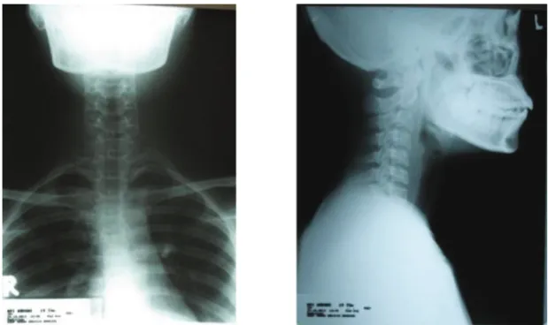
Thorax x-ray image and STL AP-Lateral pictures were examined at Dr. Hasan Sadikin Hospital. The thorax image shows no active lung tuberculosis and there are no cardiomegaly to be found. Figure 6 STL AP-Lateral pictures also appeared to be showing normal limit condition figure 7
Our patient was initially diagnosed with an infected parotid tumor suspect along with a chronic apical periodontitis including gangrenous pulp of tooth 36 with a comparative diagnosis of gland tuberculosis abscess, lymphadenopathy tuberculo-sis and parotid abscess including odontogen.
The treatment methods which was conducted to the patient at the emergency room include the administration of 3 L oxygen/minute, installation of IVFD RL with medium level of rehydration, 67 gtt/minute, installation of urine catheter with initial 150cc of urine, tea-like in color and intra vena medicine administration including one gram of ceftriaxone, infusion of 500 mg metronidazole, one ampoule of ranitidine and one ampoule of keterolac.
We admitted the patient into a regular treatment room with medium rehydration level and adminis-trated her with some intra vena medicine (including 2x1 gr ceftriaxone, 3x500 mg metronidazole infusion, 2x1 ampoule of ketorolac and 2x1 ampoule of ranitidin). We have planned to extract the patient’s teeth number 36 which was the focus point of the infection and also to conduct a FNAB examination at the treatment roomfigure 8.
Histologic examination was conducted at two separate locations. A smear was conducted at the collisinistra area while a Fine Needle Aspiration Biopsy (FNAB) was done at the parotid sinistra area. The results retrieved from the smearing include PMN lymphocyte cells, histiocytes, squa-mosal cell with a normal level core, epitheloid cell and datia langhans cell. A necrosis was discovered while no malignant tumor cell was found. The result from parotid FNAB shows necrotic mass, lymphocyte cell and epitheloid cell and no malig-nant tumor cell was found as well. The conclusion thus made was that it was a parotid a/r abscess and a tuberculous collisinistra.
Our patient were then referred to the internal disease department to receive proper treatment for her tuberculosis. The tuberculosis examination
Figure 5 Parotid sinistra ultrasound scan result: hypoechoic area appears to be benign, while the left submandibular area muscle appears to be having a suspected cystic abscess suspect which had expanded towards the neck
sequences, including PPD5TU and sputum BTA, showed a positive result. The medication which were given include I category OAT + B6 (450 mg rifampicin, 300 mg INH, 1500 mg pyrazinamide and 1000 mg ethambutol).
After receiving a month of OAT poly DOT treatment at the Dr. Hasan Sadikin Hospital in Bandung, a follow up physical examination shows that the swelling at the parotid, submandibular and collisinistra area have disappeared. The result from 3 times of sputum BTA examination showed a
Discussion
Extra pulmonary tuberculosis occurred on at least 25% of all tuberculosis morbidity cases. The most common variant of the extra pulmonary tubercu-losis is the lymph gland tubercutubercu-losis and its other forms include pleural, skeletal, central nervous system, abdominal, genitourinary, military and pericarditis tuberculosis. In developing countries where tuberculosis patients are often found, parotid gland tuberculosis often emerged as a primary parotid gland tuberculosis.5,6
Parotid gland tuberculosis is another from of the extra pulmonary tuberculosis which is rarely to be found and it could occur with two different ways of infection. The first kind is initiated through a dental, tonsil tissue or auto inoculation sputum infec-tion which reached the parenchyma or lymphatic parotid gland infection through lymphatic afferent or gland canal. The second variant is infected by pulmonary metastasis from the hematogenous or lymphatic canal.3,7
Clinically speaking, parotid gland tuberculosis infection could occur in two different ways. The first one is through an acute inflammation lesion with diffusely edema gland which can be mistaken for an acute sialadenitis or an abscess. The second form is a chronic lesion which emerged as a slow bodily mass that appears similar with a tumor and most of the time it cannot be easily differentiated with parotid tumor or the parotid gland tubercu-losis could also occur together with the parotid tumor. The diagnosis of parotid gland tuberculosis thus become difficult especially when there are no pulmonary disease clinical and systemic symptoms were found during the examination.3,5,7,8
In our case, the extra pulmonary tuberculosis displayed a complete abscess formation at the parotid, submandibular and collisinistra along with fistula drainage at the collisinistra area. There were no abnormally found during pulmonary examina-tion and no ongoing cough history was recorded as well thus the parotid tuberculosis abscess on our patient infected her through sputum autoinocula-tion which was infected by the tuberculosis and it has reached the parotid gland through lymphatic afferent or through the gland canal.
Ultrasound scan was the first option which was used as a modal to evaluate the abnormality of patient’s parotid gland.5
Ultrasound scan results on our patient showed a benign hipoechoic area while at the left submandibular muscle a cystic abscess suspect appeared and it has expanded to her neck. This finding thus became the foreground for our diagnosis saying that there is an infected parotid sinistra tumor suspect with parotid tuberculosis
Figure 7 STL AP-Lateral picture shows nothing out of ordinary
Figure 8 The extraction of tooth 36 which was the focus of the infection
In order to establish a definitive tuberculosis diagnosis tuberculin examination using Purified Protein Derivative 5 Tuberculin Units (PPD5TU), BTA and FNAB were conducted. A negative result from the tuberculin test could be retrieved from patients who have never been infected by tubercu-losis. The test result will turn positive 3 to 5 weeks after the infection occured.3
The PPD5TU result retrieved from our patient was negative because the examination was conducted too early after the infection thus the response has not transformed into a positive one. The swelling at the left parotid area was experienced by the patient approximately 18 days before her admission to the hospital. The BTA examination only resulted in one positive result from three attempts which were conducted.
Biopsy and surgical operations were the go to procedure which could be conducted in order to establish a comparative diagnosis over a chronic parotid gland disease. FNAB is a simple and effi-cient procedure to diagnose a tuberculosis infection compared to core needle biopsy or excision biopsy, and it could be conducted not only during the early stages but also as a follow up for patients who have received treatment with anti-tubercular regiment. At parotid area, FNAB possesses a degree of sensi-tivity that reaches 81 to 100% and specificity of 94 to 100% thus FNAB has always been conducted as the first method in evaluation masses at the parotid area.3,9
The final diagnosis for our patient, referring to the result of the FNAB examination, is a sinistra parotid tuberculous abscess thus the patient were given category I OAT medication which include 450 mg of rifampicin, 300 mg isoniazid, 1500 mg pyrazinamide and 1000 mg ethambutol. After receiving those medication the swelling which was previously located at parotid, submandibular and collisinistra area managed to be cured without any reoccurrence reported.
Conc
l
usion
Parotid gland abscess caused by a tuberculosis infection is a case that rarely found at a clinical level. Its symptoms which are similar to neoplasms and the non-existent symptoms of tuberculosis caused several difficulties in establishing proper diagno-sis for the patient. Parotid gland abscess caused by tuberculosis has to always be referred to as a comparative diagnosis for patients who have a soli-tary mass at their parotid glands in order to prevent unnecessary surgical operation and treatment.
C
on
fli
ct o
f I
nt
ere
st
The authors report no conflict of interest.
Re
f
erences
1. Maharjan M. Incidence of Tuberculosis in Enlarged Neck
Nodes, Our Experience. Kathmandu University Medical Journal. 2009;7(1): 54-8.
2. LaporanSubdit TB Depkes RI. SituasiEpidemiologi TB
Indonesia. Available from: http://www.tbindonesia.or.id/
pdf/ Data_tb_1_ 2010.pdf
3. Birkent H. Primary parotid tuberculosis mimicking
parotid neoplasm: a case report. Jurnal of Medical Case
Reports.2008; 2:62.Available from:
http://www.jmedical-casereports.com/content/2/1/62.
4. Bannister B, Gillespie S. Infection Microbiology and
Management. Blackwell Publishing Ltd; 2006.
5. Lin CH, Chen MK. Mycobacterium tuberculosis
infec-tion within a parotid Warthin tumor: Magnetic resonance imaging appearance. International Journal of Case Reports and Images. 2013; ISSN-(0976-3198)
6. Ghorbani GA. Primary Tuberculous Abscess of the Parotid
Gland: A Case Report. Tanaffos. 2006; 5(1): 65-8. 7. Dixit R, Shah V. Tuberculous Abscess of Parotid Gland.
Jurnal Indian Academy of Clinical Medicine. 2005; 6(2): 161-3.
8. Carlson ER, Ord R.A. Textbook and Color Atlas of Salivary
Gland Pathology Diagnosis and Management. Iowa: Wiley-Blackwell; 2008.
9. Das DK. Fine-Needle Aspiration Cytology in the Diagnosis
of Tuberculous Lesions.Laboratory Medicine. 2000; 31(11): 625-30.