Sub-bowmAn KerAtomileuSiS ComPAred
with PhotorefrACtive KerAteCtomy –
ContrAlAterAl eye Study
dean Šarić1, mirna belovari višnjić2, iva Krolo1 and Zdravko mandić11university department of ophthalmology, Sestre milosrdnice university hospital Center, Zagreb; 2Čakovec
General hospital, Čakovec, Croatia
SummAry – The aim of this study was to compare two different refractive surgery approaches in correction of myopia with or without astigmatism. in patients where one eye underwent sub-bowman keratomileusis (SbK) and the other eye photorefractive keratectomy (PrK), the objective and subjective results were retrospectively compared during the six-month follow-up. eighty four patients (168 eyes) were involved in this retrospective contralateral study. The mean preoperative spherical refraction was -3.88 diopters (d) and the mean cylinder was -0.82 d for all eyes. each patient underwent SbK on one eye and PrK on the contralateral eye. The eyes in the PrK group underwent mechanical epithelial removal, which was followed by laser treatment. mitomycin C 0.02% was used for 15 seconds if ablation was deeper than 50 microns. in the SbK group, the intended 100-µm corneal flap was created with intralase femtosecond laser. All eyes underwent customized wavefront guided laser ablation using a viSX Star S4 ir excimer laser. Preoperative and postoperative outcome measures included best spectacle-corrected visual acuity, uncorrected visual acuity, corneal topography, contrast visual acuity, and anterior optical coherence tomography ima-ging. Patients were asked to complete subjective satisfaction questionnaires at each visit. Through the first 3 months of follow up, the SbK eyes demonstrated clinically and statistically better visual results than PrK eyes, between 3 and 6 months the results in the two groups began to equalize, and after 6 months of follow up there were no clinical and statistical differences between the SbK and PrK groups. SbK seems to be more practical for the patient with less pain, faster visual recovery, fewer medications, and overall superior experience.
Key words: Sub-Bowman keratomileusis; Photorefractive keratectomy
Correspondence to: Dean Šarić, MD, university department of ophthalmology, Sestre milosrdnice university hospital Center, vinogradska c. 29, hr-10000 Zagreb, Croatia
e-mail: bbb.dean@usa.net
received february 22, 2012, accepted June 1, 2012 Introduction
in the last twenty years, two major approaches have become relevant in laser vision correction: lASiK (la-ser in situ keratomileusis) and advanced surface ablation techniques, i.e. photorefractive keratectomy (PrK), la-ser epithelial keratomileusis (lASeK) and epi-lASiK, of which the PrK method is most commonly used1-5.
lASiK offers several advantages over PrK in-cluding faster clinical and functional rehabilitation, minimal postoperative pain, subepithelial scarring avoidance, reduced need for prolonged steroid ther-apy, reduced risk of infectious keratitis, less irregular astigmatism, and stable refraction with predictable outcomes shortly after the procedure. limitations of this method include possible difficulties in the use of microkeratome, difficulties in flap repositioning and deeper corneal stromal ablations (120-140 µm greater than for PrK), which can theoretically weaken the corneal tectonics1,6-10.
Some surgeons prefer PrK method because it pro-vides better visual quality, less dry eye problems and less higher order aberrations, with the possibility of correcting refractive anomalies in patients with thin-ner cornea. Pain and discomfort of the eye are much more pronounced in PrK than in lASiK. Also, achieving final visual acuity and patient recovery is much slower11-13.
Thin flap lASiK/Sub-bowman keratomileusis (SbK) is a refractive surgical procedure that combines the advantages of lASiK and PrK, quick and pain-less recovery as in lASiK while saving tissue with thinner flap compared to conventional lASiK. Theo-retically, SbK should have more benefits for patients: a predictable thin flap, quicker visual recovery with minimal pain and discomfort, minimal biomechanical changes with fewer higher order aberrations, reduced incidence of postoperative dry eye, and a decrease in the loss of corneal sensitivity14.
The aim of this study was to compare two different refractive surgery approaches in correction of myopia with or without astigmatism. in patients where one eye underwent SbK and the other eye PrK, objective and subjective results were compared during the six-month follow-up.
Patients and Methods
A contralateral retrospective study of 84 patients (168 eyes) was conducted in the period from April 2008 until november 2010. The study included 44% of men and 56% of women. The mean patient age was 32.14±6.22 years. two groups were formed, SbK and PrK groups. each patient underwent SbK on one eye and PrK on the contralateral eye. All patients signed the informed consent form before entering the study. The inclusion criteria were spherical myopia up to -6.00 diopters (dsph) with up to -2.50 diopters of refractive astigmatism (dcyl), stable refraction for 1 year, best corrected visual acuity of at least 20/20 on each eye, and an average central corneal thickness be-tween 470 and 510 µm. The exclusion criteria were corneal topographic pattern suggestive of ectatic dis-ease, disease status that could delay the healing pro-cess of the cornea, and previous trauma or surgical procedure on the eye. Soft contact lens wearers were required to discontinue lens use for at least 10 days
be-fore surgery, whereas rigid contact lens wearers were required to discontinue use for at least 4 weeks before surgery. in the SbK group, flaps were created using an intralase femtosecond laser (60 khz, Advanced medical optics, Santa Ana, CA). intended flap thick-ness was 100 µm. in the PrK eyes, corneal epithelium was mechanically removed, subsequently undergoing laser treatment. mitomycin C 0.02% was used for 15 seconds if ablation was deeper than 50 microns. mitomycin C is an antineoplastic agent that inhib-its dnA and rnA replication and protein synthesis. when applied on the cornea, mitomycin C regulates fibroblast proliferation and myofibroblast formation, which is responsible for corneal haze after PrK15-18.
in both cases, laser correction was performed with the wavefront Customvue method using viSX Star S4 excimer laser. Preoperative and postoperative out-come measurements included the following: deter-mination of uncorrected visual acuity (uCvA) and best spectacle corrected visual acuity (bSCvA) using Snellen charts, corneal topography (oculus Penta-cam), wavefront aberrometry (Advanced Customvue wavefront wavescan), contrast visual acuity (Peli-robson letter chart), tear secretion measurement (Schirmer test) and anterior optical coherence tomog-raphy (oCt) imaging (SoCt Copernicus, optopol technology S.A., Zawiercie, Poland).
Pentacam Comprehensive eye Scanner (oculus, inc., lynwood, wA) provides an accurate three-di-mensional view of anterior eye segment and individu-alized approach to refractive surgery using rotation Scheimpflug camera in all meridians. This includes an objective determination of corneal topography (eleva-tion maps), overall pachymetry, tomographic analysis, 3d analysis of anterior chamber, measuring lens den-sity, and intraocular lens (iol) calculation after cor-neal refractive surgery. measurement is non-contact and entirely agreeable to the patient19,20.
Advanced Customvue wavefront wavescan pro-cedure allows for more precise adjustment of higher order aberrations that cannot be removed with eye-glasses or contact lenses, and have a significant impact on the quality of vision. higher order aberrations are unique to each person and include the difficulty see-ing at night, glare, halos, blurrsee-ing, starburst patterns and double vision (diplopia). waveScan-based digital technology identifies and measures imperfections in
the eye 25 times more precisely than standard meth-ods. waveScan software translates the information into a set of Customvue treatment instructions for the laser. These digital treatment instructions are then transferred to the laser, driving a new level of preci-sion and accuracy21,22.
Spectral-domain optical coherence tomography (SoCt Copernicus, optopol technology S.A., 42-400 Zawiercie, Poland) provides analysis of the eye anterior segment with a resolution of 3 microns. it is used to record pachymetry maps, measure corneal epi-thelial thickness, lASiK flap thickness, and anterior chamber angle23.
All patients were examined preoperatively and on follow-up examinations 1 day, 1 week, 1, 3 and 6 months after surgery. Patients were also required to complete a subjective questionnaire about ocular pain, eye dryness and satisfaction with vision. Pain and dry-ness were evaluated on a scale from 0 to 3, provided that 0 denotes absence of pain and dryness of eyes, 1 mild, 2 moderate, and 3 extreme soreness and dryness of the eyes. Patients were asked to assess their vision on each eye as poor, reasonable, good or excellent. The goal of all surgeries was emmetropia. no intra- or postoperative complications were encountered except for one patient who needed enhancement surgery on both eyes (one SbK and the other PrK).
Results Flap thickness
All flaps were measured with an oCt system at 1 month postoperatively. The results demonstrated that the SbK flaps had a mean thickness of 104±5 µm.
Standard deviation for each individual flap was 5 µm. Visual acuity results
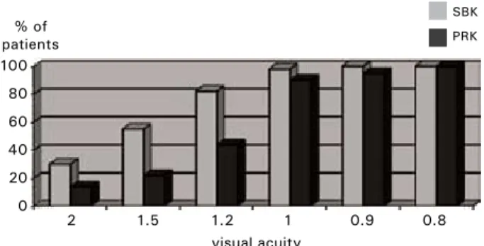
results are summarized in figures 1-3. figure 1 shows uCvA 1 month after surgery. uncorrected vi-sual acuity at 1 month showed a statistically signifi-cant difference between the SbK and PrK groups, with 98% of the SbK eyes at 1.0 or better compared to 80% of the PrK eyes (P<0.0001). figure 2 shows uCvA 2 months after surgery. uncorrected visual acuity at 2 months still showed a statistically signifi-cant difference between the SbK and PrK groups, with 98% of the SbK eyes at 1.0 or better compared to 90% of the PrK eyes (P<0.0001). figure 3 shows 100 50 0 2 1.5 1.2 1 0.9 0.8 % of patients visual acuity SBK PRK
Fig.1. Snellen visual acuity one month after surgery.
% of patients 100 80 60 40 20 0 SBK PRK 2 1.5 1.2 1 0.9 0.8 visual acuity
Fig. 2. Snellen visual acuity two months after surgery. % of patients 100 80 60 40 20 0 SBK PRK 2 1.5 1.2 1 0.9 0.8 visual acuity
Fig. 3. Snellen visual acuity three months after surgery.
90 80 70 60 50 40 30 20 10 0 % of patients
Fig. 4. Patient satisfaction with vision.
SBK PRK
1 2 3
no statistically significant difference between the two groups at visual acuity of 1.0 three months after sur-gery (P=0.4751).
Contrast visual acuity
Contrast visual acuity showed better results for SbK eyes up to 3 months after surgery, although the difference decreased over time. There was no differ-ence between the two eyes six months after surgery.
Subjective results
PrK eye was more painful for up to one month. on later examination, there was no statistically sig-nificant difference. The feeling of dryness was signifi-cantly greater on PrK eyes for up to 3 months after surgery. At 6-month follow up there was no differ-ence. Patients were more satisfied with vision on the SbK eye for up to 3 months after surgery. we found no statistically significant difference in satisfaction with vision six months after surgery (fig. 4).
Discussion
There are a great number of studies comparing PrK with SbK results, but not so many where these two methods were compared in the same patient. du-rrie et al. performed a study using different methods on each eye14. They showed advantages of SbK as a meth-od in refractive surgery over PrK. Slight differences were recorded in several measured results but the fact that SbK eyes performed better in the beginning and that the differences equalized over 3 to 6 months was the same. They also conclude that SbK provides faster visual recovery while providing end results similar to PrK. SbK seems to be more practical for the patient with less pain, faster visual recovery, fewer medica-tions, and an overall superior experience.
References
1. herSh PS, brint Sf, mAloney rK, et al. Photo-refractive keratectomy versus laser in situ keratomileusis for moderate to high myopia: a randomized, prospective study. ophthalmology 1998;105:1512-22.
2. yAnG XJ, yAn ht, nAKAhori y. evaluation of the effectiveness of laser in situ keratomileusis and photorefrac-tive keratectomy for myopia: a meta analysis. J med invest 2003;50:180-6.
3. neerACher b, Senn P, SChiPPer i. Glare sensitivity and optical side effects 1 year after photorefractive keratec-tomy and laser in situ keratomileusis. J Cataract refract Surg 2004;30:1696-701.
4. PirouZiAn A, thornton J, nGo S. one-year out-comes of a bilateral randomized prospective clinical trial comparing laser subepithelial keratomileusis and photore-fractive keratectomy. J refract Surg 2006;22:575-9. 5. durrie dS, KeZiriAn Gm. femtosecond laser versus
mechanical microkeratome flaps in wavefront-guided laser in situ keratomileusis; prospective contralateral eye study. J Cataract refract Surg 2005;31:120-6.
6. PAlliKAriS iG, SiGAnoS dS. excimer laser in situ keratomileusis and photorefractive keratectomy for correction of high myopia. J refract Corneal Surg 1994;10:498-510. 7. fArAh SG, AZAr dt, GurdAl C, wonG J. laser
in situ keratomileusis: literature review of a developing tech-nique. J Cataract refract Surg 1998;24:989-1006.
8. helmy SA, SAlAh A, bAdAwy tt, SidKy An. Photorefractive keratectomy and laser in situ keratomileusis for myopia between 6.00 and 10.00 diopters. J refract Surg 1996;12:417-21.
9. KnorZ mC, wieSinGer b, liermAnn A, et al. lASiK for moderate and high myopia and myopic astigma-tism. ophthalmology 1998;105:932-40.
10. mAldonAdo-bAS A, onniS r. results of laser in situ keratomileusis in different degrees of myopia. ophthalmol-ogy 1998;105:606-11.
11. rAJAn mS, JAyCoCK P, o’brArt d, et al. A long-term study of photorefractive keratectomy: 12-year follow-up. ophthalmology 2004;111:1813-24.
12. rAndlemAn Jb, loft eS, bAnninG CS, et al. out-comes of wavefront-optimized surface ablation. ophthalmol-ogy 2007;114:983-8.
13. ChunG Sh, lee iS, lee yG, et al. Comparison of higher-order aberrations after wavefront-guided laser in situ keratomileusis and laser-assisted subepithelial keratectomy. J Cataract refract Surg 2006;32:779-84.
14. durrie dS, SlAde SG, mArShAll J. wavefront-guided excimer laser ablation using photorefractive keratec-tomy and sub-bowman’s keratomileusis: a contralateral eye study. J refract Surg 2008;24:S77-84.
15. dAnShiitSoodol n, de Pinho CA, mAtobA y, KumAGAi t, SuGiyAmA m. The mitomycin C (mmC)-binding protein from mmC-producing microorganisms pro-tects from the lethal effect of bleomycin: crystallographic analysis to elucidate the binding mode of the antibiotic to the protein. J mol biol 2006;360:398-408.
16. tomASZ m. mitomycin C: small, fast and deadly (but very selective). Chembiol1995;2:575-9.
17. renAultJ, bAron m, mAilliet P, et al. heterocy-clic quinones.2.Quinoxaline-5,6-(and 5-8)-diones – potential antitumoral agents. eur J med Chem 1981;16:545-50.
18. mAo y, vAroGlu m, ShermAn dh. molecular characterization and analysis of the biosynthetic gene cluster for the antitumor antibiotic mitomycin C from Streptomyces lavendulae nrrl 2564. Chem biol 1999;6:251-63. 19. tAKACS Ai, mihAltZ K, nAGy ZZ. Corneal density
with the Pentacam after photorefractive keratectomy. J re-fract Surg 2011;27:269-77.
20. roChA Km, KAGAn r, Smith Sd, KrueGer rr. Thresholds for interface haze formation after thin-flap femto-second laser in situ keratomileusis for myopia. Am J ophthal-mol 2009;147:966-72.
21. hoSny m, AwAdAllA mA. Comparison of higher-or-der aberrations after lASiK using disposable microkeratome 130 and 90 micron heads. eur J ophthalmol 2008;18:332-7. 22. AlmAhmoud t, munGer r, JACKSon wb.
Ad-vanced corneal surface ablation efficacy in myopia: changes in higher order aberrations. Can J ophthalmol 2011;46:175-81. 23. KAluZny bJ, KAŁuZny JJ, SZKulmowSKA A,
GorCZyŃSKA i, SZKulmowSKi m, bAJrASZe-wSKi t, woJtKobAJrASZe-wSKi m, tArGobAJrASZe-wSKi P. Spectral optical coherence tomography: a novel technique for cornea imaging. Cornea 2006;25:960-5.
Sažetak
lASiK ultrAtAnKoG PoKloPCA (SuB-BowMan KeratoMileuSiS) ili fotorefrAKtivnA
KerAteKtomiJA
D. Šarić, M. Belovari Višnjić, i. Krolo i Z. Mandić
Cilj ove studije bio je tijekom šestomjesečnog praćenja bolesnika usporediti subjektivne i objektivne rezultate korekcije miopije s astigmatizmom očiju ili bez njega podvrgnutih dvama različitim zahvatima: refrakcijskoj kirurgiji ultratankog poklopca (sub-Bowman keratomileusis, SbK) ili fotorefraktivnoj keratektomiji (photorefractive keratectomy, PrK). osamde-set četiri bolesnika (168 očiju) su bila uključena u ovu retrospektivnu studiju. Prosječna kratkovidnost bila je -3,88 sfernih dioptrija s prosječnim astigmatizmom od -0,82 cilindrične dioptrije. u svakog bolesnika je primijenjen SbK na jednom oku i PrK na drugom oku. rožnični epitel je mehanički uklonjen na očima iz skupine PrK, nakon čega je učinjen la-serski zahvat. Zatim je apliciran mitomicin C 0,02% (vrijeme ekspozicije 15 sekunda) ako je ablacija bila dublja od 50 mikrona. Kod očiju iz skupine SbK formiran je ultratanki poklopac od 100 mikrona pomoću intralase femtosekundnog lasera. laserska korekcija je u oba slučaja izvršena metodom wavefront CustomVue laserom viSX Star S4. Prijeoperacijska i poslijeoperacijska izlazna mjerenja uključivala su određivanje vidne oštrine (nekorigirane i najbolje korigirane), rožnič-nu topografiju, aberometriju, test kontrastne osjetljivosti i optičku koherentrožnič-nu tomografiju prednjega očnog segmenta. bolesnici su na svakom kontrolnom pregledu ispunjavali upitnik o subjektivnoj procjeni rezultata. Klinički i statistički su značajno bolji rezultati bili u skupini očiju operiranih metodom SbK i to poslijeoperacijski do trećeg mjeseca. od trećeg do šestog mjeseca su se rezultati počeli izjednačavati te nakon 6 mjeseci praćenja više nije bilo statistički i klinički značajnih razlika između dviju skupina očiju. metoda SbK je objektivno i subjektivno bolja metoda u odnosu na PrK, te bolesniku omogućuje brži oporavak uz manje nuspojava.